
Abstract
Purpose
Nowadays, in order to terminate biofilm associated infections, coating of particular biomaterial surfaces with particular substances, via some nanotechnological tools, is being applied. Therefore, in the present study, investigation of anti-biofilm effects of nanometer scale silver (NmSAg) coatings on glass and polystyrene surfaces against clinical strains of Proteus mirabilis, Candida glabrata and Candida tropicalis was aimed.
Methods
In this study, glass and polystyrene slabs with 1.5 cm × 1.5 cm × 0.3 mm dimensions were cleaned by using surface plasma technology, covered with NmSAg by using a physical vapor deposition machine, and biofilm inhibition was determined by crystal violet binding assay.
Results
According to our results, 32 nm of silver layer on a glass slab decreased biofilm formation of P. mirabilis strain to a maximum amount of 88.1% and caused 20.9% inhibition in biofilm formation of C. glabrata strain. On the other hand, NmS coating of Ag on a polystyrene slab caused 34.4% and 20% inhibitions, respectively, in biofilm formations of C. glabrata and C. tropicalis strains. Although biofilm inhibition of NmSAg layer on polystyrene slab was more (34.4%) than biofilm inhibition caused by NmSAg layer on glass slab (20.9%), C. glabrata strain's biofilm formation on uncoated glass slab was lower than both uncoated and NmSAg-coated polystyrene slabs.
Conclusions
Our results show that glass surfaces with NmSAg coatings can be used as a new surface material of various indwelling devices on which P. mirabilis colonizations frequently occur and in order to avoid C. glabrata-associated biofilm infections, it is more useful to choose a surface material of glass rather than choosing a surface material of polystyrene.
Introduction
Biofilms, which play a pivotal role in healthcare-associated infections, especially in those related to the implant of medical devices such as wound dressings, intravascular catheters, urinary catheters and orthopedic implants, are composed of exopolymeric substances, synthesized by microbial communities adhering to biotic or abiotic surfaces and, by means of forming these slimy structures, microorganisms can protect themselves from both immune defense and conventional antimicrobial agents (1-2-3-4-5-6-7). Therefore, antimicrobials become ineffective due to the impermeability of biofilm-forming microorganisms and, in order to terminate these kinds of infections, the implant must be removed or some surgical operations must be applied (8). Nowadays, Proteus mirabilis emerges as one of the main microorganisms forming biofilms on indwelling medical devices and some Candida species are also known as common causes of central venous catheter (CVC)-related infections (9). The difficulties in the treatment of microorganisms embedded in biofilms show that biofilm formation should be better inhibited before it is formed. Today, by means of being used in imaging, medical apparatus designing, sensor technologies, fabrics, cosmetics, health products and water remediation technologies, nanotechnology is regarded as a tool to prevent formation of these life-threatening biofilms on life-supporting devices (9, 10). Therefore, in the present study, the anti-biofilm effect of nanometer scale silver (NmSAg) coatings on glass and polystyrene surfaces against clinical strains of P. mirabilis, C. glabrata and C. tropicalis has been investigated.
Materials and Methods
Preparation of glass and polystyrene slabs for nanometer scale silver (NmSAg) coating
In this study, glass and polystyrene slabs with 1.5 cm × 1.5 cm × 0.3 mm dimensions were used and before coating them with silver in nanometer scale, they were all cleaned carefully in order to remove impurities. Therefore, they were all immersed inside three different flasks including acetone (99.5%), ethanol (96%) and high purity deionized water separately, and flasks were placed into ultrasonic bath for 10 minutes in order to complete mechanical cleaning of glass and polystyrene slabs. Afterwards, the slabs were purged with nitrogen gas (N2) spray in order to get rid of all impurities from slab surfaces. Following this, in order to remove final griminess found on slab surfaces, surface plasma technology was used. Hence, both glass and polystyrene slabs were placed into the plasma tunnel and the cap of the device was closed tightly. Afterwards, inlet air was taken out by letting vacuum pump start working and subsequent to dropping of pressure, vacuuming started to increase. Increasing of vacuum continued until the pressure gauge showed 0.2 bars and at this point N2 gas was purged into the plasma tunnel providing starting of radio frequency (RF) generator automatically, after which plasma phenomena occurred inside the tunnel. (Exposures of RF power were adjusted to 300 W with approximately 60 W reflected power.) After a few minutes, slabs were pulled out carefully and prepared for NmSAg coating.
Nanometer scale coating of glass and polystyrene slabs with silver
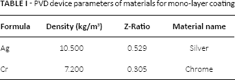
At this stage, each glass and polystyrene slab was covered with nanometer scale mono layer by the deposition of silver vapor. Hence, the cleaned slabs were completely fixed into the physical vapor deposition (PVD) machine holder and, as well as device's axis, its special holder was also revolved around the cabinet providing a homogeneous smooth surface silver coating on the slabs. Afterwards, extremely smooth and homogenous mono-layer silver (up to 10 nm) was deposited along the slab surface by PVD. In order to deposit silver on slabs by PVD machine, the deposition rate was adjusted to 0.1A°/sec and the deposition of cabin pressure was adjusted to 8 ± 2 × [10]^(-6) torr. In order to achieve stronger and smoother silver layers on glass and polystyrene slabs, chrome material was laminated between the slab surface and nanometer scaled silver layer. The parameters of materials for mono-layer coating on PVD device is given in Table I.
PVD device parameters of materials for mono-layer coating
Clinical strains
In this study, clinical strains of P. mirabilis, C. glabrata and C. tropicalis were collected from one hospital in Ankara, Turkey. Isolated strains were inoculated in to the brain heart infusion (BHI) broth media including 10% glycerol and stored at -20°C. Within these strains, clinical strain of P. mirabilis was identified in Sahal's previous study (11); whereas identification of C. glabrata and C. tropicalis strains were carried out by using CHROMagar medium.
Biofilm inhibition assay
The determination of biofilm inhibition was performed by crystal violet binding assay described by O'Toole, with some modifications (12). Briefly, microbial cells corresponding to a 2.0 McFarland optical density standard were inoculated into BHI broth medium and then were incubated at 37°C overnight. Afterwards, overnight culture was 1:100 diluted into a fresh BHI medium and NmSAg-coated slabs were added into different falcon tubes in which a particular diluted culture exists. The falcon tubes, including a particular slab and the diluted culture, were incubated for 48 h at 37°C. Following this, the medium was gently removed and the slabs were washed with distilled water. After allowing slabs to dry, each slab was stained with 1% crystal violet for 45 minutes at room temperature. Afterwards, the slabs were washed again with distilled water and by this means unbound crystal violet stain was removed. Finally, bound crystal violet in each well was solubilized by the addition of ethanol (96%) solution and solubilized crystal violet for each well was read by a spectrofotometer at 540 nm. Glass and polystyrene slabs at 1.5 cm × 1.5 cm × 0.3 mm dimensions, which are not coated with NmSAg, were used as controls and biofilm inhibition (%) was calculated using the following formula:
% of Inhibition = [(OD Control - OD Treatment)/OD Control] × 100
Results and discussion
Nowadays, increases in indwelling device-associated biofilm infections has gradually rendered traditional antimicrobial treatment ineffective and, in order to combat these infections, surfaces of some medical devices are being modified via nanotechnological tools (13). For instance, some kinds of nanoparticles (metallic, ceramic and metal) are being combined with some surface materials in order to obtain unique surfaces for biofilm inhibition (14). Besides, nanometer scale coating of surfaces with bactericidal/bacteriostatic substances can also be given as an another strategy to make biomaterial surfaces resistant to biofilm formation and, since heavy metal silver (Ag) is used as an anti-biofilm agent, coating of some particular biomaterial surfaces with Ag via some nanotechnological tools can also be used for generating new biofilm-resistant surfaces (15-16-17).
Since P. mirabilis strains are known as common causes of catheter-related urinary tract infections and glass can be used as a surface material of urinary catheters and bone substitute, anti-biofilm effect of NmSAg coatings against P. mirabilis strain were determined only on glass slab (18-19-20). Additionally, by means of extensive biofilm-forming ability of C. tropicalis strains on frequently used silicone and polystyrene latex materials, anti-biofilm effect of NmSAg coatings against C. tropicalis strain were determined only on polystyrene slabs (21-22-23), whereas anti-biofilm effect of NmSAg coatings against C. glabrata was determined both on glass and polystyrene slabs.
In this study, glass and polystyrene slabs with 1.5 cm × 1.5 cm × 0.3 mm dimensions were selected for nanometer scale (NmS) Ag coating and after the adjustment of PVD device, 32 nm of silver layer thickness was obtained on the slab surface. According to our results, NmS coating of Ag material on a glass slab decreased biofilm formation of P. mirabilis strain by a maximum amount and caused 88.1% inhibition on its biofilm formation (Fig. 1). In many studies, usages of Ag as an antimicrobial and anti-biofilm agents are being emphasized (10, 15, 18). However, a moderate benefit from Ag ions has been noted in many investigations because of their low dissolution rate (18). Therefore, this limited benefit is being improved by means of incorporating nanosilver and nanometer-sized Ag particles, because solubilized Ag ions constitute the bioactive form and nanometer-sized silver particles are indicated to increase silver dissolution in the surrounding liquid (18, 24). Similar to this information, in vitro data obtained in our study demonstrate that NmSAg coatings on glass surface have a strong biofilm inhibiting effect against the biofilm formation of P. mirabilis strains.
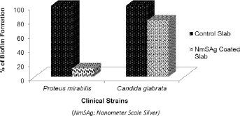
% Effect of NmSAg coated glass surfaces against biofilm formation of P. mirabilis and C. glabrata strains.
When we examined the other studies carried out on anti-biofilm activity of silver nanoparticles against different microorganisms, none were seen to have been carried out on the anti-biofilm effect of NmSAg coatings against P. mirabilis strains. However, according to one of the recent studies, inhibiting effects of silver nanoparticles (AgNPs) against the growth of Pseudomonas aeruginosa, and also lethal effects of these particles against the cells inhabiting in the matrix of biofilm were indicated. Therefore, they suggest that AgNPs can be embedded in medical devices in order to avoid microbial colonization and biofilm formation (25). In our study, in accordance with this suggestion, anti-biofilm effect of NmSAg-coated glass surfaces were used as a surface material and our results also show that NmSAg-coated glass surface can also be used as a surface material of various indwelling devices on which P. mirabilis colonizations frequently occur.
When the inhibition effect of NmSAg coating on glass slabs against C. glabrata strain's, biofilm formation was examined; 20.9% inhibition was observed (Fig. 1) and similar to our result, AgNPs are generally indicated to inhibit the adhesion of Candida to the denture surface according to the literature and are known to have an important role in preventing Candida-related biofilm formation in the oral cavity (26, 27).
In the second part of this study, the inhibition effect of NmSAg coating on polystyrene surfaces were examined and anti-biofilm effect of these surfaces against both C. glabrata and C. tropicalis strains were determined. According to our results, NmS coating of Ag material on a polystyrene slab caused 34.4% inhibition of biofilm formation in C. glabrata strains (Fig. 2). Whereas, a 20% decrease was observed in biofilm formation of C. tropicalis strain (Fig. 2).

% Effect of NmSAg coated polystyrene surfaces against biofilm formation of C. glabrata and C. tropicalis strains.
When the percentage of biofilm inhibition in C. glabrata strain is compared according to the surface material type, biofilm inhibition on NmSAg-coated polystyrene slab was more (34.4%) than the biofilm inhibition caused by NmSAg layer on glass slab (20.9%) (Fig. 3). However, although biofilm inhibition was more on NmSAg-coated polystyrene slab, biofilm formation of C. glabrata strains on uncoated polystyrene slab was stronger than its biofilm formation on uncoated glass surface (Fig. 4) confirming that less biofilm formation occurs on glass than other surfaces (28). Additionally, biofilm formation on NmSAg-coated polystyrene surface was also stronger than biofilm formation on uncoated glass surface (Fig. 4).
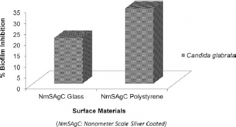
% of C. glabrata strain's biofilm inhibition on surface materials of NmSAg Coated Glass and NmSAg Coated Polystyrene.
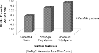
Biofilm formation of C. glabrata strain on surface materials of uncoated Glass, uncoated Polystyrene and NmSAg Coated Polystyrene.
Therefore, according to our findings, as a surface material of various indwelling devices such as urinary catheters, endotracheal tubes and prosthetic joints on which Candida colonizations frequently occur (29), the use of glass is better in order to avoid C. glabrata-associated biofilm infections. Besides, in order to increase biofilm inhibition on glass surfaces, NmSAg coatings can be applied on these surfaces. Additionally, if a surface material of polystyrene is used, application of NmSAg material on it will provide a particular inhibition in C. glabrata strains of biofilm formation.
To sum up, according to our results, a 32 nm coating of Ag layer on a glass slab decreased biofilm formation of P. mirabilis strain by 88.1% and also caused 20.9% inhibition in biofilm formation of C. glabrata strain. On the other hand, NmS coating of Ag material on a polystyrene slab caused 34.4% and 20% inhibition, respectively, in biofilm formations of C. glabrata and C. tropicalis strains. Besides, although biofilm inhibition on NmSAg-coated polystyrene slab was more (34.4 %) than the biofilm inhibition caused by NmSAg coating on glass slab (20.9%), C. glabrata strains of biofilm formation on uncoated glass slab was weaker than both uncoated and NmSAg-coated polystyrene slabs.
In conclusion, the generation of new novel biofilm-resistant biomaterials was the aim of this study and, in order to generate them, one of the most recent nanotechnological tools was used. In this respect, both glass and polystyrene slabs were coated with NmSAg layers and the anti-biofilm effects of this coating against P. mirabilis, C. glabrata and C. tropicalis strains were calculated. Our results show that, by means of inhibiting biofilm formation of P. mirabilis in huge amount, NmSAg-coated glass surfaces can be used as a new surface material of various indwelling devices on which P. mirabilis colonizations frequently occur. Additionally, the anti-biofilm effect of polystyrene surface against C. glabrata and C. tropicalis strains can be increased by the application of NmSAg coatings. Besides, glass was the best biomaterial, avoiding C. glabrata-associated biofilm formation.
Our study examined the anti-biofilm effects of NmSAg-coated biomaterials against P. mirabilis, C. glabrata and C. tropicalis strains. The findings should be of service to clinicians and scientists in many biomedical aspects.
Footnotes
Financial support: We did not have a financial support for the present study.
Conflict of interest: The authors declare that there are no conflicts of interest.
Meeting presentation: The data of this study have been presented at “20th International Symposium on Biomedical Science and Technology Meeting” on 29th of August 2014, in Koycegiz, Mugla, Turkey.