
Abstract
Purpose
The aim of this study was to investigate the color and structural changes of a maxillofacial silicone colored with 2 different pigments, after photoaging and immersion in disinfectants.
Methods
Ninety-six cylindrical specimens were fabricated and divided into 3 equal groups. The specimens of the first group consisted of unpigmented silicone (Multisil Epithetik), those of the second group consisted of unpigmented silicone, colored with red functional liquid pigment (Cosmesil Reactive 0.2% wt). The specimens of the third group were fabricated using unpigmented silicone colored with red powder pigment Cosmesil Dry at 0.2% wt. Specimens of each group were divided into 4 equal subgroups (immersed in soap solution, ethanol 95° or distilled water or placed in a photoaging apparatus for 174 hours). Structural changes were examined by infrared spectroscopy (ATR-FTIR) before and after aging. Color changes (ΔΕ*) were measured using the CIE L*a*b* system. Two-way ANOVA and Tukey's test for post hoc comparison were used at a = 0.05.
Results
Infrared spectroscopy showed no structural changes after immersion in solutions and photoaging, for all the materials tested. No statistically significant differences for ΔΕ* among the tested groups were found.
Conclusions
It can be concluded that no structural changes of pigmented and unpigmented silicone elastomers were observed among all aging procedures. Recorded color changes for the materials tested were within the limits of clinical acceptability after all aging procedures. Immersion in distilled water presented the best color stability, whereas photoaging, the poorest, for all materials.
Introduction
Maxillofacial prosthetics aim to restore missing or damaged tissues of the facial area. The destruction of facial tissues may be due to diseases, injuries or congenital deformations. Deficiencies in the neck and head may be treated using prostheses that simulate the human skin (1-2-3). The esthetic factor plays an important role, with direct effects on the psychology of the patient (4-5-6). Hence there is a need for advanced materials to fulfill the increased demands for this use. Silicones are the materials of choice in maxillofacial restorations because they can imitate human skin texture and appearance (7, 8). They can be colored intrinsically or extrinsically to simulate the color of human tissues (9). Environmental factors and habits of the patient, such as sunlight or various chemicals (cosmetics, cleaning agents etc.) can degrade the color of the prostheses (10-11-12-13). Furthermore, structural changes play a significant role in the properties of maxillofacial silicones (14).
Many studies (15-16-17-18-19-20-21) have been performed on the color stability of maxillofacial materials, investigating factors such as aging in a weathering chamber, disinfection solutions and type of pigment. Despite the fact that many research results seem controversial, the role of all these factors, separately or in combination, can be decisive for the color stability of maxillofacial silicones. Some studies (11, 22) have investigated the influence of UV radiation and accelerated aging on the structure of siloxanes.
The purpose of the present study was to investigate the color and structural changes of a maxillofacial silicone colored with 2 different types of pigment, after disinfection and photoaging. The null hypothesis tested was that no color or structural changes would occur in the different types of colored silicones after disinfection and photoaging.
Materials and Methods
Sample Preparation
An addition-type maxillofacial silicone and 2 types of red pigment were used. A total of 96 disc-shaped specimens of 16 mm in diameter and 3 mm in thickness were fabricated by placing the materials in Teflon molds, as described below. The specimens were divided into 3 equal groups (Tab. I). The specimens of group A (control group) consisted of the transparent silicone Multisil Epithetik (Bredent GmbH, Senden, Germany). The specimens of group B were fabricated by adding a liquid red silanized pigment (Cosmesil Reactive, Principality House, South Wales, UK) in the transparent silicone and the specimens of the group C by adding the red dry pigment Cosmesil Dry (Principality House, South Wales, UK) in the transparent silicone. The specimens of each group were divided into 4 equal subgroups (Tab. I) depending on the mode of disinfection and accelerating aging.
Groups and subgroups of tested materials and disinfection and photoaging procedures

For the specimens of group A, unpigmented Multisil Epithetik was mixed continuously clockwise in a plastic cup for 1 minute. Red liquid pigment (group B) and red dry pigment (group C) were added to the silicone at the mixing stage. The pigments had been previously weighted in a precision scale (AW 220, Shimadzu, Japan) to constitute 0.2% of the silicone weight.
Disinfection and Photoaging
Specimens of subgroups A1, B1 and C1 were immersed in 200-mL neutral pH soap solution (Johnson's Baby Top-to-Toe Wash; Johnson & Johnson, Italy); specimens of subgroups A2, B2 and C2 were immersed in ethanol 95°; and specimens of subgroups A3, B3 and C3 were immersed in distilled water. The specimens were immersed in the solutions for 1 week (10,080 minutes) at 23°C ± 1°C continuously, corresponding to a total time of 3 years for 10 minutes of immersion per day. Solutions were renewed daily. The specimens of subgroups A4, B4 and C4 were placed in an accelerated photoaging apparatus (Suntest CPS+; Atlas, Chicago, IL, USA) for 174 hours.
Infrared Spectroscopy
Each unset material was studied with Attenuated Total Reflectance - Fourier Transform Infra Red spectroscopy (ATR-FTIR) before mixing, employing a micro-MIR accessory (Perkin Elmer Corp., Norwalk, CT, USA) attached to an FTIR spectrometer (Spectrum GX; Perkin Elmer Corp.). Spectra were acquired under the following conditions: 4,000-400 cm−1 range, 4 cm−1 resolution, 60° para-edge KRS-5 minicrystal (10 x 5 x 1 mm) of 7 internal reflections, 50 scans coaddition at 23°C ± 1°C and ∼1 μm sampling depth at 1,000 cm−1. Spectra were acquired also from the set materials after mixing. For the degree of polymerization of silicones, peaks at wave numbers 2,158 cm−1 and 1,256 cm−1 were recorded, representing the Si-H and Si-CH3 groups, respectively (23). The peak of the Si-H group is substantially “consumed” during the polymerization, while the peak of the Si-CH3 group was changing during polymerization and therefore construed as reference group. The percentage amount of remaining Si-H groups (% RHS) in the polymerized silicone in relation to the unpolymerized material, was measured according to the equation: % RHS = 100 x (SHS x UMS / SMS x UHS), where S and U are the net peak absorbance heights of set (S) and unset (U) materials of Si-H (HS, 2,158 cm−1) and Si-CH3 (MS, 1,256 cm−1) group peaks, respectively (23). Measurements on representative specimens of every subgroup were taken using FTIR spectroscopy to determine the influence in the material structure after immersion and photoaging.
Color Measurement
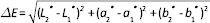
The liquid pigment contains silanes to create a stable bond with the structure of the unpigmented silicone. After polymerization, specimens were examined again by infrared spectroscopy to record any structural changes. Afterwards the color of all specimens was measured in a CIE L*a*b* system using a spectrophotometric device (PerkinElmer Lambda 35 UV/Vis Spectrometer; Shelton, USA). The apparatus consists of an integrating sphere of 50 mm, which can be used for measuring specimens under reflection angles 0° and 8°. The device emits wavelengths at 190-1,100 nm through a deuterium lamp and a tungsten-halogen lamp. Measurements were made with diffuse reflectance, to get the color parameters L*, a*, b*. It should be noted that the color of reflectance measurements corresponds to the color range perceived by the human eye. Three measurements were made for each specimen and the mean value and standard deviations were recorded. Also parameters of all specimens were measured, to calculate the color difference (ΔE*) before and after immersion and photoaging, according to the formula:
Photoaging
The photoaging apparatus has a xenon lamp emitting at 270-800 nm with an emission intensity of 765 W/m2. The average standard exposure to solar radiation (300-800 nm) in Mediterranean countries is 2,900 MJ/m2 per year. Specimens remaining in photoaging apparatus for 174 hours corresponds to 2 months of continuous exposure to sunlight (24).
Statistical Analysis
Two-way analysis of variance (ANOVA) was used for the statistical analysis of the results. Due to the failure of Kolmogorov-Smirnov normality test, data were logarithmically transformed. Tukey's test for post hoc comparison was used at a significance level of p≤0.05. Analysis was performed with the SigmaStat 3.1. (Jandel, St. Rafael, CA, USA) statistical package.
Results
Color Change
Two-way ANOVA revealed that aging significantly affected color changes (F = 2.976, p = 0.037), whereas pigmentation did not (F = 2.701, p = 0.074). No interaction between them (F = 0.772, p = 0.594) was found. The overall results are presented in Table II.
Color difference results (ΔE*) between aging procedures and materials used
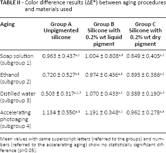
Mean values with same superscript letters (referred to the groups) and numbers (referred to the accelerating aging) show no statistically significant difference (p>0.05).
Statistical analysis for the unpigmented silicone (group A) showed no significant differences among the immersion and accelerating photoaging procedures (p>0.05). This change was mainly due to lightness reduction (-ΔL*) and yellow shift (+Δb*) (Fig. 1).
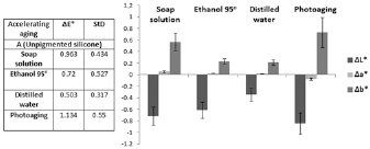
Mean values and standard deviations of ΔL*, Δa* and Δb* for unpigmented silicone specimens after disinfection and photoaging.
In general a shift to yellow and dumping of the material was observed, increasing intensively from the specimens immersed in distilled water, to ethanol 95°, to soap solution and to accelerating photoaging.
Statistical analysis for silicone with liquid pigment (group B) revealed no significant differences (p>0.05) among the tested subgroups. The color change (ΔE*) was mainly due to lightness reduction (-ΔL*), yellow shift (+Δb*) and green shift (-Δa*) (Fig. 2). In the fourth group, the reduction in lightness (-ΔL*) was accompanied by a shift to green (-Δa*) and yellow (+Δb*).
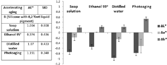
Mean values and standard deviations of ΔL*, Δa* and Δb* for silicone specimens with liquid pigment after disinfection and photoaging.
Statistical analysis for silicone with dry pigment (group C) revealed no significant differences (p>0.05) among the subgroups tested. The color change was due to reduction of all 3 color parameters (L*, a* and b*) (Fig. 3). The highest reduction of the first subgroup was mainly due to greenish shift (-Δa*). Specimens immersed in ethanol 95° presented a reduction of L* and a* (-ΔL*, -Δa*) accompanied with an increase of parameter b* (Δb*). Specimens after accelerating photoaging presented color changes that were mainly due to reduction of L* and to a smaller increase of b*.
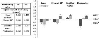
Mean values and standard deviations of ΔL*, Δa* and Δb* for silicone specimens with dry pigment after disinfection and photoaging.
The multiple comparison test for the material immersed in soap solution, in ethanol 95° and subjected to photoaging subgroups, showed no statistically significant differences for all of the materials tested (p>0.05). The only statistically significant difference was recorded in the subgroup of distilled water between the silicone with the functional pigment and the silicone with the dry pigment (p = 0.011) (Tab. II).
Structural Changes
In Figure 4a, the characteristic peaks of group Si-H (2,158 cm−1) and Si-CH3 (1,256 cm−1) are presented.
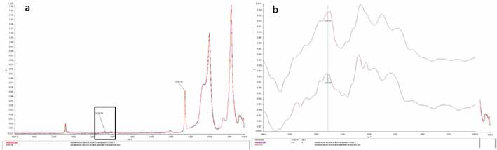
Infrared (IR) spectra of unset and set unpigmented silicone (a) and magnification of the blocked area of diagram a for calculated percentage amount of remaining Si-H groups (% RHS) (b).
For the unset and set silicone, the FTIR spectrum showed that heights of the peak 2,158 cm−1 were 0.0071 Α and 0.0046 Α, respectively, while heights for the peak 1,256 cm−1 were 0.2475 Α for the unset and 0.2429 Α for the set silicone (Fig. 4b). As mentioned above, the implementation of the formula: % RHS = 100 x (SHS x UMS/SMS x UHS) leads to a 66% RHS.
Following the same procedure, a reduction of % RHS was observed with descending intensity from photoaging to distilled water, to ethanol 95° to soap solution, for all 3 groups.
In Figure 5, comparative spectra of group A (unpigmented silicone) before and after immersion and photoaging are presented. No change in the peaks of Si-CH3 and Si-O-Si groups was recorded. The same picture is presented for group B (silicone with the liquid silanized pigment) and group C (silicone with the dry pigment).
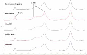
Comparative infrared (IR) spectra of unpigmented silicone before and after disinfection and aging procedures for Si-CH3 and Si-O-Si groups.
Discussion
The results of the present study showed that our null hypothesis concerning the color changes as well as the structural changes of maxillofacial silicones before and after immersion and photoaging was verified.
Emphasis was given to factors related to the daily habits of the patient, such as exposure to sunlight or/and the way patients clean the maxillofacial prosthesis. The percentage of pigment added was selected as 0.2% wt because this proportion was considered the saturation ratio for the pigment powder in a previous study (15). Although the choice of the functional liquid pigment should reasonably enhance color stability, due to the chemical bonding with the mass of silicone, no statistically greater values of ΔE* were detected in comparison with dry pigment. No significant color changes were recorded by Kiat-amnuay et al (19), who measured the color change in a RTV maxillofacial silicone, using a red dye pigment. ΔE* did not exceed the clinically acceptable limit. Also Mancuso et al (20) reported no significant color changes in a maxillofacial silicone in which 3 types of pigments were added, undergoing artificial aging (chamber UVB/condensation) until 1,000 hours.
In contrast, Eleni et al (16) tested 3 different types of polydimethylsiloxanes, after immersion in simulated body fluid and sweat at 37°C, and showed that there were significant color changes in all materials after immersion in serum and sweat. Also Polyzois et al in 2 different experiments concerning pigmentation of maxillofacial silicones under different conditions of aging (17, 18) noticed color differences (ΔE*) ranging from 2 to 3.5 units and 2.13 to 3.98 units, respectively. According to the researchers, the duration of the exposure and the type of silicone were significant factors affecting color stability.
For maxillofacial silicone prostheses, different perceptibility and clinical acceptability thresholds have been reported, ranging from 0.8 to 1.8 (25) and 1.1 to 3.0 (26), respectively. In the present study, color changes over 0.8 and up to 3.0 were considered visually perceptible and clinically acceptable, respectively. Treatment conditions induced perceivable color change (ΔE* >3) (9).
Distilled water, as expected, exhibited the smallest color change, ranging under the perceptible values (0.8) except the samples of group B were at 1.07 which is probably due to the chemical integration of the liquid color in the silicone structure, decreasing its solubility in the water. Photoaging had the greatest impact, probably due to the higher reduction of % RHS, which practically means further polymerization of the materials.
Regarding the results of infrared spectroscopy, the lack of changes in structure in the present study can be attributed to the fact that the concentration of the pigments was extremely low (0.2% wt). Changes in the structure were not qualitative but only quantitative (reduction of % RHS). The reduction in the percentage of remaining Si-H groups (% RHS) after immersion showed that solutions accelerated partially the polymerization of the silicone. Polymerization was more intense especially after photoaging probably due to the influence of radiation in the chemical bonds. In related studies, the influence of aging factors in the change of structure of siloxanes was investigated. Some researchers (11) found changes in the structural behavior of the material after prolonged exposure to radiation, whereas others (22) recorded differences in maxillofacial silicone structure when a dry and a functional pigment were added. One limitations of the present study was that only 1 type of maxillofacial silicone as well as 1 hue of colorants in one concentration was tested.
According to the results and the limitations of this research, it can be concluded that no structural changes of pigmented and unpigmented silicone elastomers were observed among all aging procedures. Recorded color changes for the materials tested were within the limits of clinical acceptability after all aging procedures. Immersion in distilled water presented the best color stability, whereas photoaging, the poorest, for all materials.
Footnotes
Financial support: None.
Conflict of interest: None.
Meeting presentions: This work was presented at the 8th convention of the Hellenic Society for Biomaterials, Athens, 15 November 2013.