
Abstract
Background
The objective of this study was to evaluate the role of autotaxin (ATX) activity and gene expression compared to soluble intercellular adhesion molecule-1 (sICAM-1) in thyroid carcinoma.
Patients and Methods
Sixty-five patients with thyroid swelling were included. There were 20 cases of simple multinodular goiter (group I), 15 cases of follicular adenoma (group II) and 30 cases of thyroid cancer (group III). Group III was further subdivided into negative and positive lymph nodes (group IIIa and IIIb; 22 and 8 cases, respectively). sICAM-1 concentration and ATX activity were measured using colorimetric enzyme-linked immunosorbent assay (ELISA), while ATX gene expression was detected by real-time polymerase chain reaction (PCR).
Results
sICAM-1 level, ATX activity and gene expression were significantly elevated in patients with thyroid carcinoma compared to other groups. The ATX activity showed significantly higher sensitivity and specificity than sICAM-1 (100% and 97.1% vs 93.3% and 88.6%, respectively). Both sICAM-1 and ATX values were significantly higher in patients with positive lymph nodes compared to those without lymph node involvement (p<0.001). Higher levels of ATX activity and gene expression were significantly correlated with larger tumor size and undifferentiated pathological subtype in thyroid carcinoma. In this respect, ATX was superior to sICAM-1.
Conclusion
Our data suggest that ATX activity and gene expression are reliable diagnostic and prognostic tools in thyroid carcinoma compared to sICAM-1.
Introduction
The incidence of thyroid cancer is increasing at a rate of 3% per year (1). Patients with thyroid cancer are detected from a much larger group who present with thyroid nodules, the majority of which are benign. Fine needle aspiration (FNA) biopsy is the recommended test for evaluation of thyroid nodules (2, 3). However, about one fifth of malignant suspicious nodules prove to be benign after many unnecessary thyroid surgeries (4, 5). Therefore, the search for reliable diagnostic markers is necessary.
Much evidence has implicated adhesins in cancer initiation and metastasis. Soluble intercellular adhesion molecule-1 (sICAM-1) or CD54 is a monomeric, transmembrane, glycoprotein molecule of the immunoglobulin superfamily (6). Elevated serum level of sICAM-1 has been observed in several malignant diseases including thyroid carcinomas, where it has been correlated with disease status and treatment response (7-10).
Cell migration mechanisms play a critical role in cancer development and metastasis (11). Autotaxin (ATX) or NPP2, an extracellular lysophospholipase D of 125 kDa, is one of the 40 most upregulated genes in invasive cancer cells (12, 13). ATX's ability to promote neoplastic invasion and metastasis could depend on its noncatalytic and/or catalytic effects (14). A noncatalytic effect of ATX was shown to occur in oligodendrocytes, where ATX acts as a matrix cellular protein through integrin-dependent focal adhesion assembly (15). On the other hand, the enzymatic activity of ATX occurs through the extracellular production of the potent bioactive phospholipid mediator lysophosphatidic acid (LPA) from lysophosphatidylcholine (LPC) (16-18).
ATX expression has been suggested to be positively correlated with the metastatic and invasive properties of a large variety of human tumors including thyroid cancers (19, 20). The aim of this study is to investigate the role of ATX activity and gene expression as diagnostic and prognostic tools in various pathological types of thyroid cancer compared to sICAM-1.
Patients and Methods
After having provided written informed consent, 65 patients with thyroid swelling were enrolled from the department of surgery, Tanta University Hospital between January 2008 and December 2010. Using FNA, 20 cases were diagnosed as simple multinodular goiter (group I); 15 were diagnosed as thyroid adenoma (group II), and 30 were diagnosed as thyroid cancer (group III). Group III was further subdivided into negative and positive lymph nodes (group IIIa and IIIb; 22 and 8 cases, respectively). Lymph node (LN) involvement was diagnosed by physical examination, neck ultrasound and confirmed by FNA and histopathological examination.
Patients with malignant swelling were divided according to the size of the tumor (≤2 or >2 cm), pathological type and stage. Staging was defined according to the international Tumor Node Metastasis (TNM) classification proposed by the American Joint Committee on Cancer (AJCC) (21).
Blood samples
Fasting blood samples were taken consecutively from all patients. The samples were centrifuged at 3000g for 10 minutes at 4°C, thereafter serum was removed and stored at −80°C. Serum ATX activity was measured according to Sajdok et al (22). This method is based on colorimetric determination of the product of ATX reaction, choline, using a choline oxidase/peroxidase system by microtitration plate at 490 nm using an enzyme-linked immunosorbent assay (ELISA) reader. The serum concentration of sICAM-1 was measured using an ELISA kit (Quantikine Human sICAM-1 immunoassay, R&D Systems) according to the manufacturer's protocol.
Tissue samples
After surgery, thyroid tissue samples were taken and snap frozen on liquid nitrogen.
RNA extraction
The tissues were thawed on ice, weighed and homogenized followed by centrifugation at 13,000 rpm for 15 minutes at 4°C. The samples were subjected to RNA extraction by Qiagen RNeasy Mini Kit according to the protocol supplied by the manufacturer. One volume of 70% ethanol was then added to the cleared lysate, mixed immediately and then transferred to Qiagen RNeasy spin column placed in a 2 mL collection tube and precessed according to the manufacturer's instructions. RNA was eluted and its concentration was measured spectrophotometrically at 280 nm. Five hundred nanograms of total RNA was used for first strand cDNA synthesis, primed with 50 pmol of random primer (Roche, Mannheim, Germany) and 0.5 μL (200 U/μL) reverse transcriptase Superscript™ II-RT (Invitrogen, Karlsruhe, Germany). Polymerase chain reaction (PCR) cycling condition was performed according to the manufacturer's instructions. A control reaction without reverse transcriptase was performed in parallel to detect genomic DNA contamination (23). The first strand cDNA was verified by running on 2% agarose gel for confirmation of the amplified band.
Real-time PCR
One microliter of the reverse transcriptase reaction mixture was added to a 20 μL reaction mixture of the QuantiTect SYBR-Green PCR kit (Qiagen) and 0.5 μM from the specific primer pair for human ATX: sense, 50-ACAACGAGGAGAGCTGCAAT-30, and anti-sense, 50-AGAAGTCCAGGCTGGTGAGA-30. Primers for glyceraldehyde 3-phosphate dehydrogenase (GAPDH) were: sense, TTGCCATC-AATGACCCCTTCA and antisense, CGCCCCACTTGATTTTGGA (24). A negative control containing the complete Master Mix without a DNA template was included.
We used thyroid tissue samples of cDNA that were assayed on the StepOne Real-Time PCR System (Applied Biosystems). Initial denaturation at 95°C for 90 seconds was followed by 40 cycles with denaturation at 95°C for 15 seconds, annealing at 60°C for 30 seconds and extension at 72°C for 20 seconds. The fluorescence intensity of SYBR-Green, specifically incorporated in the double-stranded DNA amplicon reflecting the amount of formed PCR product, was read after each extension step at 72°C (24). RNA amounts were determined with the Applied Biosystems software in mode relative to the GAPDH gene (Fig. 1).
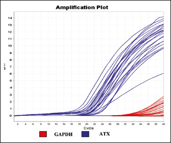
Amplification plot of autotaxin (ATX) and glyceraldehyde 3-phosphate dehydrogenase (GAPDH). It shows the log of the change in the fluorescence plotted against cycle number (representative study).
Statistical analysis
Data were analyzed with the GraphPad InStat software. Descriptive data were given as mean ± standard deviation (SD). Comparison between 2 groups was performed with the t-test, but for comparison among different groups ANOVA was used followed by the Tukey-Kramer multiple comparisons test. By using a cutoff value equal to the mean plus more than 2-fold SD, sensitivity, specificity, positive predictive value (PPV) and negative predictive value (NPP) were calculated. The Pearson correlation was performed to determine the associations between the studied parameters and conventional clinicopathological features. A p value less than 0.05 was considered statistically significant.
Results
The clinical and pathological characteristics of patients with thyroid cancer are included in Table I. The serum level of sICAM-1, ATX activity and gene expression were significantly elevated in patients with thyroid cancer (group III) compared to those with goiter (group I) or adenoma (group II) (p<0.0001). There was no significant difference between group I and II (Tab. II) (Figs. 2-4). In the thyroid cancer group, both sICAM-1 and ATX values were significantly higher in patients with LN metastasis (IIIb subgroup) compared to those without LN involvement (IIIa subgroup) (Tab. III). The s-ICAM-1 level exceeded the cutoff value of 360 ng/mL in 1 patient with goiter and 3 others with adenoma, where the serum level for 2 patients with carcinoma failed to reach such level. Consequently, the sensitivity, specificity, PPV and NPV of sICAM-1 were 93.3%, 88.6%, 87.5% and 93.9%, respectively. For ATX activity, only 1 patient with adenoma had serum activity above the cutoff value (9 nmol/mL/minute). As a result the sensitivity, specificity, PPV and NPV were 100%, 97.1%, 96.8% and 100%, respectively.
CLINICAL AND PATHOLOGICAL CHARACTERISTICS OF THYROID CANCER PATIENTS
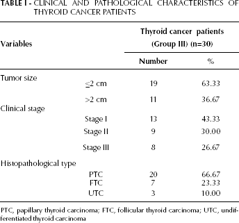
PTC, papillary thyroid carcinoma; FTC, follicular thyroid carcinoma; UTC, undifferentiated thyroid carcinoma
sICAM-1 LEVELS, ATX ACTIVITY AND GENE EXPRESSION IN THE STUDY GROUPS
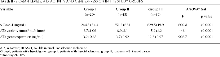
ATX, autotaxin; sICAM-1, soluble intercellular adhesion molecule-1
Group I, patients with thyroid goiter; group II, patients with thyroid adenoma; group III, patients with thyroid cancer
One-way ANOVA
CORRELATION BETWEEN BIOCHEMICAL VARIABLES AND LYMPH NODE STATUS IN THE THYROID CANCER GROUP
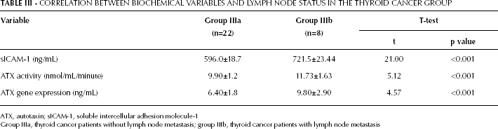
ATX, autotaxin; sICAM-1, soluble intercellular adhesion molecule-1
Group IIIa, thyroid cancer patients without lymph node metastasis; group IIIb, thyroid cancer patients with lymph node metastasis
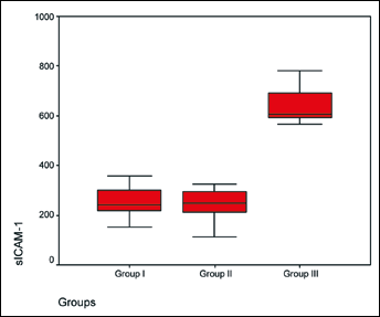
The serum level of soluble intercellular adhesion molecule-1 (sICAM-1) was significantly elevated in thyroid cancer (group III) (p<0.0001) whereas no difference was found between group I (goiter) and II (adenoma).
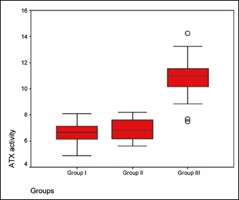
The ATX activity was significantly elevated in the thyroid cancer (group III) (p<0.0001) whereas no difference was found between group I (goiter) and II (adenoma).
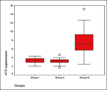
The ATX expression was significantly elevated in the thyroid cancer (group III) (p<0.0001) whereas no difference was found between group I (goiter) and II (adenoma).
ATX activity and gene expression as well as sICAM-1 level were significantly different among various stages of thyroid carcinoma with increasing values from stage I to III (Tab. IV). However, only ATX activity and expression were significantly correlated with tumor size and pathological subtype. Patients with larger tumors (>2 cm) and an undifferentiated pathological subtype had the highest level of ATX activity and expression (Tab. V).
Correlation Between Biochemical Variables and Clinicopathological Characteristics in the Thyroid Cancer Group (N=30)
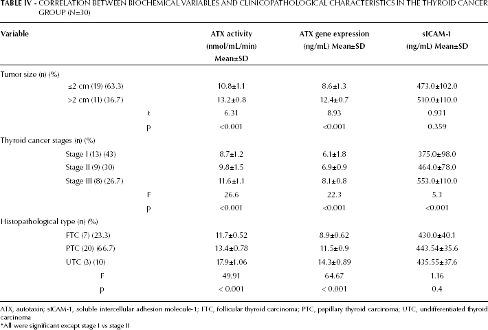
ATX, autotaxin; sICAM-1, soluble intercellular adhesion molecule-1; FTC, follicular thyroid carcinoma; PTC, papillary thyroid carcinoma; UTC, undifferentiated thyroid carcinoma
* All were significant except stage I vs stage II
Correlation Coefficient Between Biochemical Variables and Clinicopathological Characteristics in the Thyroid Cancer Group (N=30)
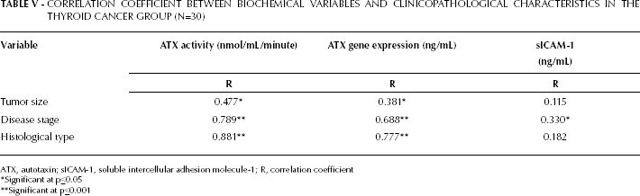
ATX, autotaxin; sICAM-1, soluble intercellular adhesion molecule-1; R, correlation coefficient
Significant at p≤0.05
Significant at p≤0.001
Discussion
ATX, a potent angiogenic factor, is linked to several malignancies (14, 19, 25, 26). Many authors have reported that ATX is one of the 40 most upregulated genes in invasive cancer cells (14, 27). In thyroid cancer, Kehlen et al demonstrated the oncogenic potential of ATX as an enhancer of proliferation and migration of malignant cells, as it augments the tumorigenic capacity and acts as an angiogenic factor (20). However, previous molecular studies of thyroid tumors have failed to define any diagnostic or prognostic markers (28-31). In our cohort, we found that ATX protein was overexpressed and its activity was elevated in thyroid cancer patients compared to goiter and adenoma cases. Furthermore, there was a significant correlation between ATX activity and tumor size as well as disease stage. The significantly elevated ATX activity was associated with larger tumor size, LN involvement and undifferentiated histopathological subtype.
The positive correlation between ATX and tumor size and histological type has also been reported by Kehlen et al (20). They quantified the ATX mRNA expression in thyroid carcinoma cell lines and in tissues of patients with thyroid carcinomas. They found that ATX gene activity was significantly higher in undifferentiated thyroid cancer compared to follicular and papillary pathological types or goiter tissues. Also, Grimm et al showed that ATX mRNAs were overexpressed in follicular thyroid cancers compared with follicular thyroid adenomas (32). Those authors concluded that increased expression of ATX correlated with the dedifferentiated state of thyroid carcinomas in patient tissues and cultured thyroid carcinoma cell lines, which may contribute to the high degree of invasiveness and metastasis. Their results were further explained by the fact that ATX markedly enhanced the motility of different human thyroid carcinoma cells (20, 27).
We found that the sICAM-1 concentration was elevated in thyroid cancer patients with reasonable sensitivity compared to those with goiter and adenoma. Therefore, sICAM-1 might be considered as a marker for diagnosis of thyroid cancer. Similiarly, Tanda et al reported an upregulation of sICAM-1 in thyroid cancer patients comparing to normal controls independently of the cancer type (33). Many other studies have confirmed the role of sICAM-1 as a powerful marker for early detection of thyroid carcinoma (34, 35).
sICAM-1 serum levels were significantly higher in thyroid cancer patients with LN metastasis than in those without LN involvement. Fernández-Real et al also reported sICAM-1 expression in distant metastases from thyroid carcinoma (36). In our cohort, the sICAM-1 concentration showed significantly increasing values from stage I to III, with no significant correlation with tumor size or histopathological type. These results support the hypothesis by Nakashima et al that the main role of sICAM-1 is in promoting cancer invasion regardless of cancer type or mass (37). Tanda et al demonstrated that all of their papillary thyroid cancer cases showed positivity for sICAM-1, whereas follicular thyroid cancer, follicular thyroid adenoma and all except 1 nodular goiters were negative (33). They suggested that the occurrence of sICAM-1 on the thyroid cell surfaces in well-differentiated papillary thyroid tumors may contribute to the understanding of their biology and could be of potential significance for diagnostic purposes.
The role of sICAM-1 in the progression of metastatic disease is dual. Although a positive correlation has been proposed between expression of sICAM-1 and progression of metastatic disease (38, 39), the increased presence of sICAM-1 in tumor cells has been associated with a decrease in the ability of these cells to migrate and spread to distant locations (40). Sawada et al reported that decreased sICAM-1 expression induced by transforming growth factor-1 (TGF-1) contributes to cancer cell escape from immunological recognition and cytotoxicity by effector cells (41). Controversially, Arnold et al found a negative correlation between sICAM-1 expression and tumor metastasis and reported that patients with sICAM-1-positive tumor cells had a better prognosis than those with cells that did not express sICAM-1 (42). Organ- or tumorspecific qualities may determine these differences (43).
The relationship between ATX and sICAM-1 in thyroid carcinoma remains elusive. Being the key enzyme with lyso-PLD activity, the aberrant expression of ATX has the potential to alter the delicate balance between LPA signaling and LPC signaling in the local microenvironment. LPC is a phospholipid with both proinflammatory activity and immunoregulatory activity by stimulating the expression of a series of genes including sICAM-1 (44, 45). Kehlen et al reported the down-regulation of sICAM-1 gene activity in a human thyroid carcinoma transfectant overexpressing human ATX (20). However, the parallel increase of sICAM-1 values and ATX expression and serum activity noticed in this study in cases with LN metastasis may underline the ATX-associated regulation of CD54 in neoplastic human thyroid cells, which could be an important step towards tumor progression to a more malignant phenotype.
In summary, the overexpression of ATX in thyroid cancer may fuel the process of carcinogenesis. Although both ATX and sICAM-1 may serve as diagnostic tools for tumor initiation, ATX activity has much better sensitivity and specificity. Also, ATX is superior to sICAM-1 as it correlates with pathological type and tumor size. ATX serum activity could be used as a simple, noninvasive reflection of ATX expression, thus providing a promising marker for pathological classification and prediction of disease progression. In addition, inhibition of ATX activity through modulation of LPA signaling could be a potential target for future anticancer drugs. Future studies are warranted to explore this area, particularly for patients with undifferentiated thyroid cancer that lacks response to conventional therapies.