
Abstract
In most reviews addressing intracellular lipid trafficking, spontaneous diffusion of lipid monomers between the cellular organelles is considered biologically irrelevant because it is thought to be far too slow to significantly contribute to organelle biogenesis. This view is based on intervesicle transfer experiments carried out in vitro with few lipids as well as on the view that lipids are highly hydrophobic and thus cannot undergo spontaneous intermembrane diffusion at a significant rate. However, besides that single-chain lipids can translocate between vesicles in seconds, it has been demonstrated that the rate of spontaneous transfer of two-chain polar lipids can vary even 1000-fold, depending on the number of carbons and double bonds in the acyl chains. In addition, the rate of spontaneous lipid transfer can strongly depend on the experimental conditions such as vesicle composition and concentration. This review examines the studies suggesting that spontaneous lipid transfer is probably more relevant to intracellular trafficking of amphipathic lipids than commonly thought.
Introduction
Why is interorganelle trafficking necessary in a eukaryotic cell? Nearly all cellular lipids are made in the endoplasmic reticulum (ER) yet are found in all other organelles, as well. Accordingly, mechanisms to transport these lipids from the ER to other organelles must exist. In principle, one or several of the following mechanisms could be involved: (i) transport in vesicles, (ii) protein-mediated transfer, (iii) monomer diffusion via the cytoplasm, and (iv) transient membrane (hemi) fusion. In most reviews on lipid trafficking, only the first two mechanisms are considered important for interorganellar lipid trafficking, while the last two are thought to be either too slow or rare to be biologically relevant. This view seems to be based on in vitro data showing that the half-time of glycerophospholipid (GPL) or sphingolipid diffusion between lipid vesicles is very slow, up to several days. However, it is very important to note that the rate of lipid intervesicle diffusion, measured in vitro, is highly dependent on the structure of the lipid as well as the experimental conditions. 1 Another (possible) reason for downplaying spontaneous lipid transfer (SLT) is that this process is thought to be incompatible with lipid compositional gradients existing between organelles. However, SLT would not necessarily abolish such gradients since the other processes contributing to organelle lipid compositions, such as transbilayer movement and metabolic processes, are likely to be much faster than SLT. The purpose of this review is to briefly discuss the data that seem pertinent regarding the role of SLT in cellular physiology.
Definition of SLT
Traditionally, SLT has been defined as a process where a lipid molecule moves without the assistance of a protein carrier (a lipid transport protein) from one membrane to another. At low membrane concentrations, typically used in in vitro experiments, the rate-limiting step in this process is the efflux of a lipid molecule from a donor membrane, while at high membrane concentrations, membrane collisions can dominate S LT. 1 Further mechanistic details can be found in previous publications.2–5 In this review, SLT is also considered to be the mechanism of transfer when proteins are indirectly involved in the transfer process, ie, when they tether two membranes, thereby increasing the probability of intermembrane lipid translocation. 6 In addition, transient hemifusion (ie, only the outer membrane leaflets fuse) of two membranes is considered as an alternative mechanism of SLT as it allows for intermembrane lipid translocation.
Factors Influencing SLT
There are several factors influencing the rate of lipid monomer diffusion between lipid vesicles (SLT), such as the structure of the diffusing lipid, vesicle size (=bilayer curvature), vesicle concentration, and bilayer composition, as will be discussed below.
Lipid structure
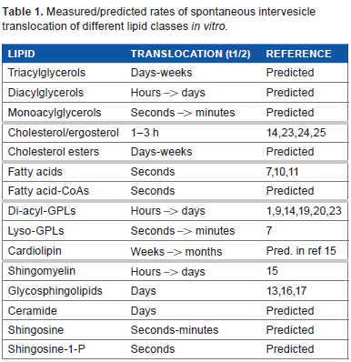
It is well established that the number of alkyl chains, their length, and degree of unsaturation each have a remarkable effect on SLT. Single-chain lipids, such as fatty acids and lyso-GPLs, monoacylglycerol, sphingosine, and sphingosine 1-phosphate, have been shown or predicted to diffuse in seconds from a vesicle population to another,7–12 while this process takes hours or even days for typical two-chain GPLs and sphingolipids.13–17 Importantly, however, the rates of diffusion of two-chain GPLs differ remarkably, depending on the length and the degree of unsaturation of their alkyl chains. Thus, it has been shown that removing two methylene units from or adding a double bond to an acyl chain of a diacyl GPL increases the rate of diffusion 5- to 10-fold.18–20 Consequently, the rate of intervesicle diffusion of two-chain GPLs can differ by more than 1000-fold (ie, the half-time varies from minutes to days), depending on the number of alkyl chain carbons and double bonds. On the other hand, the polar head group structure has only a modest effect on SLT.21,22
The rate of spontaneous translocation of cholesterol is much faster than that of GPLs as expected from its structure containing fewer hydrophobic carbons. For instance, McLean and Phillips found that the half-time for cholesterol translocation between vesicles is only 2.4 hours, 23 which is an order of magnitude less than the corresponding value for intact GPLs (see above). A similar value has been found by others,24,25 and even faster rates (~1 hour) have been reported. 14 The rate of the spontaneous transfer of ergosterol (the cholesterol equivalent in yeast) has not been determined, but it is probably even faster than that of cholesterol due to the presence of two additional double bonds, albeit there is an extra methyl group in ergosterol. It is worthy to note here that the intermembrane translocation rate of the fluorescent cholesterol analog dehydroergosterol (and probably cholestatrienol as well) is significantly faster than that of cholesterol 26 due to the additional double bonds that decrease the molecular hydrophobicity significantly. In contrast to GPLs and other polar lipids, triglycerides and cholesteryl esters are too hydrophobic to move spontaneously between membranes by the aqueous monomer diffusion mechanism. This applies to cardiolipin as well, which is intriguing as it may explain why this GPL remains fully confined to its site of synthesis, ie, mitochondria. 19 Table 1 summarizes the measured or predicted rates of SLT for the main mammalian lipid classes.
Measured/predicted rates of spontaneous intervesicle translocation of different lipid classes in vitro.
Bilayer curvature and vesicle concentration
It has been demonstrated that SLT is markedly dependent on the donor surface curvature in vitro, ie, the rate of transfer (efflux) increases markedly with increasing curvature of the donor surface. Thus, transfer of phosphatidylcholine (PC) or cholesterol is significantly faster from small vesicles with a high bilayer curvature vs. large vesicles with low bilayer curvature.23,24 The effect of interphase curvature is due to the lipid–lipid interactions that become weaker with increasing interphase curvature, which increases the chemical activity of the lipids and thus their efflux propensity. PC and cholesterol transfer from lipoprotein particles also increases markedly with increasing particle curvature,27,28 albeit the apoproteins could also contribute to the faster transfer (see below). Vesicle concentration can also have marked effect on SLT as was demonstrated by Jones and Thompson 1 who found that intervesicle transfer of PC increased six fold when the vesicle concentration was increased from 0.1 to 40 mM, which is similar to the estimated concentration on membranes in cells. Kinetic data analyses indicated that the increase of SLT at high vesicle concentrations is due to enhancement of lipid translocation by intervesicle collisions. 2
Lipid composition
Also the lipid composition of the (donor) bilayer can have a marked effect on SLT as anticipated based on the fact that the lipid composition can influence bilayer packing remarkably. Thus, cholesterol efflux is much faster from bilayers consisting of unsaturated vs. saturated lipids 29 and, strikingly, Wimley and Thompson found that inclusion of 30 mol% of PE in PC vesicles enhanced PC translocation by 100-fold at high vesicle concentrations. 30 Other studies have shown that fatty acids and other single-chain amphiphiles can markedly increase the efflux of cholesterol from membranes.31,32 It has also been reported that the composition of the acceptor vesicles can influence SLT. 33 However, in this study, the transfer of an unnatural, short-chain fluorescent PC was studied, and thus, this result needs to be verified by using probe-free, long-chain lipids.
Lange et al have found that the amount of cholesterol relative to phospholipids can be critical for cholesterol efflux and transfer.34,35 It was proposed that when the cholesterol to phospholipid ratio exceeds a critical value, the cholesterol molecules in excess (ie, not complexed with the phospholipids) will have an increased chemical activity, and that the active cholesterol molecules have an increased tendency to efflux from the membrane.34–37 However, there is yet no direct experimental evidence for such cholesterol–phospholipid complexes, first proposed by Radhakrishnan and McConnell. 38 In fact, the several critical cholesterol/phospholipid stoichiometric ratios observed in the experiments are more readily explained by the superlattice (regular distribution) model, which does not involve complex formation but proposes that (i) each critical ratio corresponds to a specific, regular lateral arrangement of cholesterol and phospholipids and (ii) and those specific arrangements (or cholesterol to phospholipid ratios) are more stable than random arrangements because they correspond to local minima in the membrane-free energy.39–41 Notably, the superlattice model also predicts several critical phospholipid/ phospholipid ratios for multicomponent membranes. 41 Thus, an excess of a particular phospholipid can exist when the composition deviates from a critical ratio (Fig. 1). Similar to the case of cholesterol, the phospholipid molecules in excess should have an increased chemical activity and thus an increased efflux propensity. 42
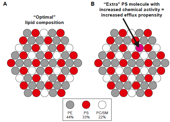
Lateral arrangement of the different GPLs in the inner leaflet on the human erythrocyte membrane as predicted by the superlattice model. (
Membrane proteins
Membrane proteins are highly abundant in biological membranes and, consequently, a major fraction of membrane lipids must reside next to a membrane protein molecule. The intrabilayer sequences of these proteins contain multiple protruding aminoacyl chains, which make the protein surface rough. Because of this, the lipid molecules in the protein boundary cannot pack as well as those outside the boundary, and thus, they would have an increased chemical activity. Such protein-induced packing perturbations are probably responsible for some transmembrane peptides that act as nonspecific lipid scramblases in vitro, ie, they greatly enhance transbilayer movement of phospholipids. 43 Also some cellular membrane proteins, such as cytochrome b5 44 and opsins, members of the ubiquitous family of G-protein-coupled receptors,45,46 act as highly efficient phospholipid scramblases in vitro. Besides such passive protein-induced lipid scrambling, active lipid scrambling also seems to exist as in the case of the ABCA1 transporter. 5
The anticipated increased chemical activity of the lipids in protein boundaries probably translates to their increased propensity of efflux from the membrane. Since efflux is the rate-limiting step in SLT (see above), membrane proteins could thus significantly enhance SLT. In line with this prediction, the rate of cholesterol transfer between liposomes was considerably increased upon addition of proteins to lipid vesicles. 25 Also, the fact that GPL transfer from small reconstituted lipoproteins is an order of magnitude faster than from neat lipid vesicles 25 is consistent with this model. However, the smaller size of the lipoproteins may also contribute to the faster rate of transfer from these particles (see above). Besides proteins, small amphipathic molecules, such as fatty acids and sphingosine, can also increase the chemical potential of a membrane lipid and thus its efflux, as shown for cholesterol by Lange et al. 32
SLT in Cells
Membrane contact sites
There are yet no data demonstrating that SLT contributes significantly to interorganelle lipid translocation. However, there is little evidence against this either. We will now discuss where in a cell could SLT be particularly relevant. The most obvious sites are the intermembrane contacts thought to be present between the ER (the site of lipid synthesis) and several other organelles such as the mitochondria, Golgi apparatus, plasma membrane (PM), peroxisomes, and endosomes.47–52 Such membrane contacts should increase SLT significantly because the closeness of the acceptor and donor membranes increases the likelihood that a lipid molecule, after its efflux from the donor membrane, inserts into the acceptor membrane. 6
The best studied membrane contact sites are those between the mitochondria-associated ER membranes (MAMs) and the outer mitochondrial membranes (OMMs).49,53–55 Several studies have proposed that these contact sites are involved in lipid transfer between the ER and mitochondria and that specific proteins play a role there. However, despite that this issue has been studied for more than two decades, 56 no protein directly involved in lipid transfer between these membranes has been identified.48,49,53 This makes one wonder whether such proteins indeed exist? Another reason underlying this, perhaps provocative question is that the half-time of phosphatidylserine (PS) translocation from MAM (where it is synthetized) to mitochondria is slow, ie, the half-time of translocation was 7–15 hours for typical PS species.57,58 Notably, such long half-times are similar to those predicted for spontaneous transfer of PS between membranes, and thus do not support the involvement of lipid-transfer protein(s).
Besides SLT, lipids might move between MAMs and the OMM upon a transient hemifusion of the respective membranes. Thus, Ca2+ could cross-link PS molecules via their head groups, 59 thereby creating PS-rich domains in MAM. Such domains could act as nucleation sites for a transient hemifusion of MAM with the outer leaflet of the OMM, which then would allow translocation of PS and other lipids between the two organelles (Fig. 2). Consistent with this speculative model, (i) PS synthesis requires a relatively high Ca2+ concentration in vitro, 60 (ii) Ca2+ is released from MAM lumen into the intermembrane space between the MAM and the OMMs,61,62 and (iii) Ca2+ can induce (hemi)fusion of PS-rich membranes in vitro. 63 An alternative model suggests that the localized synthesis of PS in MAM 49 increases the concentration of this GPL beyond its optimal concentration (Fig. 1), thus creating a pool of active PS molecules prone to efflux from MAM to the OMM. The latter, but not the former, model is consistent with the slower translocation of the more hydrophobic PS molecules from MAM to mitochondria.57,58,64 However, in both models, tethering proteins would enhance (indirectly) PS translocation from MAM to mitochondria by keeping these organelles close to each other.
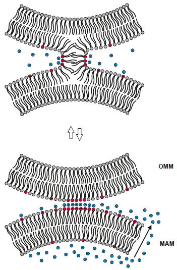
Hypothetical model for Ca2+-induced hemifusion of MAM and the OMM. The concentration of Ca2+ in the lumen of the MAM is of orders of magnitude higher than that in the cytoplasm (see text). When Ca2+ is released (via a protein channel, not shown) from the lumen of MAM (arrow) to the space separating MAM and OMM, this should strongly stimulate the synthesis of PS (red), which is Ca2+-dependent. The PS molecules in the cytosolic surface of MAM attract Ca2+ ions (blue), which leads to the formation of PS-rich domains and subsequent bridging of the PS domains with negatively charged lipids in OMM by Ca2+. This destabilizes the contacting membranes, thus promoting their hemifusion that allows the lipids move from MAM to OMM and vice versa by lateral diffusion. The double arrows indicate that the process could be reversible, ie, when the concentration of Ca2+ decreases, the membranes detach from each other. An alternative model suggests that the rapid Ca2+-induced synthesis of PS creates an excess of this lipid in MAM (Fig. 1). The PS molecules in excess have an increased chemical activity, which makes them prone to efflux from MAM to OMM.
Since contact sites also seem to exist between the ER and the PM, it is feasible that aqueous diffusion or membrane hemifusion could be involved in lipid translocation between these membranes. Regarding possible hemifusion, it is notable that the concentration of PS in the PM inner leaflet is very high, ie, 10–20 mol%. 65 Such a high concentration of PS could drive hemifusion of the ER and PM membranes, thus promoting lipid transfer via the contact sites. However, we have shown that the rate of transfer of fluorescent PS molecules from the PM to mitochondria decreases exponentially with increasing chain length (hydrophobicity) of the lipid. 66 This result is inconsistent with the hemifusion mechanism (which should be insensitive to lipid hydrophobicity), but consistent with spontaneous diffusion via the cytoplasm being the rate-limiting step in the transport of PS from the PM to mitochondria.
A recent study addressed the relevance of SLT between PM/ER contact sites in vitro by employing a rather involved approach based on various fluorescent probes. 67 It was concluded that neither PC nor cholesterol translocates spontaneously through contact sites between PM and ER membranes. In particular, the lack of cholesterol transfer is puzzling as cholesterol translocates quite rapidly between lipid vesicles in vitro (see above). Also, it has been shown that intracellular cholesterol in glutaraldehyde-fixed, nonleaky cells could be rapidly and completely oxidized by extracellular cholesterol oxidase. 68 Since fixing should stop both vesicle- and protein-mediated cholesterol transfer to the PM, 68 these data imply that spontaneous translocation of cholesterol, possibly via membrane contact sites, could be involved.
Is the deacylation/reacylation cycle involved in lipid translocation?
In mammalian cells, GPLs are rapidly remodeled after their synthesis, ie, their acyl chains are exchanged for others. 69 This so-called Lands pathway 70 involves removal of an acyl chain by an A-type phospholipase followed by acylation of the lyso-GPL with another fatty acid. Since the spontaneous diffusion of lyso-GPLs is very fast (see above), it should rapidly distribute to other membranes where it could be reacylated to an intact GPL. If so, spontaneous intermembrane transfer of a GPL would have been achieved in effect. 71 However, there is as yet no experimental proof for this putative mechanism of intermembrane lipid translocation.
Future Perspectives
There are very few experimental studies addressing the importance of SLT in vivo. Besides the presumption that SLT is irrelevant for organelle biogenesis, there is a lack of methods suitable to investigate the importance of this mechanism. First of all, there is, or will be, no specific method to inhibit or eliminate SLT, such as knockdown of gene products, which has been highly useful in studies on the other translocation mechanisms. Thus, the evidence for or against the importance of SLT is likely to be indirect, ie, will derive from experiments where the other mechanisms of translocation have been eliminated. Also studies where the hydrophobicity of a lipid under study is varied in a systematic manner would be useful since the different transport modes respond differently to varying lipid hydrophobicity. 57 Of note, there is substantial evidence that particular lipid-binding/-transfer proteins are enriched at membrane contact sites, where they appear to facilitate the interorganelle transport of specific lipids.72–74 This observation is not discordant with the SLT mechanisms discussed here since one can envision that transfer of protein-dependent and -independent mechanisms complement each other in the transfer of distinct lipid species with particular acyl chain length and degree of saturation.
Abbreviations
endoplasmic reticulum;
glycerophospholipid;
plasma membrane;
phosphatidylcholine;
phosphatidylserine;
spontaneous lipid transfer.
Author Contributions
Conceived the concepts: PS. Wrote the first draft of the manuscript: PS. Developed the structure and arguments for the paper: PS. Made critical revisions: PS. The author reviewed and approved of the final manuscript.
Footnotes
Acknowledgments
I am grateful to Dr. Vesa Olkkonen and the members of my group for critical reading of the manuscript.