
Abstract
In epilepsy, novel pharmacological and nonpharmacological treatment approaches are commonly assessed in model systems of acute motor and often generalized seizures. We developed a rodent model with short-term electrical stimulation of the perforant path resulting in stereotyped limbic seizures. Limbic structures play a major role in human intractable epilepsy. In 10 rats, single electrical 5-second and 20-Hz stimuli to the perforant path reliably produced limbic seizures characterized by resting behavior and subtle motor signs. Electrophysiological recordings from the dentate gyrus demonstrated a seizure pattern with 4-Hz to 5-Hz discharges. Multiple inductions of seizures within 72 hours did not alter behavioral and electrophysiological seizure characteristics. Electrophysiological excitatory and inhibitory parameters assessed by evoked single and paired pulses did not change with increasing number of seizures. We present preliminary findings on a new model of electrically induced limbic seizures of mesiotemporal origin. This model may represent a reliable screening tool for new treatment approaches such as deep brain stimulation.
Introduction
Animal models of chronic epilepsy assessing novel treatment options are complex and laborious in daily practice. Recurrent spontaneous seizures have to be recorded for several weeks to examine the potential anti-ictal effects of therapeutic interventions. Thus, these chronic model systems are rather ineligible as screening tools for new anti-ictal treatments.
In the 1930s and 1940s, two acute seizure models were proposed. With only minor modifications, these models are still used as screening tools for novel anti-ictal substances. The maximal electroshock(MES) model was developed by Putnam and Merritt 1 in 1937 to identify the phenyl derivative phenytoin as an efficacious anti-ictal drug. The MES model basically tests positively for sodium channel blockers such as phenytoin, carbamazepine, or lacosamide. These substances are known to be effective in prevention of partial-onset seizures. Some years later, the chemoconvulsant pentylenetetrazole (PTZ) was introduced to induce acute seizures. 2 The PTZ model selects substances such as phenobarbital or ethosuximide, both of which are effective against primary generalized seizures.
The development of new seizure and epilepsy models well mirroring the human condition has recently been proposed by the Joint American Epilepsy Society and International League Against Epilepsy Translational Workshop. 3
As the majority of human intractable epilepsies are confined to the mesial temporal lobe, 4 at least some model systems ideally should reflect seizures arising from these structures. In rodents, electrically induced self-sustaining status epilepticus (SSSE) results in chronic epilepsy and behavioral seizures, as well as electrophysiological and histopathological features that closely resemble human mesial temporal lobe epilepsy. 5
The primary aim of the present study was to modify the SSSE model characterized by later spontaneous, but in terms of time points of occurrence, unpredictable seizures. We intended to develop a model of inducible stereotyped limbic seizures in naïve animals. A subordinate question was whether induction of multiple seizures with shorter (1 hour) or longer (24 hours) interseizure intervals left behavioral and electrophysiological seizure characteristics unchanged. This would allow assessment of different stimulation parameters of therapeutic deep brain stimulation, a novel nonpharmacological approach in the treatment of epilepsies.
Methods
Synopsis
After implantation of electrodes into limbic structures, animals were allowed to recover for 1 week before experiments were started. Before the first induced seizure, single and paired pulses were applied in order to test for neuronal excitability. The delays between seizure induction and the time points for single and paired pulse measurements are demonstrated as timeline in Figure 1. Experiments were conducted in accordance with the German Animal Protection Act and had been approved by the regional authority (“LAGeSo–-Landesamt für Gesundheit und Soziales”).

Experimental protocol.
Animals
Male Wistar rats weighing 2 80–320 g were used for the experiments (n = 7 in pilot and n = 10 in experimental blocks). Before surgery, animals were housed in groups of four to six under a 12-hour light-dark cycle in an air-conditioned animal care facility with temperature ranging between 21 and 23°C and relative humidity of 45–55%. Food and water were available ad libitum. After surgery, animals were housed under the same conditions. To avoid mutual biting into the electrode pedestals, the animals were kept alone. Induction of seizures and electrophysiological measurements were performed in special open-topped plastic cages (30 × 40 × 50 cm) in an animal behavior laboratory room. At the end of the experiment, all animals were sacrificed by decapitation 5 minutes after intraperitoneal (ip) administration of 30 mg/kg pentobarbital.
Electrode implantation
Animals were anesthetized with 52 mg/kg ip pentobarbital (Synopharm, Barsbüttel, Germany) and analgized with 0.05 mg/kg buprenorphine subcutaneously (sc) (Temgesic®, Essex Pharma GmbH, Germany). As bipolar stimulation electrode, two twisted monopolar electrodes (PlasticsOne, Roanoke, USA) were stereotactically implanted into the right perforant path (6.9 mm posterior and 4.1 mm lateral of bregma) 6 and a monopolar recording electrode was placed into the granule cell layer of the ipsilateral dentate gyrus (3.1 mm posterior and 1.9 mm lateral of bregma). 6 The depth of electrodes was adjusted following the maximum population spike (PS) after a single electrical stimulus (150 μs, 4 mA). Field potentials were amplified and filtered (0.3–1000 Hz bandpass) via a NeuroLog amplifier (Digitimer, Welwyn Garden City, UK) and visualized onto an oscilloscope. Intracerebral electrodes were joined in an electrode pedestal (PlasticsOne, Roanoke, USA), which was fixed to the skull with dental acrylic. At the end of the procedure, animals were administered another dose of 0.05 mg/kg buprenorphine sc to relieve postsurgery pain.
Electrophysiological measurements
For each animal, an input-output curve was established to determine the lowest current that would elicit the maximum PS response. The current was identified by single perforant path stimuli of 150-μs duration in 0.5-mA steps from 1.0 to 5.0 mA. The identified current strength was maintained constant for all electrophysiological measurements and for induction of seizures.
The extent of excitation and inhibition in the dentate gyrus was assessed electrophysiologically by using the paired pulse paradigm as described previously. 7 In brief, two identical bipolar pulses of 150-μs duration were applied to the perforant path at interpulse intervals (IPIs) of 20, 25, and 100 ms, respectively. PS latency, PS amplitude, slope of the excitatory postsynaptic potential (EPSP), and paired pulse ratio (PPR) were assessed at predefined time points 10 minutes before induction of some of the seizures (Fig. 1). Latency was defined as time from the first stimulus artifact to the negative PS peak, amplitude as the mean of the descending and ascending parts of the first PS, and slope as the gradient of the inclining part of the first EPSP (Fig. 2). For determining the PPR, the amplitude of the PS following the second stimulus was related to the PS amplitude following the first stimulus (Fig. 3); PPR <1 indicates inhibition of the second PS and PPR >1, facilitation.

Excitatory postsynaptic potential.

Maintained inhibition.
Electrophysiological data were recorded via WinTIDA (HEKA Electronic, Lambrecht, Germany) after amplification with a NeuroLog system (Digitimer, Welwyn Garden City, UK) (0.5 Hz high-pass filter; 1000 Hz low-pass filter) and digitalization with analog-to-digital interface (Micro1401–3, Cambridge Electronic Design Ltd, UK). Analyses of single and paired pulse measurements were performed in Signal 2 (Version 3.07, Cambridge Electronic Design Ltd).
Intracerebral recordings of induced seizures from the dentate gyrus were amplified (CED1902, Cambridge Electronic Design Ltd) and digitalized via an analog-to-digital interface (Micro1401–3, Cambridge Electronic Design Ltd). Data were then transferred to a personal computer and recorded with Spike 2 (Version 7.00, Cambridge Electronic Design Ltd).
Seizure induction
In freely moving rats, seizures were induced by 20 Hz bipolar electrical stimulation of the perforant path; the pulse duration was 150 μs. Seizure activity was defined electrographically if, after the end of stimulation, rhythmic discharges occurred at a frequency of ≥2 Hz for at least 5 seconds (Fig. 4). Seizures were analyzed regarding latency (time from last stimulus artifact to first discharge), duration (period of first to the last discharge with an amplitude of at least 0.5 mV, where the last discharge was defined by an interval of <500 ms relative to the previous discharge), and frequency of discharges (Fig. 4). Discharges incorporated spikes, polyspikes, spike waves, and polyspike waves. Furthermore, the motor behavioral changes during seizures were typified by the Racine classification. 8 Racine score 0: electrographic seizure without motor manifestation but potentially resting behavior with sniffing; Racine score 1: mild orofacial automatisms with stereotyped sniffing; Racine score 2: orofacial automatisms with chewing and head nodding; Racine score 3: uni- or bilateral forelimb cloni; Racine score 4: forelimb cloni with rearing; Racine score 5: forelimb cloni with rearing and loss of postural stability. In pilot experiments (n = 7 rats), different current strengths (0.5–5.0 mA) and durations of stimulation (1–10 seconds) were applied in order to identify optimal stimulation parameters resulting in reproducible limbic seizures (Racine stages 0–1).

Electrographic seizure.
Data analysis
Kolmogorov–Smirnov test was performed to test for Gaussian distribution and homoscedasticity, and it was negative. Subsequently, analysis of variance with repeated measures (Pillai's Trace) was performed. Continuous data are given as mean ± standard deviation. Statistical procedures were performed with PASW 18.0 for Mac.
Results
In pilot experiments, a 5-second electrical stimulus with 150 μs pulse and 20 Hz frequency applied to the perforant path with the lowest current strength necessary to elicit an EPSP with a maximum dentate gyrus PS response was optimal for induction of reproducible limbic seizures.
Seizure characteristics
In the dentate gyrus, electrical seizure activity starts with short latency after the end of ipsilateral perforant path stimulation. Seizures characteristically consist of two epochs (Fig. 4). In the first epoch of seizure activity, initial irregular spiking is followed by a phase of rhythmic discharges; assessment of discharge frequency refers to the latter phase. Typically, after 49 ± 6 seconds of postictal field potential depression (f PD; with amplitudes of 0.2–0.5 mV compared to 1.0–2.0 mV before the seizure), a shorter second epoch of regular discharges appears (Fig. 4).
In a nutshell, increase in the number of evoked seizures over a period of 72 hours did not result in significant changes of the ictal electroencephalographic characteristics, as summarized in Table 1.
Electrical and behavioral characteristics of induced seizures.

If animals were lying with closed eyes, presumably sleeping, the short perforant path stimulation primarily resulted in a short arousal reaction, with sudden opening of eyes and “getting up.” A short (<5 seconds) freezing was seen, if the stimulus was applied during grooming, scoring, or moving around.
Analysis of ictal signs revealed that all evoked seizures (first epoch) were characterized by resting behavior with sniffing (Racine 0) and as far as distinguishable with additional mild orofacial automatisms (therefore classified as Racine 0–1). We did not observe any aggravation in behavioral severity of seizures over time (Table 1). Between both seizure epochs and with onset of fPD, the animals were moving around in larger circles, seemingly sniffing. In the meantime, wet-dog shakes occurred occasionally. Apart from wet-dog shakes, no further behavioral signs were observed. The second seizure epoch was determined only electrographically without any behavioral correlate.
Electrophysiology–-excitatory and inhibitory parameters
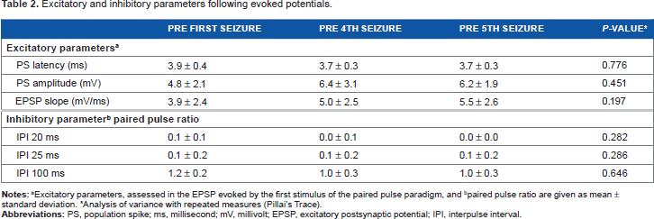
Excitatory parameters, ie, PS latency, PS amplitude, and EPSP slope, showed no significant change with the number of induced seizures (Table 2). Preseizure PPRs demonstrated an almost complete inhibition of the second PS at short IPIs of 20 ms (PPR = 0.1 ± 0.1) and 25 ms (PPR = 0.1 ± 0.2), as well as the typical slight facilitation at an IPI of 100 ms (PPR = 1.2 ± 0.2). Inhibition (IPI: 20 and 25 ms) and facilitation (IPI = 100 ms) in the dentate gyrus are robust in terms of the number of previous electrically induced seizures (Table 2).
Excitatory and inhibitory parameters following evoked potentials.
Discussion
The primary aim of this experimental study was to develop an acute model of electrically induced seizures that are confined to mesiotemporal structures. A subordinate aim was to assess whether seizures can be induced multiple times over a couple of days in the same animal with unchanged behavioral and electrophysiological seizure characteristics.
We have demonstrated that a single short train (5 seconds) of stimuli applied to the right perforant path results in a stereotyped limbic seizure lacking overt motor signs. These limbic seizures resemble complex partial seizures in humans. The current model is in contrast to the 120-minute perforant path stimulation resulting in SSSE. This induced seizure can be reproduced over a period of 72 hours with variable interseizure intervals in the same animal without changes in seizure characteristics. We did not observe significant ictal changes in behavior and in electrophysiological parameters such as latency between end of electrical stimulation and first discharge, as well as the duration and frequency of discharges over the study period. In addition, dentate gyrus excitability, as assessed by the paired pulse paradigm, remained unchanged even after repeated induction of seizures. We cannot exclude the possibility that further-induced seizures over longer periods of time result in altered brain excitability, but this was not within the scope of the current set of experiments.
Epilepsy is a severe medical condition, and around 35% of cases are pharmacoresistant. 9 Only a minority of those suffering may benefit from resective epilepsy surgery. 10 Thus, novel treatment strategies, in particular various modes of neuro-stimulation, are urgently needed. This aim has been proposed by the International League Against Epilepsy as one of the pivotal domains in epilepsy research. 11
Ideally, new treatment approaches are tested in experimental animal models first. Commonly, new anti-ictal drugs or other treatments such as deep brain stimulation are assessed in naïve animals by using acute seizure models. The general idea is to screen a wide range of substances or electrical stimulation parameters in a rather simple experimental setting and, if proven positive, to further assess this therapeutic approach in more laborious chronic models of epilepsy. In acute rodent models, seizures are induced by 50-Hz electrical stimulation of the cornea or by systemic administration of chemoconvulsants such as PTZ.12,13 All these models evoke ictal motor manifestations ranging from tonic hindlimb seizures (MES) to partial or generalized clonic or tonic seizures (PTZ). 14 In contrast, the seizures in our model have none or at least subtle motor features, the main behavioral characteristic is resting behavior with sniffing when awake and short arousal reaction when asleep. This semiology indicates limbic seizures, strongly arguing that the ictal electrophysiological pattern as recorded from the dentate gyrus indeed is restricted to mesiotemporal structures. This mostly resembles the 6-Hz psychomotor seizure model that is increasingly used again after abandonment for decades shortly after its description in the early 1950s because of its lack of sensitivity to phenytoin. 15 After 6-Hz electrical stimulation of the cornea, a minimal clonic phase is followed by stereotyped, automatistic behaviors that are reminiscent of human patients with limbic epilepsy. 16
In the current model, we have demonstrated that multiple seizures can be induced in the same animal without changes in behavioral and electrophysiological seizure characteristics and in dentate gyrus excitability as assessed by single and paired pulses. While in previously established acute seizure models, animals can be used for one experiment only (probably due to severity of the induced motor seizure),16,17 the current model may allow multiple uses. A variant of the PTZ model presents the “timed intravenous infusion PTZ test,” in which the chemoconvulsant can be precisely titrated to produce just a short myoclonic twitch. Initially, it was believed that the severity of seizures remains unchanged and thus serveral experiments can be performed in the same animal.18,19 However, recent data on the timed intravenous infusion PTZ model demonstrated that after three to five induced seizures, severity becomes increasingly intense. 20
The possibility of inducing multiple seizures in the same animal without alteration of seizure characteristics allows for intraindividual comparison of anti-ictal effects of selected treatments. In previous models, other animals had to serve as interindividual controls. In addition to assessment of pharmacological approaches, the current model system is ideal to screen for targets and optimal stimulation parameters in deep brain stimulation. Our paradigm, illustrated in Figure 1, allows for assessment of short-term (up to 1 hour) and long-term (up to 24 hours) stimulation of various brain structures in the context of prevention of mesiotemporal seizures. If defined brain targets and stimulation parameters prove to have anti-ictal properties during screening, they can be translated to and applied in model systems of chronic epilepsy with spontaneous recurrent seizures.
This preliminary experimental study is limited by the current lack of data on pharmacological and neuromodulative anti-ictal treatments. For the time being, this was beyond the scope of the current pilot experiments that, in a first step, aimed to establish an acute seizure model with data based on electrophysiology and behavioral alterations. Furthermore, we did not address possible neuronal loss in the hippocampal formation. Published data have indicated that hippocampal neurodegeneration depends on the duration of perforant path stimulation, with a cutoff point of at least 40 minutes. 21 Because we applied only an ultrashort 5-second stimulus to the perforant path, we assume that neuronal loss is not an issue in this seizure model.
Conclusion
We present preliminary behavioral and electrophysiological data on a new model system of acute seizures induced by short-term electrical stimulation of the perforant path. In contrast to most other acute models in epilepsy, seizures in this model are limbic, lacking overt motor features and thus closely resemble seizures in human mesial temporal lobe epilepsy. Furthermore, we demonstrated that seizures could multiply be induced in the same animal without hints of increased brain excitability. Further studies are needed to characterize this model in the context of pharmacological and neuromodulative treatments and histopathological consequences.
Author Contributions
Conceived and designed the experiments: AK and MH. Analyzed the data: AK. Wrote the first draft of the manuscript: AK and MH. Contributed to the writing of the manuscript: AK and MH. Jointly developed the structure and arguments for the paper: AK and MH. Made critical revisions and approved final version: AK and MH. Both authors reviewed and approved of the final manuscript.