
Abstract
Background
Endometriosis is one of the most common gynecological diseases associated with infertility. Endometriosis may affect the androgen receptor (AR) mRNA expression in human granulosa cells and the methylation of the promoter region of AR. We investigated 28 patients with endometriosis and 47 subjects without endometriosis undertaking IVF treatment.
Methods
Granulosa cells were obtained from 28 patients with endometriosis and 47 subjects without endometriosis as a control. Expressions of AR and FSHR mRNA were then evaluated by OneStep real-time PCR analysis, and the level of methylation of the promoter region was qualified by methylation-specified PCR (MSP).
Results
The expression of AR mRNA in the endometriosis group was statistically lower than that in the control group. As well, FSHR mRNA expression in the control group showed a positive correlation with AR mRNA expression; however, there was no such correlation in endometriosis patients. In the control group, AR mRNA expression was statistically higher in pregnant subjects compared with non-pregnant subjects; however, in the endometriosis group, no significant difference was identified. The promoter of AR was heavily methylated in all endometriosis cases; however, only 5 (45.4%) were methylated in the control group.
Conclusion
Lower AR mRNA expression and methylation of the AR promoter region might affect the expression of AR and FSHR the presence of endometriosis, thus leading to a disturbance in the regulation of AR and FSHR.
Introduction
Endometriosis is one of the most common gynecological diseases associated with pelvic pain and infertility. 1 An association between endometriosis and infertility has often been reported; however, the mechanism of endometriosis-associated infertility is not well understood. Lower oocyte quality, pregnancy rates and implantation rates in patients with endometriosis undertaking IVF-ET treatment were compared with patients without endometriosis.2–4 We reported that mitochondrial gene expression in granulosa cells might serve as a marker of oocyte quality and IVF-ET outcomes. 5 Furthermore, in 2011, we reported that a higher PR-A gene expression in granulosa cells might have a negative effect on both clinical outcomes and the embryo quality of patients with endometriosis undertaking IVF-ET. 6
Androgen either enhances or inhibits granulosa cell steroidogenesis, depending on the stage of follicular development. Thus, granulosa cells of dominant follicles show stronger nuclear staining for androgen receptor (AR) than in theca cells. In the luteal phase, the staining intensity of AR is strongest in the early luteal phase just after ovulation and then gradually declines. 7 Subsequent studies have demonstrated interactions between FSH and androgen in the follicular development and have revealed that follicles demonstrate a highly significant positive correlation between FSH receptor (FSHR) and AR mRNA levels when comparing the mean expression from each group of follicles in individual test monkeys. 8 It has also been suggested that androgen promotes follicular growth by sensitizing granulosa cells to FSH. 8
The mechanism by which DNA methylation exerts its respective function has not been completely defined. Many studies associated with gene inactivation with methylation depend on an analysis at one or several CpG sites typically using a methylation-sensitive restriction enzyme. Sodium bisulfite treatment has also been used to permit the examination of methylation at each CpG site within a CpG island.
In endometriosis cases, there is a possible involvement of epigenetic anomalies such as the methylation of steroidogenic factor-1 (SF-1), ER and the deacetylation of the promoter region of histones 3 and 4. Recently, Bulun mentioned genetic predisposition, or the exposure to environmental toxins, of fetal progenitor cells destined to form adult female pelvic organs may result in epigenetic events, including promoter hypomethylation and overexpression of SF-1 and ER-β, which can play critical roles in the pathogenesis of endometriosis. 9 Androstenedione (A4) induces aromatase in human endometrial cells, which might lead to a pathogenesis of endometriosis, as A4 is the predominant sex steroid in the peritoneal fluid in all phases of the menstrual cycle.10,11 Additionally, A4 has been shown to increase the proliferation and survival of endometrial cells and explants collected in the early secretory phase. 12 Thus, the influence of endometriosis on androgen activity via AR in human endometrial cells has gradually become better understood. Until now, however, there has been no report as to the expression of AR and the possible involvement of the methylation of AR in follicle development in human granulosa cells of endometriosis patients.
To evaluate how endometriosis affects the expression of AR mRNA and FSHR mRNA in granulosa cells, our study evaluated the mRNA expression of both in granulosa cells of mature follicles and compared it between endometriosis and non-endometriosis cases.
Afterwards, genomic DNA from the granulosa cells of endometriosis patients was secured, and methylation-specific PCR (MSP) was carried out in order to investigate the epigenetic modification of promoter regions of AR.
Material and Methods
Patient population
Twenty-eight patients with r-AFS stage IV endometriosis and 47 subjects without endometriosis, as a control group, were investigated to quantify the mRNA expression of AR and FSHR. Moreover, genomic DNA was collected from 11 patients with endometriosis and 11 without endometriosis for MSP. All of them undertook IVF-ET from October, 2009 to June, 2011 at Osaka Medical College. Our institutional review board approved this protocol and its consent form. Moreover, we received informed consent for this study from all participants.
Protocol for Controlled Ovarian Hyperstimulation(Coh)
Controlled ovarian hyperstimulation, hormone assays, follicle monitoring, oocyte retrieval, insemination, embryo culture and embryo transfers were performed as previously reported. 6 Embryo quality was evaluated based on Veek's classification, 13 and pregnancy was confirmed during ultrasound examination by the identification of an intrauterine gestational sac.
Isolation of Rna, Cdna Preparation and Genomic Dna Preparation from Granulosa cell
Isolation of RNA and cDNA preparation of granulosa cells was carried out as previously described. 14
Genomic DNA was extracted from granulosa cell using DNeasy Tissue kit (Qiagen, Valenica, CA) in accordance with the manual.
Real-Time PCR
Oligonucleotide primers for TaqMan probes were designed using Primer Express (version 1.0, Perkin-Elmer Applied Biosystems) using the GeneBank database, based on published sequences of mitochondrial DNA. Human glyceraldehyde-3-phosphate dehydrogenase (GAPDH) was used as an internal standard. Table 1 shows the primers used in this study and the expected sizes from the reported cDNA sequences. Quantitative analysis of AR mRNA and FSHR mRNA levels was in a 20 μL reaction containing 1× TaqMan Universal PCR Master Mix (Perkin-Elmer Applied Biosystems, Tokyo, Japan), 200nM each forward primer and reverse primer corresponding to 100 nM of TaqMan probe (Perkin-Elmer Applied Biosystems, Tokyo, Japan). PCR conditions were 95 °C for 15 seconds, followed by 60 °C for 1 minute, for 40 cycles on OneStep realtime PCR (Perkin-Elmer Applied Biosystems, Tokyo, Japan). Amplification of AR and FSHR mRNA relative to GAPDH was compared using AACt method.
Primers used in realtime PCR.
Bisulfite Treatment and ms-PCR
Bisulfite treatment of genomic DNA was performed with the EZ DNA Methylation-Gold Kit (Zymo Research), and MS-PCR was carried out for DNA of granulosa cell. The primers used for MS-PCR are shown in Table 1.
Statistical analysis
All experiments were performed in duplicates. Statistical calculations were performed by using Stat View statistical software (SAS Institute Inc., Cary, NC, USA). The statistical significance of each difference was determined using Mann-Whitney U test or χ 2 tests. The Spearman's rank correlation coefficient was also used to analyze the relation between two different values. A value of P 0.05 was considered statistically significant.
Results
Patient age
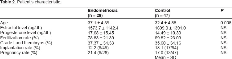
The mean age of patients with endometriosis was significantly older than that of the control group (P < 0.008) (Table 2). However, no correlation of AR and FSHR mRNA expression with the patient's age was identified in either group (Fig. 1).
Patient's characteristic.
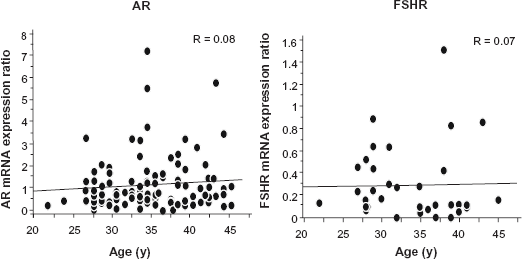
There was no correlation between age and mRNA expression of AR and FSHR.
Clinical outcomes
Serum E2 and P4 levels at oocyte retrieval did not reveal a significant difference and, furthermore, fertilization rates, high-quality embryo rates, and pregnancy rates showed no statistical difference between the two groups. Implantation rates tended to be lower in endometriosis patients, compared with those in the control group, and no significant difference was identified in either group (Table 2).
AR and FSHR mRNA Expression in Granulosa cell
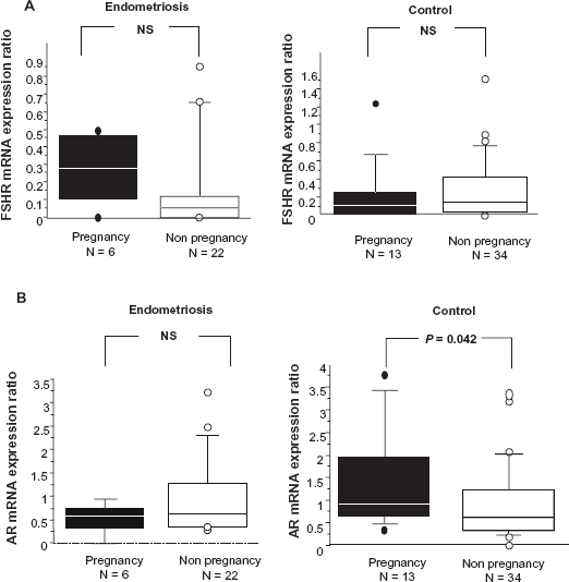
In the comparison of the AR and FSHR mRNA expression ratios between pregnant and non-pregnant subjects, FSHR mRNA expression revealed no significant difference (Fig. 2A). However, the AR mRNA of pregnant courses in the control group showed a higher expression ratio than in the non-pregnant group, whereas, such a significant difference was not identified in the endometriosis group (Fig. 2B). Moreover, FSHR mRNA expression showed no statistical difference between the two groups. On the other hand, the AR mRNA expression ratio in endometriosis patients was statistically lower than that in the control group (Fig. 3A). Additionally, FSHR mRNA expression in the control group indicated a positive correlation with AR mRNA expression; however, there was no such correlation between them in the endometriosis group (Fig. 3B).

MSP for genomic DNA secured from granulosa cell
Methylation of HRE (Hormone Response Elements) was verified in all patients (11/11) with endometriosis; however, only 45.4% (5/11) of subjects in the control group revealed methylation via MSP. Statistical difference of the methylation ratio in the endometriosis group was higher than that in the control group (P = 0.02) (Table 3). On the other hand, the fertilization, implantation, and high-quality embryo ratios in the methylated group revealed no statistical difference compared with those in the non-methylated group (data not shown).
Results of MSP.

Discussion
The present study comprises material from human granulosa cells and lymphatic cells. The data revealed that, in the presence of endometriosis, the AR mRNA expression ratio was statistically lower and the AR promoter region was heavily methylated as well.
This suggests that the methylation of the AR promoter region appears to cause a deregulation between FSHR and AR, thus leading to infertility in endometriosis patients.
Classic theories have failed to propose a precise pathogenetic mechanism for endometriosis. 15 It is considered that the poor outcome of IVF in cases of endometriosis is caused by altered folliculogenesis, 16 embryo toxicity against early embryotic development, 17 and impaired fertilization. 18 The androgen receptor is the only sex hormone receptor gene on the X chromosome, exists in 11q2-12 consisting of 8 exons, and is localized in granulosa cells. The most particular stimulatory action of androgen is the ability to enhance FSH-stimulated follicular differentiation,19–24 whereas androgen has been thought to inhibit follicular differentiation and induce follicular atresia.25,26 Ageing is also thought to induce a lower AR expression in grannulosa cells, although there have been no conclusive reports so far concerning the correlation between age and AR expression in the ovary. In our study as well, AR expression showed no correlation with patient's age. The genetic variants of AR, such as the CAG repeat alleles of the exon 1 have been correlated with the risk of developing endometriosis; however it is unclear whether endometriosis affects AR mRNA expression in granulosa cells. Recently it has been reported that AR knock-out mice exhibited longer estrous cycles and fewer ovulations, and it has also been concluded that AR promotes pre-antral follicle growth and prevents follicular atresia. 15 Transient expression of AR and FSHR mRNA in granulosa cells isolated from preovulatory follicles induced by an LH surge has also been identified27–29 and, furthermore, AR-mediated androgen signaling is considered to play an important role in the female reproductive system, such as follicular development and luteinization. 7 It has also been reported that most ovarian cells, including oocyte, granulosa, thecal, stomal and luteal cells, have the ability to process AR. In particular, granulosa cells appear to be the main site of AR expression in the ovary in several species, humans included.19,30 AR expression is highest in pre and early-antral follicles and gradually decreases as the follicle matures.7,21,22 However, AR is clearly detected in granulosa cells, as well as in theca cells, of dominant follicles even in the mid and late follicular phase. 7 Androgen is mainly utilized as a substrate for estrogen synthesis in preovulatory follicles, 20 and ovaries from rhesus monkeys showed that the AR gene was most abundantly expressed in granulosa cells in the ovary and augmented in healthy follicles. 31 Because it is unclear how endometriosis affects AR gene expression in granulosa cells, we carried out real-time PCR to confirm this. In our study, the results revealed that a higher expression level of AR mRNA leads to higher pregnancy rates in the control group, thus indicating that AR plays an important role in the process of oocyte maturation and fertilization; however, such functions of AR are thought to be disturbed in the presence of endometriosis. In studies of follicles with a diameter of 3–9 mm, a highly significant association between the expression of AR and FSHR in granulosa cells has been identified. 32 Vendola reported that AR probably does not regulate the FSHR gene because no androgen response element on the FSHR gene promoter has yet been identified. 33 The enhancement of local production of IGF1 or inhibin B might be induced by an indirect influence of AR and FSHR with AMHRII, 33 and Nielsen noted that the AMH signaling system might have an impact on the interaction of AR and FSHR expression. Our study showed the expression of AR mRNA is positively correlated with FSHR mRNA in non-endometriosis cases; however, such correlation was not confirmed in endometriosis patients. These results may imply that endometriosis has a negative effect on the interaction between AR and FSHR in granulosa cell and, therefore, we hypothesized that the HRE of AR might be methylated in the presence of endometriosis. Several studies have revealed links between DNA methylation and gene expression.34,35 Sequences near silent genes generally are methylated, whereas those near active regions are not. Hypermethylation of cytosine-rich areas (CpG island) in the promoter region of steroid receptor genes has been associated with transcriptional inactivation of genes and has been considered to be functionally equivalent to an inactivation mutation. 36 An AR promoter contains specific CpG island methylation hot spots which are markers of gene silencing. AR gene encodes a ligand dependent transcription factor involved in regulating the expression of an array of androgen-responsive genes. Recently aberrant DNA methylation patterns have been identified in a variety of human diseases, particularly cancer. In cases of prostate cancer, Takahashi et al reported that AR methylation might represent a phenotype important in the development of hormone independence in a subset of advanced prostate cancers in which AR expression is not found. 37 Although DNA demethylation of the aromatase in endometriosis patients has already been reported, 38 the epigenetic change of AR remains unknown. Our results imply that the methylation of the AR promoter region might be an important cause of the deregulation between AR and FSHR in the presence of endometriosis. In this study most subjects in the control group had partners being treated for male infertility, unlike those patients in the endometriosis group. Therefore, it would have been quite difficult to show how the methylation of HRE in endometriosis patients actually affects post-fertilization embryo development.
In conclusion, by using cDNA from granulosa cells and genomic DNA from granulosa cells, the present study revealed that endometriosis might cause the methylation of the AR promoter region, thus leading to a disturbance in the regulation of AR and FSHR. These results encourage further studies focusing on the epigenetic change of the AR promoter region by quantitative MSP in order to reveal the effect endometriosis has on the methylation of the AR promoter region.
Footnotes
Acknowledgement
The authors thank the laboratory technicians Junko Hayashi and Kumiko Sato of the Department of Obstetrics and Gynecology for their skilful and excellent work.
Author contributions
MH performed the experiments and contributed to the study design, compiled and interpreted data. YY advised and reviewed. MH and YY wrote this manuscript. AH and YY were involved in the experiments to collect samples. YT carried out laparoscopy to confirm the diagnosis of endometriosis prior to IVF. HK helped MH retrieving oocyte and granulosa cells. MO instructed the study and reviewed.
Funding
Author(s) disclose no funding sources.
Competing Interests
Author(s) disclose no potential conflicts of interest.
Disclosures and Ethics
As a requirement of publication author(s) have provided to the publisher signed confirmation of compliance with legal and ethical obligations including but not limited to the following: authorship and contributorship, conflicts of interest, privacy and confidentiality and (where applicable) protection of human and animal research subjects. The authors have read and confirmed their agreement with the ICMJE authorship and conflict of interest criteria. The authors have also confirmed that this article is unique and not under consideration or published in any other publication, and that they have permission from rights holders to reproduce any copyrighted material. Any disclosures are made in this section. The external blind peer reviewers report no conflicts of interest.