
Abstract
Introduction
Fine needle aspiration cytology (FNAC) is a reliable diagnostic tool used to diagnose breast lesions preoperatively. However, FNAC is also associated with diagnostic pitfalls. Further studies are needed to improve its diagnostic efficacy. We noticed ovoid, bare nuclei arranged in closely touching diads (benign pairs) in a significant number of cytology smears. This prompted us to assess their diagnostic utility.
Materials and Methods
This was a prospective study conducted in Sarojini Naidu Medical College, Agra, India. Quantitative estimation of benign pairs per 1000 ductal cells in at least 20 high power field was attempted in cytology smears of 128 cases.
Results
The average number of pairs in benign and malignant lesions was calculated as 7.07 ∓ 5.96 and 0.28 ∓ 0.78, respectively. Statistical analysis showed a significant difference between the number of pairs in benign and malignant cases (
Conclusions
Quantitative estimation of benign pairs is helpful in distinguishing benign from malignant cases.
Introduction
Fine needle aspiration cytology (FNAC) is a simple and effective tool used to distinguish benign from malignant lesions.1–3 The presence of small, naked bipolar cells in breast FNAC has proved to be an important cytomorphological feature.3–5 Naked nuclei often appear as pairs in close apposition or gently touching each other. Trott 6 reports that these pairs are almost exclusively found in benign lesions. This observation led him to label these pairs as benign pairs. In this study, we studied the differences in numbers of benign pairs in breast lump aspirates from breast lesions and their diagnostic utility along with histological correlation.
Materials and Methods
This was a prospective study conducted in Sarojini Naidu Medical College, Agra, India. Before conducting this study, permission from the Institutional Ethical Committee was obtained. A total of 128 cases, who were advised fine needle aspiration cytology (FNAC) by the surgery department, were recruited using simple random sampling methods from 2008 through 2009 (December 1, 2008 to August 31, 2010). All the patients who participated in this research gave their written, informed consent. The breast tissue was aspirated using blue-hub 23-G needle (EROSE Glass Agencies, Ambala, India) 7 and cytology smears were subjected to May-Grunwald Giemsa stain (BioLab Diagnostics, Mumbai, India). 8
Quantitative estimation of benign pairs per 1000 ductal cells was done by 3 different observers (2 pathology residents and 1 pathology professor with 35 years of experience in the field of pathology) and their average was calculated. At least 20 High Power Field (HPF) with least overlapping of cells were selected.
The benign pairs are defined as small, ovoid, bare nuclei with a hyperchromatic homogenous chromatin pattern that are arranged in closely touching diads. 7
Results
A total of 128 cases were subjected to FNAC. Six cases were inadequate. The distribution of cases according to diagnosis is shown in Table 1.
Distribution of cases according to diagnosis.

Table 1 shows that the maximum number of cases were benign (101/122 or 82.79%) and the maximum number of cases were of fibroadenoma (44/122 or 36.10%). *Six cases were inadequate.
Qualitative estimation of benign pairs in various breast lesions is shown in Table 2.
Qualitative estimation of benign pairs in various breast lesions.
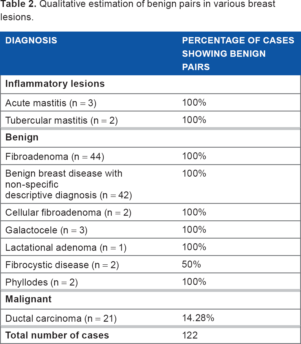
Thus benign pairs were seen in all 100% cases of fibroadenoma, benign breast disease with nonspecific descriptive diagnosis, cellular fibroadenoma, and phyllodes tumor and in 50% of cases of fibrocystic disease and 14.28% of malignant cases.
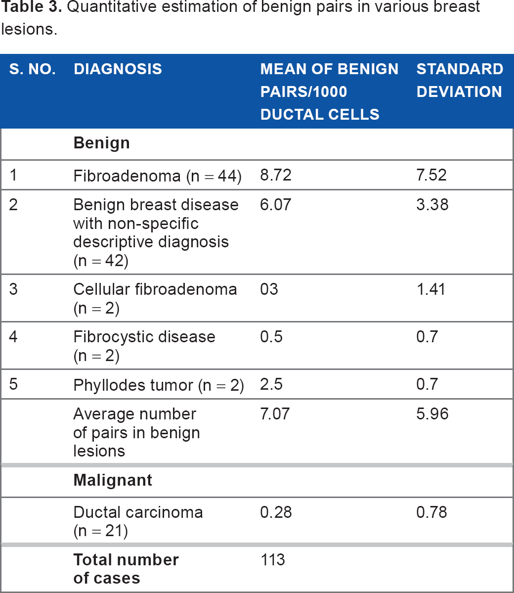
Quantitative estimation of benign pairs in various breast lesions is shown in Table 3.
Quantitative estimation of benign pairs in various breast lesions.
It is important to note that though benign pairs were present in 100% cases of acute mastitis, tubercular mastitis, galactocele, and lactational adenoma (Table 2), it was difficult to assess the number of benign pairs in at least 20 HPF and the number per 1000 ductal cells in these conditions.
The number of pairs was higher in benign lesions with an average of 7.07 ∓ 5.96 (Figs. 1A-1D). The maximum number of pairs was seen in fibroadenoma (8.72 ∓ 7.52). The average number of pairs was significantly reduced in ductal carcinomas and was just 0.28 ∓ 0.78 (Figs. 2A-2D).

Cytology of benign lesions of breast showing benign pairs.

Cytology of ductal carcinoma of breast showing benign pairs and pseudopair.
Sometimes an entity known as pseudopairs was also seen, wherein in cases of ductal carcinoma, 2 malignant epithelial cells showing prominent nucleoli were present in diads (Fig. 2D). Such diads were carefully omitted.
Considering benign pairs as the index of benignancy, the sensitivity, specificity, false positive rate, false negative rate, negative predictive value, and positive predictive value to detect a benign case were calculated.
The following values were obtained:
Sensitivity = a/(a + c) × 100 = 98.91%
Specificity = d/(b+d) × 100 = 85.71%
False positive rate = b/(b+d) × 100 = 14.28%
False negative rate = c/(a+c) × 100 = 1.08%
Positive predictive value = a/(a+b) × 100 = 96.80%
Negative predictive value = d/(c+d) × 100 = 94.73%
The nonparametric Mann-Whitney
In the present study, out of 92 benign cases, histology was available for 34 cases (36.95%), and, out of total malignant cases (21), histology was available for 14 cases (66.66%). No discrepancy was noted. However, those cases in which histology was not available, showed cytologically unequivocal malignant or benign features.
Discussion
We studied 128 cases, out of which 6 cases (4.69%) were inadequate considering the adequacy criterion specified by Eckert 9 and Lester et al. 10
One hundred and one cases (82.79%) were benign with the majority of cases being fibroadenoma (n = 44) and benign breast lesions with nonspecific descriptive diagnosis (n = 42). Twenty-one cases (17.21%) were malignant (ductal carcinoma).
Benign pairs were found in 100% of cases of fibroadenoma, benign breast disease, cellular fibroadenoma, and phyllodes tumour; in 50% of cases of fibrocystic disease; and in 14.28% of cases of ductal carcinoma.
All the cases of acute mastitis, tubercular mastitis, and galactocele and lactational adenoma (13 cases in this study) showed benign pairs in one field or the other.
However, quantitative estimation of the average number of benign pairs/HPF per 1000 ductal cells could not be done in these conditions. In acute mastitis, the number of ductal cells itself was less than 1000; in tubercular mastitis, the necrotic background and necrosed cells hindered in counting, and in galactocele and lactational adenoma, there was obscuring of cell morphology by lipoproteinaeous material and overlapping of ductal cells at most places. Although quantitave estimation of benign pairs could not be done in these cases, 100% of these cases showed benign pairs in one field or the other.
Benign pairs were counted per 1000 ductal cells by 3 different observers, and the average value was calculated to improve accuracy.
The average number of benign pairs per 1000 ductal cells were highest in fibroadenoma, corresponding to 8.72 ∓ 7.52, followed by benign breast lesions with nonspecific descriptive diagnosis (6.07 ∓ 3.38), cellular fibroadenoma (3 ∓ 1.41), phyllodes tumor (2.5 ∓ 0.7), and fibrocystic disease (0.5 ∓ 0.7). Few cases of ductal carcinoma showed pairs; the average number was 0.28 ∓ 0.78.
The nonparametric Mann-Whitney
The differences in the average number of benign pairs among the various benign conditions was not significant (
Benign pairs as an index of benignancy of a case.

The sensitivity, specificity, false positive rate, false negative rate, false negative rate, positive predictive value, and negative predictive value of benign pairs to detect a benign breast disease were calculated to be 98.91%, 85.71%, 14.28%, 1.08%, 96.80%, 94.73%, respectively.
Sturgis et al. 11 also found the highest number of pairs in cases of fibroadenoma (7.3 benign pairs/10 HPF) followed by fibrocystic disease (3.5 benign pains/10HPF). They found 1 to 2 benign pairs in carcinoma, which were thought to be derived from adjacent nonneoplastic breast tissues. They reported such benign pairs in 68% of benign lesions and 3.8% of cases of carcinoma. These authors did not comment upon the statistical significance of these differences.
These authors also said that the presence of benign pairs appears to be a useful discriminating feature in subclassification of benign lesions, particularly in the differential diagnosis of fibroadenoma and fibrosis/fibrocystic disease. They found benign pairs in 89% of fibroadenomas and 53% of fibrocystic disease. In the present study, 50% of the cases of fibrocystic disease showed pairs whereas 100% cases of fibroadenoma showed pairs.
Yu et al. 12 reported pairs in 70% benign lesions and 1% cases of carcinoma. They also found that such pairs were a more specific indicator of benign entity when compared with single nuclei alone.
Pattari et al. 13 did not find pairing of myoepithelial cells as a significant observation even in benign lesions. 12 Our study also reconfirms the findings of Sturgis et al. 11 and Yu et al. 12
Thus, we conclude that the number of benign pairs on cytology smears is an important criterion to distinguish benign from malignant lesions. However, it does not help us in subclassification of benign lesions. We sincerely hope that further larger and independent studies will help us in understanding the significance of benign pairs in cytology smears of the breast.
Footnotes
Author Contributions
Conceived and designed the experiments: MR, PK, AG, SKB, HA. Analyzed the data: MR, PK, AG, SKB, HA. Wrote the first draft of the manuscript: MR. Contributed to the writing of the manuscript: MR, PK, AG, SKB, HA. Agree with manuscript results and conclusions: MR, PK, AG, SKB, HA. Jointly developed the structure and arguments for the paper: MR, PK, AG, SKB, HA. Made critical revisions and approved final version: MR, PK, AG, SKB, HA. All authors reviewed and approved of the final manuscript.
Disclosures and Ethics
As a requirement of publication the authors have provided signed confirmation of their compliance with ethical and legal obligations including but not limited to compliance with ICMJE authorship and competing interests guidelines, that the article is neither under consideration for publication nor published elsewhere, of their compliance with legal and ethical guidelines concerning human and animal research participants (if applicable), and that permission has been obtained for reproduction of any copyrighted material. This article was subject to blind, independent, expert peer review. The reviewers reported no competing interests.