
Abstract
Apoptosis is an active process of programmed cell death. Fas is a cell-surface protein which is expressed on activated lymphocytes and known as CD95, TNFRSF6 or APO-1. Fas-L is ligand of Fas and known as CD95 LG or TNFSF6. Apoptosis or cell death is a result of binding of Fas-L to Fas which is expressed on the surfaces of these cells. Cancer cells escape this binding by overexpression of Fas-L or down expression of Fas. Fas and Fas-L exist in membrane bound and soluble forms. The serum level of sFas and sFas-L can be evaluated by immunostaining, immunohistochemical methods, immunofluorescence, flow cytometry and Western blotting. Head and neck squamous cell carcinoma diagnosis, staging and prognosis can be evaluated early and accurately by sFas and sFas-L expression levels detection.
Short Commentary
Apoptosis is an active process of programmed cell death which was first described by Kerr et al in 1972. 1 It is a process of destruction and elimination of aged or damaged cells with reusage of their contents by macrophages. 2 This process is genetically controlled.
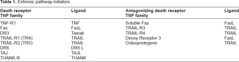
Apoptosis can be stimulated through two main pathways: the intrinsic and extrinsic death pathways. 3 The intrinsic pathway produces damage within the cell by the effect of different apoptotic stimuli including anticancer agents, oxidative damage, UV irradiation, and is mediated through the mitochondria. The extrinsic pathway is initiated by the tumour necrosis factor receptor family members which have extracellular ligands inducing oligomerization of death receptors such as Fas. 4 Table 1 shows the different extrinsic pathway initiators. 5
Extrinsic pathway initiators.
Fas is a cell-surface protein which is expressed on activated lymphocytes and known as CD95, TNFRSF6 or APO-1. Fas-L is ligand of Fas which causes transudation of the apoptotic signal from the cell surface into the cytoplasm, thereby triggering cell death and known as CD95LG or TNFSF6. Apoptosis or cell death is a result of binding of Fas-L to Fas which is expressed on the surfaces of these cells.
Cancer cells escape this binding by overexpression of Fas-L or down expression of Fas and this mechanism of immune escape has been reported in squamous cell carcinoma (SCC) of the head and neck.6–12 By this escaping process, malignant cells escape the apoptosis process. So, the ability of the tumour cells to produce Fas-L opposes the ability of lymphocytes to produce Fas playing an important role in escaping the patient's immune system helping tumour growth. Fas and Fas-L exists in membrane bound and soluble forms. 13
It has been found that the loss of Fas expression in primary tumours correlates with disease progression or metastasis. 14 Patients with Fas positive tumors exhibited significantly longer survival than patients with Fas-negative tumours in various types of carcinomas.
High serum sFas-L (soluble Fas-L) levels in cancer patients may represent a further mechanism of immune escape as a result of an attack against immune cytotoxicity cells15–17 and subsequent tumour survival advantage. 16
The serum level of sFas and sFas-L can be evaluated by immunostaining, immunohistochemical methods, immunofluorescence, flow cytometry and Western blotting.18,19
The human Fas gene is located on chromosome 10q24.1, and consists of nine exons and eight intons, and encodes 334 amino acids. 20 The human Fas-L gene is located on chromosome 1q23, and consists of four exons and encodes 281 amino acids. 21
Tumour cells with decreased levels of Fas may be resistant to cytotoxic drugs because the Fas/Fas-L signaling system plays an important role in chemotherapy-induced apoptosis. 22 Recently, it has been shown that apoptosis induced by c-myc oncoprotein is dependent on Fas expression. 23 Accordingly, the loss of Fas expression may result in the resistance of c-myc-transformed tumour cells to apoptosis and contribute to tumour growth.
Various types of malignant tumour cells are known to acquire resistance to Fas receptor (Fas)-mediated apoptosis. In Fas-sensitive cells, Fas-mediated apoptosis is observed when anti-Fas antibody is bound to Fas. Bcl-2 and Bcl-XL are representative anti-apoptosis proteins reported to be capable of suppressing Fas-mediated apoptosis. 24 Malignant tumour cells that acquire resistance to apoptosis are also resistant to chemotherapy or radiotherapy. 25
Fas and Fas-L expression was detected in oral and oropharyngeal SCCs. A significant correlation was found between tumour staging and Fas/Fas-L expression frequency. 25
Fas-L expression is highest in T3 SCCs. SCC is the most common malignancy of head and neck regions. It is important that these lesions be diagnosed early and accurately by sFas and sFas-L expression levels detection.
El-Badry et al found increasing level of serum sFAS in patients with an increased risk of laryngeal cancer. 26 In a study of postsurgical removal of T2 laryngeal SCC, serum level of sFas and sFas-L was significantly decreased indicating that there is a link between the serum level of sFas and sFas-L and laryngeal SCC. 27
In another study, the combined FAS genotypes were associated with a statistically significantly increased risk of pharyngeal cancer particularly among older women and drinkers. 28
Fas also was found to be highly expressed in carcinoma of the tongue. 29
Disclosures
This manuscript has been read and approved by the author. This paper is unique and is not under consideration by any other publication and has not been published elsewhere. The author and peer reviewers of this paper report no conflicts of interest. The author confirms that they have permission to reproduce any copyrighted material.