
Abstract
Piroxicam is one of the important therapeutic nonsteroidal anti-inflammatory class of drugs used mainly to suppress pain and inflammation in arthritis and other musculoskeletal disorders. Besides being anti-inflammatory, these drugs are analgesic and antipyretic often used for the relief of nonspecific fever condition. Recently, piroxicam has also gained attention as an effective therapy for tumors, colorectal, and invasive bladder cancers. The objective of the current study is to evaluate the protective effects of the alcoholic leaf extract of Hibiscus rosa-sinensis (AEH), Malvaceae, against piroxicam-induced toxicity in mice. Sixty adult Swiss albino mice (Mus musculus) were divided into four groups (n = 10), which included a control group, a group treated orally with AEH (30 mg kg–1 b.w.) for 15 days, a group treated orally with piroxicam (6.6 mg kg–1 b.w.) for 15 days, and another group treated orally with piroxicam and AEH for 15 days. The results indicated that treatment with piroxicam alone resulted in a significant increase in the activities of serum marker enzymes, namely, aspartate transaminase, alanine transaminase, and alkaline phosphatase with profound hepatic lipid peroxidation as evidenced by a marked increment in the level of thoibarbituric acid reactive substances along with a distinct diminution in reduced glutathoine content and various antioxidant enzymes such as superoxide dismutase, catalase, and glutathione peroxidase in the liver. However, treatment with AEH during piroxicam treatment retrieved or partially antagonized the effects induced by piroxicam toward the normal values of controls. Histopathological observations also corroborate with the above findings. It can be concluded that AEH exhibited a protective action against piroxicam toxicity and effective in combating oxidative stress-induced hepatic damage.
Introduction
The liver is a vital organ and plays amazing functions such as protein synthesis, production of biochemicals, including fight against disease, removal of toxic substances from the body, thus maintaining and regulating homeostasis of the body. Further, the main functions that can be attributed to this organ are the metabolism of the body, including the regulation of glycogen, the synthesis of plasma protein, the production of hormone, bile secretion, vitamin storage, and the essential task of detoxification. The hepatocytes, a highly specialized tissue of the liver, also help in controlling high volume biochemical reactions necessary for normal vital functions. The liver is very much susceptible to the toxicity from the agent such as drugs when taken in overdoses. Even if the drugs are introduced within the therapeutic ranges, it may cause an injury to the organ and thus inducing hepatotoxicity.
Drugs can induce oxidative stress by generating free radicals that are mostly available as by-products or as an aerobic metabolic product. These free radicals when generated excessively at cellular level may cause damage to tissue proteins, nucleic acids, and membrane lipids, and are associated with many age-related problems.1,2 The balance between the production and scavenging of reactive oxygen species (ROS) or free radicals determines the susceptibility of the body to the oxidative damage. The self-antioxidant defense mechanisms of an organism minimize the production of free radicals, thus protecting the oxidative damage, but may not be sufficient to prevent the damage entirely. The level of these defense mechanisms may not be altered through the introduction of the drugs and there is ineffective scavenging of free radicals that may cause tissue injury.
Nonsteroidal anti-inflammatory drugs (NSAIDs) are important therapeutic class of drugs used to suppress pain and inflammation in case of rheumatoid arthritis, osteoarthritis, and other inflammatory diseases. 3 Besides being anti-inflammatory, these drugs are analgesic and antipyretic and are often used for the relief of nonspecific fever conditions. 4 More than 100 million NSAIDs are prescribed throughout the world5,6 and are associated with liver injury.7–10 Piroxicam (an acidic carboxamide), which belongs to a chemical subgroup of NSAIDs that are oxicam derivatives, a class of enolic acids are advocated for use in various painful and inflammatory conditions, specially as single largest group of NSAIDs associated with the palliation of symptoms rheumatoid arthritis, ankylosing spondylitis, and musculoskeletal disorders. 11 Recently, piroxicam (a nonspecific COX inhibitor) has also gained attention as an effective therapy for tumors, colorectal, and invasive bladder cancers. 12 Despite its widespread use, it can cause many adverse effects such as severe gastrointestinal toxicity, ulcerogenic gastropathy, renal hemostatic abnormalities, 13 proteoglycan synthesis from chondrocytes, foetotoxicity and other processes depending on prostaglandins. 14 Like other NSAIDs, the mechanism of action of piroxicam involves reduction of prostaglandin synthesis by inhibiting cyclooxygenase enzyme through competitive antagonism for arachidonic acid. 15 Accordingly, by inhibiting the prostaglandin synthesis, indirectly reducing gastro protective mucin secretion and an increased risk of ulceration arises. The central roles in liver in drug metabolism predispose them to toxic injury. Since piroxicam is metabolized in the liver, there is a possibility of injury in the liver. The toxicity developed due to piroxicam is mediated through oxidative stress, which leads to lipid peroxidation (LPO) and free radical generation. Accordingly, there occurs hepatic dysfunction and failure.
With this in view, much attention has been paid on the protective effect of some naturally occurring antioxidants on the living system. In ayurvedic treatment, different parts of the plant have been prescribed for different ailments. The active principle from the plants has been identified and become useful in curing various diseases.16,17 The present investigation was undertaken to study the protective effect of the alcoholic leaf extract of Hibiscus rosa-sinensis (AEH) on antioxidant status against piroxicam-induced hepatotoxicity in mice.
Materials and Methods
Plant material
H. rosa-sinensis (Malvaceae) was identified by a plant taxonomist of Botany Department, Kalyani University. The matured fresh green leaves were collected, shed, dried and powdered (about 500 g), and later subjected to extraction with 70% ethanol (1.5 L), then made them into a semisolid mass under reduced pressure following the methods described by Srinivasan et al and Essa et al.18,19 The extract was dissolved in a double distilled sterile water and was used in the investigation.
Experimental animals
Swiss albino male mice (Mus musculus) (20–25 g) were purchased from the supplier and were acclimatized in the laboratory for 7 days. They were maintained and housed in polypropylene cages in the departmental animal house under room temperature (25 ± 1 °C) with 12 h light and dark cycle and provided with standard pellets and water ad libitum. All experiments were approved by the ethical committee (vide No 892/ac/05) constituted through CPESCA. The chemicals used were of AR grade.
Experimental design
The mice were selected randomly and divided into four groups of 10 each and fed with normal diet. The extract, the dissolved drug, and double distilled sterile water were administered orally.
Group A: Control (only normal diet)
Group B: Mice treated with AEH (30 mg kg–1 b.w.) for 15 days
Group C: Mice treated with piroxicam (6.6 mg kg–1 b.w.) for 15 days
Group D: Mice treated with piroxicam and AEH for 15 days
Hepatoprotectivity
At the end of the scheduled treatment, the blood samples were collected from sacrificed mice by cardiac puncture under ether anesthesia and allowed to clot at room temperature for 45 minutes. Serum was separated by centrifugation at 4,000 rpm at 4 °C for 15 minutes and was used for the assay of serum marker enzymes, for example, aspartate aminotransferase (AST), alanine aminotransferase (ALT), following the method of Reitman and Frankel, 20 and alkaline phosphatase (ALP) was determined by the method of Kind and Kings. 21 It was well known that the activities of serum transaminases and phosphatases generally represented the functional status of the liver. Liver samples were taken and immediately washed in ice-cold saline for removal of blood as much as possible. It was weighed, and 10% (W/V) tissue homogenate was prepared in phosphate-buffered saline (pH 7.2) to measure LPO in terms of thiobarbituric acid reactive substances (TBARS) following the method of Neihaus and Samuelsson. 22 Glutathione (GSH) (reduced glutathione [GSH]) and superoxide dismutase (SOD) were measured by the method of Eillman 23 and Kakkar et al. 24 , respectively. However, the other two hepatoprotective enzymes catalase (CAT) and glutathione peroxidase (GPx) were measured as per the methods of Sinha 25 and Rotruck et al. 26
Histopathology
For histopathological studies, a portion of the liver was taken and fixed in 10% formalin. After the scheduled period of fixation and dehydration, routine histological technique was followed for section cutting at 6 nm. The tissue sections were thoroughly stained with hematoxylene and eosin stains and observed under microscopes, and good stained sections were photographed. 27
Statistical analysis
Results of biochemical estimations were presented as mean ± standard error of mean of five repeated determination for 10 mice in each of the 4 groups of mice. The significance of difference in the means of all parameters was determined using one-way analysis of variance. Difference was considered to be significant when P < 0.05.
Results
Table 1 shows the activity of serum enzymes in the normal and experimental groups. The enzyme activity was significantly higher in piroxicam-treated mice. Mice coadministered with piroxicam and AEH showed significantly lower activity when compared to corresponding piroxicam-treated group. Mice treated with AEH alone did not alter the enzyme activity when compared to the normal values.
Table 2 shows that the level of LPO was higher, whereas the levels of SOD, CAT, GSH peroxidase (GSH-Px), and GSH were significantly low in the liver of piroxicam-treated mice. Mice treated with piroxicam and AEH showed significantly (P < 0.05) low levels of LPO and significantly (P < 0.05) elevated levels of SOD, CAT, GSH-Px, and GSH when compared with the corresponding piroxicam-treated group.
Effect of AEH on changes on serum marker enzymes of normal and treated mice.

P < 0.01 compared with the control group.
P < 0.05 compared with the treated group. @Activities are expressed as units/ml.
Effect of AEH on changes on oxidative stress related enzymes of normal and treated mice.

P < 0.01 compared with the control group.
P < 0.05 compared with the treated group.
Histopathological observations
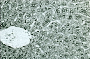
Liver section of the controlled mice showed normal histology at the centrilobular and periportal regions (Fig. 1). No significant alterations were observed only in AEH-treated mice. Histopathological observation of piroxicam-treated mice liver showed some abnormalities compared to the tissue sections of the control/normal liver. Some of the abnormalities encountered as a fatty degeneration, vacuolations, and sinusoidal dilations (Figs. 2–4). Pycnotic and hypertrophied nuclei were also some prominent features available in the section of liver of mice treated with piroxicam (Fig. 5). Administration of the AEH concurrently with the drug helps to maintain the normal architecture of the liver, except few mild irregularities (Fig. 6).

T.S. of Liver of control mice showing normal histological architecture (H & E, 200 X).

T.S. of Liver of piroxicam treated mice showing sinusoidal dilation (H & E, 200 X).

T.S. of Liver of piroxicam treated mice showing fatty changes (H & E, 200 X).
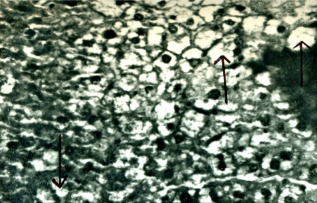
Magnified view of T.S. of Liver of piroxicam treated mice showing vacuolations (solid arrow) in the cord cells. (H & E 400 X).
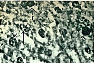
Magnified view of T.S. of Liver of piroxicam treated mice showing pycnotic (solid arrow) and hypertrophic (broken arrow) nuclei. (H & E 600 X).
T.S. of Liver showing normal arrangement of cord cells in piroxicam and AEH co-administered mice (H & E, 400 X).
Discussion
It is a well established fact that the oxidative stress developed due to the introduction of the xenobiotics, causing damage to the cells via oxidative stress-mediated LPO.28,29 The use of various dietary antioxidant treatments in terminating or reducing free radical attacks30–32 that are involved in various diseases are also an important part of the antioxidant mechanism. The antioxidant may act as free radical scavengers, reducing agents, and activators of antioxidative defense enzymes system to suppress the radical damage in biological system. 33
Piroxicam is the most commonly used drug causing hepatotoxicity in the experimental study. The peroxidative degradation of the lipid membrane is one of the principal causes of hepatoxicity. Studies have revealed that the mechanism of piroxicam heoatotoxicity relates both to impairment of adenosine triphosphate synthesis by mitochondria and production of active metabolites, particularly 5-hydroxy piroxicam, which causes direct cytotoxicity. 34 Induction of mitochondrial membrane permeability transition has also been shown to be important in NSAID-induced liver injury, resulting in the generation of ROS, mitochondrial swelling, and oxidation of nicotinamide adenine dinucleotide phosphate and protein thiols. Similar events might also occur in this case. The present study shows that the levels of serum marker enzymes, eg, AST, ALT, and ALP in the experimental Group C mice become elevated, which may be due to the liver damage caused by drug-induced free radical generation. The review made by Pandit et al. 35 also shows an elevated level of liver enzymes in patients regularly taking diclofenac. The elevated levels of transaminases have also been described by Sokolove et al. 36 with the use of TNF drugs, for example, adalimumab, etanercept, and infliximab. The type of liver injury is associated with the relative rise of ALT and ALP is documented by Hussaini and Farrington37,38 and that too confirm the results of the present study. The administration of AEH in Group D has significantly reduced these liver enzyme levels.
The present experiment also shows the elevated levels of LPO in piroxicam-treated (Group C) animals. The increase in TBARS levels in liver suggests the enhancement of LPO generating free radical, which is deleterious for the cell membrane. Increased LPO damage the membrane function considerably by decreasing membrane fluidity and changing the activities of membrane bound enzymes, leading to oxidative stress. This phenomenon also suggests the failure of antioxidant defense mechanism to some extent. Treatment with AEH (Group D) significantly prevents these changes by suppressing LPO level. This may be due to the free radical scavenging properties of AEH.39,40 Since, AEH in Group D animals has significantly increased the SOD, CAT, GSH, and GSH-Px contents of the liver, it may also be important in preventing hepatotoxicity caused by the drug. The drug decreased the antioxidant level in Group C, whereas AEH-treated group (Group B) is almost similar to the normal group (Group A).
For the cellular antioxidant defense system, reduced GSH can be considered as one of the most important agents, thus protecting the cell against damage from exposure to oxidizing agents. 41 During cellular metabolism, ROS are formed continuously, which are normally prevented or scavenged by a host of antioxidants.42–44
The SOD and CAT may play an important role in detoxification of superoxide anion and hydrogen peroxide, respectively, thus protecting the ROS-induced damage. GSH in conjunction with GSH-Px helps protection against free radicals and the toxic compounds.45,46 The increased level of liver antioxidant enzyme activities in piroxicam and AEH treated mice may be due to the presence of chemical compounds notably flavonoids in AEH, which may have a positive role in reducing the oxidative stress by inducing cellular antioxidant enzymes.
Histopathological studies showed that the drug induces fatty degeneration and necrosis in the liver tissue. The results of the present histopathological study are an agreement with the work of Bessone 8 who noticed liver injury in the form of necrosis by the use of nimesulide. Treatment of AEH shows the reversibility of the original condition in liver tissue, thus indicating the protection against drug-induced liver toxicity. Further studies are needed to investigate and isolate the active ingredients for possible mechanism of action of the extract in controlling toxicity. This will also add to our understanding of the role of H. rosa-sinensis L. in ameliorating chemical carcinogenesis due to the prolonged use of chemicals/drugs.
Author Contributions
Conceived and designed the experiments: CS. Analyzed the data: CS. Wrote the first draft of the manuscript: CS. Contributed to the writing of the manuscript: CS. Agree with manuscript results and conclusions: CS. Jointly developed the structure and arguments for the paper: CS. Made critical revisions and approved final version: CS. The author reviewed and approved of the final manuscript.