
Abstract
Background
The impact of infiltrating macrophages on tumor progression in malignant gliomas has been studied extensively. However, there is a lack of animal models for studying the role of infiltrating macrophages in malignant gliomas.
Results
BT4C malignant gliomas are highly vascularized tumors with an infiltrative behavior. BT4C gliomas demonstrated a high infiltration rate of macrophages. Particularly, a CD68/VEGFR-1 positive subtype of macrophages was detected at the edges of malignant gliomas. Also, CD133 positive cells were located mainly at the infiltrative edges of gliomas, whereas VEGFR-2 was highly expressed throughout the malignant glioma.
Conclusion
The immunocompetent BT4C rat malignant glioma model shows features similar to its human counterpart, which makes it a valuable model to study the impact of tumor associated macrophages in the pathology of malignant gliomas.
Introduction
Malignant gliomas account for more than 70% of all brain tumors. The most frequent and at the same time most malignant type of glioma, ie, glioblastoma multiforme (GBM), accounts for 65% of all cases. 1 The treatment of these tumors has been particularly challenging because of their infiltrative behavior into the surrounding healthy brain tissue. Despite the recent advances in surgical techniques, curative resection of the tumor is considered virtually impossible, depicting the need to develop novel therapeutic modalities. 2 In recent years the impact of the tumor microenvironment on tumor cells has become a topic of intense research. Even though much knowledge has been gained regarding the molecular biology of cancers, there is still an unsatisfactory understanding about the interaction between the tumor cells and their microenvironment.3,4 For this reason, much attention is currently being directed towards studying the tumor microenvironment, as to understand its impact on the pathology of cancers and their metastasis. 3 One of the distinct characteristics of malignant gliomas is the profound infiltration of macrophages into the tumors. In humans, up to 30% of the tumor mass can be composed of macrophages. 5 Their contribution in promoting angiogenesis and supporting tumor cell invasion and metastasis has been reported in several studies.6,7 In gliomas, the infiltration of microglia cells and/or macrophages has been shown to correlate with tumor progression, histological grade and poor prognosis. 8 The paradoxical role of macrophages has been explained by the functional plasticity of these cells, being able to express different sets of cytokines depending on the stimuli coming from the microenvironment.
In order to better understand the interactions between tumor cells and tumor microenvironment, appropriate animal models are required. However, there is a lack of valuable immunocompetent animal models that would bear similar features to that of the human disease. Traditionally, ectopic models (ie, subcutaneous implantation of malignant glioma cells) have been used to study the in vivo efficacy of therapeutics. However, these models have little relevance with respect to the human disease. Alternatively, orthotropic models are more appropriate, but also in this case most of the commonly used models that are immunocompromised models. The aim of this study was to characterize the immunocompetent BT4C malignant rat glioma model in order to evaluate its relevance in studying the interaction between the tumor microenvironment and tumor cells.
Materials and Methods
Animal model
Inbred male BDIX rats (n = 8) (Charles Rivers Laboratories, France) weighing between 175–200 g (6–8 weeks of age) were inoculated with 10,000 BT4C cells in 5 μl into the right corpus callosum at a depth of 2.5–3.0 mm over a time period of 2–3 minutes. A 27G 25 μl Hamilton-syringe (Hamilton, Bonaduz Ab, Switzerland) attached to a stereotactic apparatus (David Kopf instruments) was used for cell transplantation. 9 The presence of the tumor was verified on day 14 post tumor cell inoculation with Magnetic resonance imaging (MRI). For immunohistochemical analysis purposes, animals were perfused transcardially with PBS and sacrificed at the end of a survival study, after which brains were resected and embedded for further analysis. All protocols were approved by the Ethics Committee for Animal Experiments, University of Eastern Finland.
Histology
Immunohistochemical staining was performed on paraffin embedded tissue samples using primary antibodies as follows: goat anti-rat CD34 (R&D Systems), goat anti-rat VEGFR-1 and 2 (1:200, Santa Cruz), mouse anti-rat CD68 (1:500, AbD Serotec), goat anti-rat CD163 (1:500, Abcam) and mouse anti-rat CD133 (1:50, Abcam). Primary antibodies were incubated overnight at 4 °C. Secondary antibodies (anti-goat and anti-mouse) were used in a 1:200 dilution. The sections were counterstained with hematoxylin. In the double staining procedure, tissues samples were first incubated with the CD68 antibody overnight at 4°, after which slides were washed three times with PBS for 5 min. and incubated with the secondary antibody for 30 min. An avidin-biotin-HRP system (Vector Elite, Vector Laboratories) was used for signal detection. Then, slides were incubated overnight at 4° with the anti-VEGFR1 antibody. The following day slides were washed with PBS three times for 5 minutes and incubated with the secondary antibody for 30 minutes. An avidin-biotin-AP system (Vector Laboratories, Burlingame, CA) was used for signal detection.
Results
BT4C gliomas demonstrate an infiltrative and invasive behavior within BDIX rats
In this study we characterized BT4C malignant gliomas by immunohistochemical analysis in order to evaluate its value as a model to study the interaction between the tumor microenvironment and tumor cells. In all animals, H&E staining revealed a profound infiltrative behavior of BT4C malignant gliomas into the surrounding healthy brain tissue, in addition to the formation of microsatellite lesions in close proximity to the tumor edges (Fig. 1A and B). Furthermore, when the malignant gliomas were stained with the stem cell marker CD133, positive cells were predominantly seen at the edges of the tumor, at their infiltrative arms (Fig. 1C), but also offsite the tumor (Fig. 1D). Additionally, occasionally BT4C malignant gliomas have shown to metastasize occasionally to distant brain areas, as a result to anti-angiogenic therapy (Fig. 1E). Of note, there is currently an intense discussion about the possibility of anti-angiogenic therapy to induce invasion and metastasis in cancer, as similar results have also been observed in the clinics.10–12
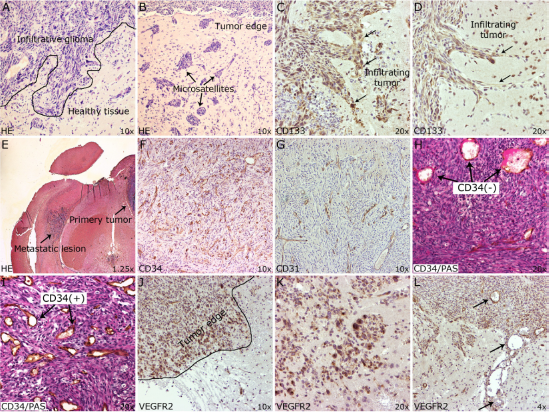
H&E staining of BT4C malignant gliomas. The staining demonstrates an infiltrative nature of these tumors (
BT4C gliomas are highly angiogenic tumors
In our study, BT4C malignant gliomas proved to be highly angiogenic tumors, visualized by CD34 and CD31 staining (Fig. 1F and G). CD34 and CD31 positive vessels were detected throughout the tumor in all animlas (Fig. 1F and G). Furthermore, CD34/PAS double staining, commonly used as a marker for vasculogenic mimicry, revealed some positive vessels without endothelial lining (Fig. 1H and I). Interestingly, vasculogenic mimicry was observed more often in the center of the tumor than at its infiltrative edges. When the BT4C malignant gliomas were further stained against the vascular endothelial growth factor receptor (VEGFR) 2, we found high expression of the protein (Fig. 1J and K). Interestingly, the protein seemed to be expressed mainly by the malignant glioma cells themselves (Fig. 2J and K). Also, VEGFR-2+ and VEGFR-2– blood vessels were found within the malignant gliomas (Fig. 1L).
BT4C malignant gliomas were highly infiltrated by macrophages/microglia cells
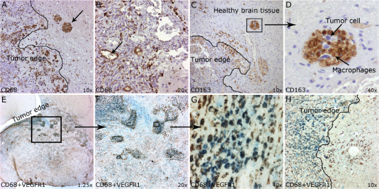
CD68 is a common marker used for the detection of macrophages within tumors.8,13,14 When BT4C malignant gliomas were stained for CD68, we found abundant expression of the protein throughout the tumor (Fig. 2A). Up to 35% of the tissue slides examined were positive for CD68, which correlated with the quantity that has been described in human malignant gliomas. 5 Interestingly, we found also clusters (in 3 out of 8 animals) offsite the tumor that stained positive for this marker (Fig. 2A, arrow). Furthermore, we detected blood vessels positive for CD68 (Fig. 2B). Even though these vessels were low in numbers, they contained erythrocytes signifying their functionality (Fig. 2B, arrows pointing at erythrocytes). Additionally, we performed a CD68/VEGFR-1 double staining, as it has been shown that particularly VEGFR-1 positive macrophages are involved in promoting tumor cell invasion. 15 The results revealed that only a subset of CD68 positive cells expressed VEGFR-1, appearing as individual clusters, which were mainly concentrated at the infiltrative edges (Fig. 2C and D), whereas the centers of the malignant gliomas were mainly positive for CD68 (Fig. 2D). No CD68/VEGFR-1 positive cells could be detected offsite the tumors (Fig. 2D).
(
Finally, we stained the malignant gliomas against CD163, which is commonly used as a marker for the M2 phenotype of macrophages.8,16 Also with CD163, similar results were observed (Fig. 2E and F). CD163 was abundantly expressed throughout the tumor and like in the CD68 staining, we observed clusters of cells offsite the tumors that stained positive for CD163 (Fig. 2E and F). However, no blood vessels with CD163 positive endothelium were found.
Discussion
Chronic inflammation plays a major role in the pathogenesis of many cancers. 17 Numerous cancers (including malignant gliomas) are strongly infiltrated by inflammatory cell, such as macrophages.3,18–20 Although, these cells have in general the ability to kill tumor cells, recent studies have shown that these cells may also support tumor growth and metastasis.21,22 Macrophages are the primary inflammatory cells in tumors. At least two different subpopulations of macrophages have been described in tumors, the M1 (classically activated) or M2 (alternatively activated) phenotypes, each of them expressing a different set of cytokines. 23 Tumor-associated macrophages often express genes typical for the M2 phenotype. Even though useful, the classification of macrophages into M1 and M2 is an oversimplification and does not necessarily represent the real plasticity of these cells.24,25 In any case, what is of interest is the fact that in cancer patients macrophages have been shown to contribute up to 50% of the tumor mass. 18 The paradoxical role of macrophages in tumors has been explained by their functional plasticity that can result in a polarized expression of cytokines and growth factors. In tumors, macrophages characteristically express genes such as IL-10, Il-6, TGF-β and MMP's and they are typically compromised by the tumor resulting in the impaired surface expression of MHC class II proteins. 26 They have been shown to have profound influence on tumor angiogenesis and to influence tumor progression and metastasis.14,27,28
In this study we characterized the BT4C rat malignant glioma model and evaluated, whether this model would be suitable to study the interaction between tumor associated macrophages and tumor cells. Human malignant gliomas are considered to be one of the most vascularized tumors with profound angiogenesis. 29 Microvessel density and positivity for vasculogenic mimicry have been correlated with tumor grade and poor prognosis.30,31 In this study we demonstrate that the BT4C malignant glioma is a highly vascularized tumor with an infiltrative behavior into the surrounding brain tissue. Furthermore, we show that BT4C malignant gliomas form microsatellitic lesions offsite the tumors, which is also a characteristic of human malignant gliomas. 2 In patients, the diffuse infiltrative behavior of malignant gliomas is a major factor for therapeutic failures. 2 Malignant gliomas tend to invade either as individual cells or in small groups into the surrounding brain. Particularly, the manifestation of microsatellite lesions is a feature that has been connected with a poor prognosis. 2 Because these microsatellite lesions are not visible with current imaging methods complete resection is virtually impossible. Furthermore, CD133 positive cells were found at the infiltrative edges, as well offsite of BT4C tumors. Even though controversial as a marker, CD 133 has commonly been used to identify brain tumor stem cells and its expression in human malignant gliomas has been described in numerous articles. 32 However, the origin of tumors from cancer stem cells is a topic of intense discussion. Interestingly, it has been demonstrated that CD133 positive cancer stem cells derived from malignant gliomas were able to transdifferentiate into endothelial-like cells contributing to angiogenesis in malignant gliomas (Ref.). In patients, the CD133 gene signature has been identified as an aggressive subtype of malignant gliomas with a poor prognosis. 33 Its expression has been correlated with resistance to radiotherapy and increased invasiveness.34,35
Human malignant gliomas are known to express vascular endothelial growth factor-A (VEGF-A), a main angiogenic factor, which binds to VEGFR-1 and 2. 29 VEGFR-1 is also involved in the recruitment of hematopoietic progenitor cells and monocytes.14,36 VEGFR-2 staining on BT4C malignant glioma, revealed a profound expression of this protein within the tumors. Interestingly, the amplification of the VEGFR-2 has been connected to malignant gliomas, indicating an autocrine loop around the VEGF-A/VEGFR-2 axis. 37 The existence of an autocrine loop around the VEGFR-2/VEGF-A axis is supported by previous studies done by Knizetova et al, where they demonstrated that autocrine VEGF signaling enhanced growth and viability of astrocytoma cells. 38 Furthermore, interference with the VEGF-VEGFR2 signaling potentiated the ionizing radiation-induced tumor cell death. 38 Recently, VEGFR-2 on tumor associated macrophages has also been implied as an important chemotactic receptor essential for VEGF-stimulated migration of tumor associated macrophages into tumors. 39 In our model, BT4C malignant gliomas were highly infiltrated by CD68 positive cells, contributing up to 35% of the tumor mass. Furthermore, clusters of macrophages were detected surrounding infiltrating tumor cells. This implies that macrophages initially are attracted by these tumor cells; however, the recruitment of these immune cells may not result in the removal of the tumor cells, but on the contrary, may support the invasion and metastasis of these cells. Of interest, Hiratsuka et al, demonstrated in a recent study that the invasion and metastasis of tumor cells was promoted by VEGFR-1 expressing macrophages. Therein, activation of the VEGFR-1 on macrophages resulted in the expression of MMP9, consequently promoting tumor cell invasion and metastasis. 15 Similarly, we observed in our study CD68(+)/VEGFR-1(+) cells particularly at the edges of the tumors, suggesting that they may be involved in the infiltration of glioma cells into the surrounding brain parenchyma. Furthermore, blood vessels with CD68 positive endothelium were detected. The concept that macrophages may be functioning as endothelial-like cells is not new. 14 In fact, studies indicate that macrophages play an important role in angiogenesis and that angiogenesis can be abolished by depleting these cells from the target tissue.14,40 For example, Fantin et al demonstrating that macrophages orchestrate angiogenesis by functioning as a bridge between two tip cells and depletion of macrophages resulted in an impaired angiogenesis. 41 Nishie et al demonstrated a correlation between the number of CD68 positive microglia/macrophages and the grade of histological malignancy, as well as an association between CD68 positive microglia/macrophage infiltration and neovascularization in human malignant gliomas. 13 In vitro, monocytes have been shown to have the capability to transdifferentiate into endothelial-like cells under sustained angiogenic stimulation. 42 These results demonstrate the plasticity of these cells, whose function is driven by the stimuli coming from the microenvironment.
In summary, inflammation and macrophage infiltration in malignant gliomas has been a subject of intense research. We evaluated the suitability of the BT4C glioma model to study the tumor micro environment, growth and invasion. Particularly, we believe that this model is particularly useful to study the role of inflammation and macrophage infiltration in malignant gliomas, as it behaves in many aspects similar to its human counterpart.
Funding
This study was supported by the European Union's Seventh Framework Programme, the Finnish Academy, the Kuopio University Hospital (EVO grant), North-Savo Cancer Foundation, Sigrid Juselius Foundation and the Antti Heikkosen and Anne-Liisa Rasasen grants from the Kuopio University Foundation.
Author Contributions
Conceived and designed the experiments: TW, FA, AP, HS. Analysed the data: TW, HS. Wrote the first draft of the manuscript: TW. Contributed to the writing of the manuscript: TW. Agree with manuscript results and conclusions: TW, FA, AP, HS, SYH. Jointly developed the structure and arguments for the paper: TW, FA, AP, HS, SYH. Made critical revisions and approved final version: SYH. All authors reviewed and approved of the final manuscript.
Competing Interests
Authors declare no potential conflicts of interest.
Disclosures
This manuscript has been read and approved by all authors. This paper is unique and is not under consideration by any other publication and has not been published elsewhere. The authors and peer reviewers of this paper report no conflicts of interest. The authors confirm that they have permission to reproduce any copyrighted material.