
Abstract
The possible protective effect of Piper aduncum capsule on DMBA (dimethylbenz[α]anthracene)-induced breast cancer in rats was assessed by monitoring the tumor and lung metastases incidence and recording hematological and biochemical parameters and frequency of micronuclei. Mammary carcinogenesis was induced in 36 female Holtzman rats by providing a single subcutaneous injection of DMBA. Oral administration of P. aduncum capsule lowered adenocarcinoma and lymph node metastases incidence. Pulmonary metastasis was significantly lowered (P < 0.05). Hematological indicators showed that the triglyceride level was significantly lowered (P < 0.01) and high-density lipoprotein (HDL) level was significantly increased (P < 0.01). Also, P. aduncum capsule significantly lowered the C reactive protein (CRP) level (P < 0.01) and malondialdehyde level (P < 0.05). There was a significant decrease in the frequency of DMBA-induced micronucleated polychromatic erythrocyte (P < 0.01). Considering the antitumorigenic, hypolipidemic, anti-inflammatory, antioxidant, and antigenotoxic properties of P. aduncum capsule, we conclude that it has a protective effect on DMBA-induced breast cancer in rats.
Introduction
Breast cancer is the most common malignant disease and the leading cause of cancer death among women in the world. 1 In the United States, breast cancer is the second leading cause of cancer death among women.2,3 The American Cancer Society has estimated that 89% of women survive 5 years after invasive breast cancer diagnosis. 4
Endocrine therapy targets estrogen receptor-positive breast cancers and represents the most effective treatment for them. In the last four decades, several hormonal agents have been used in palliative settings for advanced cancer and as adjuvant therapy to prevent the recurrence. However, there are several proposed molecular mechanisms that explain endocrine resistance in a large proportion of patients, which limit the effectiveness of endocrine treatments. 5
Eighty percent of world's population uses plants to cure disease, so the WHO (World Health Organization) recommends the use of medicinal plants in primary health care systems but based on scientific evidence to assure the safety, effectiveness, and quality required.6,7 It has been reported that flavonoids inhibit the breast cancer resistance protein (BCRP) encoded by the ABCG2 gene. 8
The Piperaceae family consists of 14 genera and over 1,900 species. Piper is the largest genus with over 1,000 species. 9 It has been demonstrated that some species of Piper have antitumor action. The crude extract and compounds isolated from Piper claussenianum, especially 2′,6′-dihydroxy-4-methoxychalcone, could inhibit cell growth in the breast cancer cell line MCF7. 10 The alcoholic extract of the fruits of P. longum and one of its compounds, the “piperine”, inhibit the solid and ascites tumor development in mice induced by Dalton's lymphoma ascites (DLA) cells and increase life span of EAC (Ehrlich ascites carcinoma)-bearing mice. 11 P. nigrum shows in vitro immunomodulatory activity, which could prevent or reduce carcinogenesis. 12 Also, apigenin has been identified in the aerial parts of P. capense Lf. 13 Being a flavonoid, it could have antioxidant activity and a protective effect against breast cancer. 14
Phytoestrogens such as isoflavones, daidzein, genistein, coumestrol, resveratrol, and glycitein are safer alternatives than hormone replacement therapy (HRT) and breast cancer protective agents because they regulate the cell cycle, expression of cyclin D1, apoptosis, Bcl-2/Bax ratio, and the transcriptional activity of NF-κB in the breast cancer cell line MCF7. 15
Previous studies have reported that P. aduncum has activity against Tetranychus urticae, 16 Aedes aegypti (L.), A. albopictus, 17 and Leishmania. 18 P. aduncum was also proved to have antihypertensive, 19 gastroprotective, 20 and hepatoprotective 21 effects. However, the effect of P. aduncum in breast cancer has not been studied yet. The aim of this study was to determine the protective effect of P. aduncum capsules on DMBA (dimethylbenz[α]anthracene)-induced breast cancer in rats.
Materials and Methods
Chemicals
DMBA was purchased from Sigma Chemical Company. All other chemicals and solvents used were of analytical grade and highest purity.
Preparation of P. aduncum capsules
Leaves of P. aduncum were collected in Huariaca, Cerro de Pasco, Peru. Taxonomic identification was made at the Museum of Natural History, National University of San Marcos, Lima, Peru. The leaves were ground and macerated in ethanol at 96°C. A qualitative phytochemical screening of the resulting extract was carried out. The extract was filtered and concentrated to obtain the solid residue, its final weight was noted, and it was kept refrigerated until further use. The capsules were filled with 180 mg of the ethanol extract of the leaves of P. aduncum and 300 mg of excipients (Hv veegum and Aerosil 200). The excipients were added according to good manufacturing practices and under quality control. 22 The capsules obtained were subjected to stability studies by storing in sealed containers at 40°C/65%RH and 30°C/75%RH storage conditions for a period of 6 months (accelerated test) and 24 months (real-time test), respectively. 23
Qualitative phytochemical screening
Determination of alkaloids
Dragendroff's test: A few drops of Dragendroff's reagent were added to the extract. Reddish brown precipitate indicated the presence of alkaloids. 24
Mayer's test: A few drops of Mayer's reagent were added to extract solution. Creamy white precipitate indicated presence of alkaloids. 24
Determination of anthraquinones
Borntrager's test: A small amount of the powder was mixed with small quantity of ether. If pink, red, or violet color appeared in aqueous layer, it indicated the presence of anthraquinone derivatives. 25
Determination of fat and oil
Paper test: A small amount of powdered drug was taken on filter paper and was pressed between the folds of a filter paper. The presence of an oily stain indicated the presence of fats and fixed oil. 26
Determination of flavonoids
Shinoda's test: A few drops of sodium hydroxide solution was mixed with the extract; the formation of yellow red precipitate indicated the presence of flavonoids. 24
Determination of phenols
Ferric chloride test: A few drops of FeCl3 were added to the extract. If blue-green color appeared in the solution, it indicated the presence of tannin compounds. 26
Determination of proteins
Ninhydrin test: The extract solution was boiled with 0.2% solution of ninhydrin. The appearance of violet color indicated the presence of proteins. 25
Determination of saponin
Frothing test: The powdered drug was taken in a test tube half-filled with water and vigorously shaken. Frothing indicated the presence of saponin. 26
Determination of steroids and terpenes
Liebermann-Burchard's test: To 0.2 g of each portion, 2 mL of acetic acid was added and the solution was cooled well in ice, and conc. H2SO4 was added carefully. Color change from violet to blue or bluish-green indicated the presence of a steroidal ring, ie, aglycone portion of cardiac glycoside. 27
Determination of tannins
Gelatin test: A 1% gelatin solution containing 10% sodium chloride was added to each leaf and bark (mixture) extract. Formation of a precipitate indicated the presence of tannins and phenolic compounds. 28
GC-MS analysis
The major compounds of the ethanolic extract of the leaves of P. aduncum were determined on a Shimadzu QP-5050A mass spectrometer system. Compounds were separated on a PTE-5 column (30 m × 0.25 mm, 0.25 μM film thickness). A sample of 1.0 μL was injected in the split mode with a split ratio 1:10. Helium was used as carrier gas at a flow rate of 22.3 mL/min. The column oven temperature was programmed from 80°C to 220°C at the rate of 4°C/min; initial and final temperatures were held for three and five minutes, respectively. The mass scanning range was 50–550 atomic mass unit, at a rate of two scans per second. Data acquisition and handling was done using the Shimadzu CLASS 5000 software. The resulting raw data files were processed by AMDIS v. 2.1. 29 The major constituents, compounds whose concentration was higher than 3%, and their concentrations are given in Table 1.
Phytochemical constituents of ethanolic extract of leaves of P. aduncum.

Animals
Thirty-six female Holtzman rats weighing 76 ± 14 g were procured from the National Institute of Health of Peru (INS). The animals were housed in well-ventilated, large, spacious cages in the bioterium of the Faculty of Medicine, National University of San Marcos. The animals received a balanced diet of commercially available pellet rat feed and water ad libitum. The rats were kept at 12 hours light/dark cycle and a temperature of 21 ± 2°C. The experiment began with a 7-day preconditioning period.
Tumor induction
In all groups, except negative control and PA200 groups, tumor induction was carried out by following the method of Barroso et al. 30 DMBA was used as a carcinogen for breast cancer induction. 31 The animals received a single dose of 20 mg of DMBA dissolved in negative control subcutaneously by intraperitoneal injection.
Experimental design
The 36 Hotlzman rats were divided into six groups of 6 rats each. Groups were named according to the treatment and dose in mg/kg. PA refers to P. aduncum capsule. The negative control group received capsules of Veegum Hv and Aerosil 200 (200 mg/kg body weight) orally for 15 weeks. Group PA200 received capsules of P. aduncum (200 mg/kg body weight) as drug control. Group DMBA were induced with DMBA on the first day and received capsules of Hv veegum and Aerosil 200 (200 mg/kg body weight) orally for 15 weeks. The groups DMBA + PA50, DMBA + PA150, and DMBA + PA300 received capsules of P. aduncum (50, 150, and 300 mg/kg body weight, respectively) orally for 15 weeks after DMBA induction.
At the end of the experimental period, the rats were weighed. Blood samples were obtained to assess the biochemical parameters, hematological indicators, and nuclear abnormalities.
The animals were sacrificed under pentobarbital anesthesia (100 mg/kg). The protective effect was determined through weights as well as biochemical, hematological, and pathological markers; the antigenotoxic capacity through micronucleus test; the antioxidant capacity by superoxide dismutase (SOD), malondialdehyde (MDA), and nitric oxide (NO); and the anti-inflammatory capacity by the level of C-reactive protein (CRP).
The rats were palpated to detect any mammary tumors. Sites and approximate size of mammary tumors were recorded. Breasts were removed and fixed in 10% phosphate-buffered formalin. They were stained with hematoxylin and eosin (H&E) for histological examination. Kidneys, liver, heart, and lungs were carefully examined grossly for metastases. Since pulmonary metastases were found, lungs also were fixed and stained.
Hematological parameters
Hct was measured from centrifuged arterial blood samples taken in heparinized capillary tubes. Hb content was determined spectrophotometrically (B-hemoglobin, Hemocue). The total leukocyte count was performed in a Neubauer chamber. Blood glucose was quantitated using a commercial enzymatic kit (Wiener Lab, Argentina) obtained from fasted rats. Total cholesterol was estimated by the modified Roeschlau's method. 32 HDL-cholesterol level was determined based on the method of Trinder. 33 Tryglicerides were estimated by enzymatic GPO-PAP method, as described by Annoni. 34 Alanine aminotransferase (ALT) was determined using the Reitman and Frankel method. 35 Alkaline phosphatase (ALP) activity was assessed according to the method of King. 36 Urea determination was based upon the cleavage of urea with urease (Berthelot's reaction) according to Fawcett and Scott. 37
Biochemical parameters
SOD was assayed as described by Beauchamp and Fridovich 38 based on the reduction of nitroblue tetrazolium (NBT) to water-insoluble blue formazan. Lipid peroxidation was detected by the determination of MDA production determined by the method of Begue and Aust.39,40 Nitric oxide scavenging assay was performed using the Griess reagent method. 41 The levels of CRP were determined using a Biochemistry VITROS and Integrated system VITROS 5600. 42
Micronucleus test
The micronucleus test was carried out following the Schmid method. 43 Peripheral blood was obtained by cardiac puncture to prepare a blood film. The slides were fixed with absolute methanol and stained with 3% Giemsa. The frequency of micronucleated polychromatic erythrocyte (MNPCE), based on the observation of 1000 polychromatic erythrocytes (PCE) per animal, was recorded.
Histological analysis
The abdominal-inguinal and contralateral mammary glands and lungs were collected from euthanized rats. They were fixed in 10% formalin, processed, and embedded into paraffin blocks. Sections were cut at 5 μm thickness and stained with H&E. The slides were examined under a light microscope (Olympus BX51) to observe for adenocarcinoma, lymph node, and pulmonary metastases, and confirmed by an experienced histopathologist.
Statistical analysis
Data are presented as mean ± standard deviation (SD). Data were analyzed for homogeneity of variance by the Levene test and normality by the Wilk-Shapiro W statistics. One-way analysis of variance (ANOVA) followed by Tukey multiple comparison method was carried out to compare the mean value of different groups. Fisher's exact test was used to evaluate the differences in adenocarcinoma and lymph node and pulmonary metastases incidence. The F-test was performed to analyze the variability intra and intergroups. A P-value of <0.05 was considered statistically significant in all cases. Data were analyzed by using SPSS v. 18.
Ethical considerations
During the entire experimental process, international ethical principles for research using laboratory animals were respected. The rats were euthanized by intravenous injection of pentobarbital (100 mg/kg). This method leads to a quick and peaceful death, acceptable for rats according to Recommendations for Euthanasia of Experimental Animals of the European Commission. 44 The protocol was approved by the Institute for Ethics in Health of the National University of San Marcos (01414-R-12-UNMSM).
Results
Table 1 shows the phytochemical constituents present in the ethanolic extract of leaves of P. aduncum used to make the capsules. The ethanol extract exhibited a high amount of total flavonoid content, followed by phenols and alkaloids. Sesquiterpenes are the major compounds of the extract, according to Table 2.
Major compounds of ethanolic extract of leaves of P. aduncum.

The morphology of the rat mammary glands was observed by microscopy. Adenocarcinoma was found in all groups treated with DMBA. However, DMBA + PA50 had the lowest incidence. All the groups treated with PA had lower incidence of lymph node metastases. Pulmonary metastases incidence was significantly lowered to 50% by the administration of P. aduncum capsules at the dose of 150 mg/kg (P < 0.05) (Table 3).
Effect of P. aduncum capsule on histological parameters.

(P < 0.05) versus group DMBA.
H&E stained sections of mammary tissue from the group negative control showed normal structure. PA200 presented a histological profile almost similar to the normal mammary tissue but with no gross vascular congestion. The tumors of group DMBA were histopathologically confirmed as differentiated adenocarcinoma. Mediastinal infiltration, fibrosis, and big tubular structures were found. The cellular architecture was altered, and the epithelial cells had enlarged hyperchromatic nuclei. It was observed that the group DMBA + PA 50 showed mild reduction of tubular structures with surrounding fibrosis. In the group DMBA + PA150, ductular proliferation was very little and nuclei were in different shapes and sizes. The group DMBA + PA300 showed tubular structures and vessels with great desmoplasia (Fig. 1).

H&E staining of mammary glands of control and experimental animals. (
Hematological indicators show that the triglyceride level was significantly lower in group DMBA + PA50 and group PA200 (P < 0.05 and P < 0.01) than in group DMBA. Administration of P. aduncum at doses of 200 mg/kg significantly increased the level of HDL (P < 0.01) (Table 4).
Effect of P. aduncum capsule on hematological parameters.
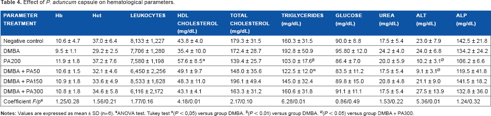
ANOVA test. Tukey test
(P < 0,05) versus group DMBA.
(P < 0.01) versus group DMBA.
(P < 0.05) versus group DMBA + PA300.
Biochemical indicators showed that the CRP level was significantly lower in group DMBA + PA50 and group DMBA + PA150 (P < 0.05 and P < 0.01) than in group DMBA. Administration of P. aduncum at the dose of 50 mg/kg significantly reduced the MDA level (P < 0.05) as compared with group DMBA (Table 5).
Effect of P. auduncum capsule on biochemical parameters.

ANOVA test. Tukey test
(P < 0.05) versus group DMBA;
(P < 0.01) versus group DMBA.
The effects of P. aduncum capsule on the frequency of MNPCE in rat's peripheral blood after exposure to DMBA are shown in Figure 2. Examination of the antimutagenicity profile revealed significant decrease in the frequency of DMBA-induced MNPCE in groups DMBA + PA50, DMBA + PA150, and DMBA + PA300 (P < 0.05, P < 0.01, and P < 0.05) as compared with group DMBA.

Effect of Piper aduncum capsule on the frequency of micronucleated polychromatic erythrocytes (MN PCE). Each value represents mean ± SD (n = 6). ANOVA followed by Tukey's post hoc test. *(P < 0.01) versus group DMBA. **(P < 0.05) versus group DMBA.
Discussion
DMBA is a polycyclic aromatic hydrocarbon used to induce breast cancer. This potent carcinogen induces DNA damage. In the cell, the reactive metabolite DMBA-3,4-dihydrodiol-1,2-epoxide (DMBA-DE) adds adenine and guanine residues to DNA. The conversion of genotoxic metabolites as DMBA-DE is promoted by the action of cytochrome P450 family. CYPlAl and CYP1B1 are identified as the enzymes that metabolize DMBA to produce DMBA-DE. 45
Breast cancer is a complex and multifactorial disease. Age, early menarche, delayed menopause, use of contraceptives, hormone therapy, family history, history of benign breast disease, obesity, and excess weight are the risk factors and show their effects through oxidative stress.46–48 In this study, P. aduncum capsule reduced the incidence of adenocarcinoma and lymph node metastases. Lung metastases incidence was significantly lowered. Also, significant reduction was observed in triglyceride level, and HDL level was significantly increased, indicating a hypolipidemic effect. A significantly low level of MDA implicates potent antioxidant effect, and a significantly decreased CRP level indicates a strong anti-inflammatory effect. The significant reduction in MNPCE frequency indicates potent antigenotoxic effect. The results suggest that P. aduncum could have inhibited the abnormal cell growth during DMBA-induced breast carcinogenesis. These findings are consistent with those reported by Rao et al, who found that the administration of P. bettle aqueous extract inhibited the DMBA-induced mammary carcinogenesis in rats. 49 The anticarcinogenic action of P. bettle was due to its antioxidant and antiproliferative effects against MCF-7 cells. 50
P. a duncum capsule contains saponins, poly phenols, tannins, alkaloids, and flavonoids in significant quantities. 51 The effects of the capsule could be due to its ability to reduce oxidative stress and free-radical formation since it contains flavonoids. Also, flavonoids have an anti-inflammatory effect52,53 by the inhibition of the formation of leukotriene B4, release of nitric oxide, and formation of prostaglandin E2. 54 Several flavonoids have inhibitory effects on aromatase, a mitochondrial cytochrome P450 family enzyme produced at high levels in breast tissues that catalyzes the conversion of androgens to estrogens.55–57 Flavonoids have protective effects in estrogen-dependent breast cancer by binding to estrogen receptor and modulating estrogen metabolism by selective inhibition of CYP1B1 activities without affecting CYP1A1 or CYP1A2. 58 Since flavonoids inhibit DNA binding, they have antimutagenic, anticlastogenic, antitumor, and anti-carcinogenesis effects.52,54,59,60
According to the analysis of P. aduncum, its chemical composition was dominated by terpenes. These components can contribute to the protective effect of the P. aduncum capsule. β-Caryophyllene oxide inhibits growth and induces apoptosis in human prostate and breast cancers, 61 and linalool has a well-known cytotoxic effect. It activates antitumor immunity and has a pro-oxidant effect in tumor tissue and an antioxidant effect in liver.62,63 (E)-Nerolidol inhibits carcinogenesis on azoxymethane-induced neoplasia in rats. 64 Finally, β-Elemene has an antiproliferative effect and antineoplastic activity because it induces cell cycle arrest and apoptotic cell death.65–68
The lipid-lowering effect of P. auduncum capsule could directly influence the development of cancer. Alikhani et al proved in a mouse model that a hyperlipidemic status can enhance mammary tumor growth and pulmonary metastasis. 69 And there is an association between increased TC, LDL-C, and decreased HDL-C levels with increasing tumor. 70 Also from in vitro studies, exogenous LDL-C has been found to promote proliferation and migration of cancer cells. 71 High-cholesterol levels induce a pro-inflammatory microenvironment that contributes to BC initiation and progression. The use of statins before cancer diagnosis reduces cancer-related mortality because of its lipid-lowering effect. 72 P. auduncum capsule has not only a hypolipidemic effect but also has antioxidant, anti-inflammatory, and antigenotoxic properties. This can explain its protective effect on DMBA-induced breast cancer.
It is important to mention that a dose-dependent effect was not found. PA150 has better results in lymph node and lung metastases compared to PA300. The latter has a higher concentration of tannins, which could have an antinutritional effect. They form complexes with dietary components and alter digestive enzymes, preventing the absorption of other beneficial components. 73
The limitations of this study include the lack of assessment of basal and induced CYP1 activity and determination of the active ingredient. Although the exact mechanism of the protective effect of P. aduncum capsule is unclear, it would be based on its hypolipidemic, antioxidant, anti-inflammatory, and antigenotoxic properties. However, is possible that the effect on enzymes activity could also play a role.
Conclusion
From the above results, it can be inferred that P. aduncum capsule attenuated DMBA-induced oxidative stress because of the antioxidant activity of its bioactive compounds. Considering the antitumorigenic, hypolipidemic, anti-inflammatory, antioxidant, and antigenotoxic properties of P. aduncum capsule, we conclude that it has a protective effect on DMBA-induced breast cancer in rats. There is a great potential to develop P. aduncum capsule as a chemotherapeutic agent in breast cancer treatment, and hence further studies are needed, particularly clinical studies, to further evaluate this effect.
Author Contributions
Conceived and designed the experiments: JAA, RJCA, RD. Analyzed the data: RJCA. Wrote the first draft of the manuscript. JAA, RJCA, RD. Contributed to the writing of the manuscript: RJCA, JAA, AAG, JRG. Agree with manuscript results and conclusions: RJCA, JAA, JRG. Jointly developed the structure and arguments for the paper: AAG. Made critical revisions and approved final version: RJCA, JAA, AAG. All authors reviewed and approved of the final manuscript.