
Abstract
Macrolides, one of the most commonly used class of antibiotics, are a group of drugs produced by Streptomyces species. They belong to the polyketide class of natural products. Their activity is due to the presence of a large macrolide lactone ring with deoxy sugar moieties. They are protein synthesis inhibitors and broad-spectrum antibiotics, active against both gram-positive and gram-negative bacteria. Different analytical techniques have been reported for the determination of macrolides such as chromatographic methods, flow injection methods, spectrofluorometric methods, spectrophotometric methods, and capillary electrophoresis methods. Among these methods, spectrophotometric methods are sensitive and cost effective for the analysis of various antibiotics in pharmaceutical formulations as well as biological samples. This article reviews different spectrophotometric methods for the determination of macrolide antibiotics.
Introduction
The term “antibiotic” was put forward by Vuillemin in 1889 to designate the active component involved in the process of antibiosis. The Greek word “anti” means against and “bios” means life. Benedict and Langlykke coined a general and acceptable definition of antibiotic, which states that “antibiotic is a chemical compound derived from or produced by a living organism, which is capable, in small concentration, of inhibiting the life processes of micro-organism.” 1 Antibiotics are used to treat infections caused by bacteria, the microscopic organisms, some of which may cause illness. Antibiotics can save lives either by killing bacteria or by inhibiting their reproduction. Antibiotics can be classified on the basis of their chemical structures as macrolides, fluoroquinolones, beta lactams, tetracyclines, amino glycosides, nitro furans, and so on.
Macrolide antibiotics are widely employed in human and veterinary medicines. Macrolides have 14-, 15-, or 16-membered large lactone rings with one or more sugar moieties, generally desosamine and cladinose. 2 The presence of another sugar moiety containing a dimethylamine group extends the basic behavior of macrolides. 3 These antibiotics are used to treat a range of problems such as allergic reactions, gastrointestinal disturbance, slow bactericidal action, and hepatotoxic effects.4,5
Classification of Macrolide Antibiotics
Based on the number of atoms in the lactone ring, clinically useful macrolide antibiotics are classified into three groups as 14-membered, 15-membered, and 16-membered antibiotics.
Fourteen-membered macrolide antibiotics
Erythromycin, clarithromycin, and roxithromycin are 14-membered macrolide antibiotics.
Erythromycin.
Erythromycin (Fig. 1A) is the first macrolide antibiotic. In 1949, some Fillipino scientists isolated erythromycin from a strain of Streptomyces erythreus from soil sample. It is available in the form of tablets, capsules, oral suspensions, ophthalmic solutions, ointments, gels, and injections. It is a white or slightly yellow crystal or powder with a melting point of 191°C and a dissociation constant pKa = 8.9. 6 It is freely soluble in alcohols, acetone, chloroform, acetonitrile, and ethyl acetate and moderately soluble in ether, ethylene dichloride, and amyl acetate.
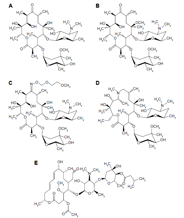
Structures of (A) erythromycin, (B) clarithromycin, (C) roxithromycin, (D) azithromycin, and (E) josamycin.
Clarithromycin.
In 1970, researchers of a Japanese drug company, Taisho Pharmaceutical, invented clarithromycin (6-O-methyl erythromycin; Fig. 1B). Taisho filed a patent for clarithromycin in 1980. It is available in the form of tablets, oral suspensions, gel, or lotion. It is a colorless crystalline solid with a melting point of 217-220°C and a dissociation constant pKa = 8.99. It is soluble in acetone and slightly soluble in ethanol, methanol, and acetonitrile.
Roxithromycin.
Roxithromycin (Fig. 1C) is a semi-synthetic macrolide antibiotic. In 1987, a German pharmaceutical company, Hoechst Uclaf, introduced roxithromycin. It is available in the form of tablets and oral suspensions. It is derived from erythromycin with N-oxime side chain attached to the lactone ring. It is a white solid having a melting point of 111°C. It is soluble in ethanol, methanol, acetonitrile, and acetone.
Fifteen-membered macrolide antibiotics
Azithromycin is a 15-membered macrolide antibiotic.
Azithromycin.
In 1980, Pliva, a Croatian pharmaceutical company, discovered azithromycin (Fig. 1D). It is one of the most successful antibiotics. 7 It is derived from erythromycin, with methyl-substituted nitrogen atom included in the lactone ring, which makes it a 15-membered lactone ring. It is available in the form of tablets, oral suspensions, and injections. It is a white solid having a melting point of 113-115°C with a dissociation constant pKa = 8.74. 6 It is soluble in ethanol, methanol, acetonitrile, and acetone.
Sixteen-membered macrolide antibiotics
Josamycin is a 16-membered macrolide antibiotic.
Josamycin.
Josamycin (Fig. 1E) is synthesized from the strains of Streptomyces narbonensis var. josamyceticus. It is a yellowish crystalline powder with a melting point of 130-133°C. It is available in the form of tablets and dry syrup. It is soluble in ethanol, chloroform, acetone, ether, benzene, and toluene and partly soluble in water. Unlike the 14- and 15-membered macrolide antibiotics, josamycin is not commonly used.
Pharmacology of Macrolides
Macrolides bind to the 50S subunit of the bacterial ribosome. They inhibit the bacterial protein synthesis. At lower concentrations of bacteria, these antibiotics act as bacteriostatic but may become bactericidal at high concentrations or depending on the type of microorganism. Macrolides assemble within leukocytes by which they are transported to the site of infection. 8
Therapeutic Applications
The antimicrobial spectrum of macrolides is similar to that of penicillins. However, in contrast to penicillins, macrolides are also active against Legionella pneumophila, Mycoplasma pneumoniae, and some Rickettsias and Chlamydias.9,10 Clarithromycin is used to treat gastric ulcers as a component of multidrug combinations. 11
Generally, macrolide antibiotics are prescribed for people who are allergic to penicillin antibiotics in the treatment of urinary tract infections, upper as well as lower respiratory tract infections, skin and soft tissue infections, ear infections, mouth infections, eye infections, intestinal infections, and tetanus infections.
Use of Macrolides in Pediatrics
Azithromycin, erythromycin, and clarithromycin have been widely used for pediatric infections. Azithromycin and clarithromycin are more stable and better absorbed than erythromycin. They are used for acute otitis media caused by Streptococcus pneumonia, Haemophilus influenza, and Moraxella catarrhalis and for tonsillitis caused by Streptococcus pyogenes. Azithromycin is more active against H. influenza. Oral suspension of azithromycin is used against pneumonia caused by Chlamydophila pneumonia, H. influenza, M. pneumonia, and S. pneumonia. Clarithromycin is used to treat acute maxillary sinusitis, skin structure infections, pneumonia, and disseminated mycobacterial infections, as well as asthma.12,13 Children suffering from soft tissue infections, skin infections, and respiratory tract infections are treated with roxithromycin. Erythromycin is used to treat gastrointestinal disorders in children.
Adverse Effects
High intravenous administration of macrolide antibiotics generally causes thrombophlebitis; sometimes, it may cause skin rashes. 14 The most common adverse reactions of this class of antibiotics are gastrointestinal disturbances, nausea, diarrhea, abdominal pain, and headache. 15 Macrolide antibiotics can also produce acute cholestatic hepatitis as a hypertensive reaction.16,17
Analysis of Macrolide Antibiotics
Different methods have been reported to determine macrolides separately as well as simultaneously. Some of these methods are spectrophotometry, high-performance liquid chromatography, voltammetry, spectrofluorometry, capillary electrophoresis, and titrimetry. The most widely used cost-effective and sensitive methods are spectrophotometric methods, mainly based on charge transfer and ion-pair interactions.
Spectrophotometric Methods
Spectrophotometric methods are based on the formation of a complex between the drug and the reagent. The color intensity is used as a measure of drug concentration. The complex formed between the drug and reagent is of either charge transfer or ion-pair type. The charge transfer complex is also known as electron donor–acceptor complex in which a fraction of electronic charge is transferred between the molecules. In the ion-pair complex, ions of opposite electric charge are held together in solution by Coulomb attraction. Spectrophotometric methods have been reported for the analysis of some antibiotics (other than macrolide antibiotics), including tetracycline, doxycycline, cefixime trihydrate, streptomycin sulfate, gentamicin sulfate, and amoxicillin.18–22
Spectrophotometric Methods for the Analysis of Macrolide Antibiotics
Different spectrophotometric methods of analysis of macrolide antibiotics are discussed below.
Paula et al 23 reported a method for the determination of azithromycin using quinalizarin as a charge transfer reagent. The complex shows the maximum absorbance at 564 nm and obeys Beer's law over a narrow concentration range of 4-20 mg L-1. A fairly low detection limit of 0.35 mg L-1 has been reported for azithromycin estimation. This method was successfully applied to the analysis of tablets without any interference from other ingredients.
Rachidi et al 24 proposed a method based on the extraction of the ionic-pair formed between azithromycin and Mo (V)–SCN complex in dichloroethane medium. The measurements were performed at 469 nm against a blank solution prepared analogously to the standard solutions.
Huang et al 25 employed two reagents 7,7,8,8,-tetracyanoquinodimethane (TCNQ) and chloranilic acid (CL) for the estimation of azithromycin in tablets. After the reaction with azithromycin, both the reagents produced charge transfer complexes with a maximum absorbance at 743 and 842 nm, respectively. The molar absorptivities of these complexes have been found to be 2.7 × 10 4 and 5.0 × 10 4 L mol-1 cm-1, respectively. Wide linearity between concentration and absorbance has been reported for azithromycin–CL complex ranging from 5 to 225 µg mL-1.
Liu et al 26 established a method based on charge transfer complex for the determination of azithromycin with 2,4-dinitrophenol having a linear range of 5-30 µg mL-1 at 364 nm; this method was used to analyze azithromycin tablets.
Charge transfer complex between azithromycin and alizarin red was studied by Li et al 27 in alcohol–water medium. The complex shows the maximum absorbance at 525 nm with a linear range of 5-55 mg L-1 and molar absorptivity of 1.26 × 10 4 L mol-1 cm-1.
Spectrophotometric estimation of azithromycin in tablets with potassium permanganate in alkaline medium at 547 nm was studied by Jayanna et al. 28 The method was used to determine azithromycin between 2 and 20 µg mL-1 in the final measured solution with no interference from the ingredients commonly found in azithromycin tablets.
Li et al 29 studied the charge transfer spectra of azithromycin and alizarin in ethanol medium at 546 nm with molar absorptivity of 5.79 × 10 3 L mol-1 cm-1 and Beer's law limit of 5-120 mg L-1, showing a wider linear range.
The method reported by Li et al 30 to study the charge transfer complex between azithromycin and TCNQ used acetone medium. The complete complex formation required an elevated temperature of 50°C for a period of 30 minutes. The molar absorptivity of the complex at 745 nm has been reported to be 1.44 × 10 3 L mol-1 cm-1, and the method was used to analyze azithromycin tablets.
Ashour and Bayram31 developed and validated a method for the assay of two macrolides, azithromycin and erythromycin, in pure and pharmaceutical formulations. It was based on the reaction of these two drugs with sodium 1,2-napthoquinone-4-sulfonate in alkaline medium at 25°C. The maximum absorbance was found to be at 425 nm with linear ranges of 1.5-33.0 and 0.92-8.0 µg mL-1, respectively. The limits of detection 0.026 and 0.063 µg mL-1 and molar absorptivity values 4.3 × 10 4 and 12.3 × 10 4 L mol-1 cm-1 have been reported for azithromycin and erythromycin, respectively. The method has a narrow linear range in the case of erythromycin.
Kelani et al 32 described a method for the determination of azithromycin with 2,3-dichloro-5,6-dicyano-p-benzoquinone (DDQ), and 588 nm wavelength was chosen to give the maximum sensitivity. This method was applied for reference materials as well as dosage forms.
Simple and rapid methods have been developed by Keskar and Jugade 33 for azithromycin, roxithromycin, and erythromycin by using bromocresol green as a reagent. These complexes were formed at 630, 620, and 625 nm with linear ranges of 4-46, 3-53, and 7-73 µg mL-1 for azithromycin, roxithromycin, and erythromycin, respectively. Compositions of the complexes were found to be 2:1. The values of detection limit were found to be 0.19, 0.56, and 0.30 µg mL-1, respectively, for the three drugs. Stability constant values were found to be 2.78 ± 0.03, 4.73 ± 0.02, and 4.86 ± 0.06 with molar absorptivity values of 1.485 × 10 4 , 2.312 × 10 3 , and 3.090 × 10 3 L mol-1 cm-1, respectively, for the three drugs. These methods are applied to determine the three drugs in pharmaceutical formulations and spiked human urine samples.
Walash et al 34 reported Eosin Y as an efficient reagent for the estimation of four macrolides, namely, erythromycin, azithromycin, clarithromycin, and roxithromycin. All the four drugs gave the maximum absorbance between 542 and 544 nm. The linear working ranges for the four drugs were 2-20, 1-10, 3-30, and 2-20, respectively. This method has been applied for the analysis of these macrolides in bulk, pharmaceutical formulations and spiked human urine and plasma samples.
Sayed et al 35 studied a spectrophotometric method for the determination of azithromycin dihydrate, erythromycin thiocyanate, and clarithromycin using a combination of rose bengal and copper. The complexes formed are extractable with methylene chloride and found to give the maximum absorbance at 560, 558, and 557 nm, respectively. Ringbom optimum concentration ranges for the three drugs were found to be 9-16, 20-50, and 0-35 µg mL-1, respectively. Sandell's sensitivity values were found to be 0.02, 0.07, and 0.05 µg cm-2, respectively. This method was successfully applied to tablets, capsules, and suspension forms of the respective drugs.
Jugade and Keskar 36 developed a new spectrophotometric method for the determination of azithromycin in bulk and pharmaceutical formulations with bromophenol blue as an ion-pair reagent. The maximum absorbance of the complex was found to be at 595 nm. Composition of the complex was found to be 2:1. Calibration curve was found to be linear over the range 0-50 µg mL-1, with the limit of detection 0.10 µg mL-1. The molar absorptivity was found to be 1.369 × 10 4 L mol-1 cm-1 with a stability constant of 6.19 ± 0.04, indicating high stability of the complex.
Spectrophotometric determination of roxithromycin based on charge transfer reaction with cresol red was studied by Zhao and Li. 37 The reaction conditions include interaction between the drug and reagent at 35°C for 10 minutes in alcohol–acetone medium. The complex formed has a maximum absorptivity of 1.05 × 10 4 L mol-1 cm-1 at 456 nm. The wide linear range of 0-80 mg L-1 is the most significant feature of this method.
Sastry et al 38 reported an ion-pair complex formation of roxithromycin with supracen violet 3B and tropaeolin 000. Regression analysis of the Beer's plot showed good correlation in the concentration ranges 5-60 and 5-40 µg mL-1, respectively.
Li et al 39 developed a method for the determination of roxithromycin using TCNQ. The reaction was completed in acetone medium within 30 minutes at room temperature, with a wide linear range of 20.93-418.5 mg L-1 at 848 nm.
Two spectrophotometric methods have been reported for roxithromycin and alizarin red as a charge transfer reagent. The method developed by Chen et al 40 uses hydrochloride medium and has a linear range of 20-120 mg L-1. The method developed by Bai et al 41 uses alcohol–water medium and has a linear range of 10-110 mg L-1 at 525 nm.
The reaction between roxithromycin and methylene blue was studied by Peng 42 in alcohol–HCl medium at 666 nm. Beer's law was obeyed in the range of 30.14-66.30 mg L-1, and molar absorptivity was found to be 2.01 × 10 3 L mol-1 cm-1. This method was applied to tablets and capsules with satisfactory results.
Charge transfer reaction of roxithromycin and purpurin was studied by Li et al 43 at 544 nm. The stability constant, molar absorptivity, and Beer's law linear range were found to be 3 × 10 3 , 6.56 × 10 3 L mol-1 cm-1, and 0-120 mg L-1, respectively. This method was used to determine roxithromycin in capsules.
Zhao 44 determined roxithromycin with 1,2,5,8-tetrahydroxyanthraquinone in ethanol–acetone medium. The maximum absorption wavelength was found to be 570 nm with a molar absorption coefficient of 2.56 × 10 3 L mol-1 cm-1 and a stability constant of 6.59 × 10 5 . This complex was reported to have 1:2 composition.
Sultana et al 45 studied charge transfer spectra of roxithromycin, clarithromycin, and erythromycin with CL reagent. The absorption maxima were found to be 496, 491, and 498 nm with linear ranges of 4-40, 8-40, and 3-36 µg mL-1, respectively. Stoichiometry was found to be 1:1 for all the three complexes. Molar absorptivity values were determined as 1.81 × 10 4 , 1.67 × 10 4 , and 2.07 × 10 4 L mol-1 cm-1, respectively.
Determination of erythromycin with methyl violet by heating at 50°C for 10 minutes in a water bath has been reported by Xu et al 46 at 583 nm with molar absorptivity of 1.61 × 10 4 L mol-1 cm-1.
Charge transfer reaction between erythromycin and methylene blue was studied by Xu et al 47 in water medium. Molar absorptivity was found to be 1.59 × 10 4 L mol-1 cm-1 with Beer's law range of 0.0008-0.025 mg mL-1.
An ion-pair complex formation of erythromycin ethyl succinate using bromothymol blue was studied by Dikran et al. 48 The complex absorbs at 414.5 nm at pH 4.0 (phthalate buffer), with a linear range of 0.5-50 µg mL-1. Sandell's sensitivity was found to be 47.620 µg cm-2. This method was successfully applied to tablet assays.
A charge transfer spectrum of erythromycin with alizarin red in water–ethanol medium was studied by Sun 49 at 580 nm. The composition of the complex was found to be 1:1 with molar absorptivity of 8.70 × 10 3 L mol-1 cm-1 and a stability constant of 1.6 × 10 4 .
Yanqing et al 50 studied charge transfer spectra of erythromycin and quinalizarin at 570 nm. Stoichiometry of the complex was found to be 1:1 with molar absorptivity of 1.14 × 10 4 L mol-1 cm-1 and a stability constant of 1.8 × 10 5 .
Ion-pair formation between erythromycin and bromothymol blue, methylthymol blue, and thymol blue was studied by Dabrowska et al. 51 The associates were extractable with chloroform at the maximum absorption wavelengths of 415, 430, and 550 nm, respectively.
Li et al 52 studied charge transfer spectra of erythromycin ethyl succinate with TCNQ. Determination of erythromycin and its stearate and succinate esters with gentian violet in alkaline medium with the maximum absorbance at 633 nm has been described by Amin and Issa. 53 This method has been reported to be highly specific for the estimation of erythromycin.
Erythromycin forms an association with methyl orange as described by Smith et al, 54 while charge transfer determination of erythromycin with purpurin was described by Kan and Kun 55 in ethanol–water medium. The composition of the complex was found to be 1:1 with a stability constant of 1.9 × 10 5 and molar absorptivity of 9.18 × 10 3 L mol-1 cm-1.
Complex formation of clarithromycin with iron (III) and Folin–Ciocalteu reagent has been described by Rao et al 56 with the maximum absorbance at 750 and 775 nm, respectively. These methods were applied to tablets.
Charge transfer complex between clarithromycin and DDQ was studied by Darwish et al. 57 An important advantage of this method is the wide linear range of 20-850 µg mL-1.
Charge transfer reaction between clarithromycin and 2,4-dinitrophenol was studied by Zhao 58 at 364 nm with molar absorptivity 1.55 × 10 4 L mol-1 cm-1, linear range 5-45 mg L-1, and the composition of the complex was found to be 1:1.
Charge transfer reaction between clarithromycin and alizarin red was studied by Li et al 59 in alcohol–water medium. Molar absorptivity of the complex was found to be 7.31 × 10 3 L mol-1 cm-1 at 546 nm, while the stability constant and linear range were found to be 3.4 × 10 4 and 1-100 mg L-1, respectively.
Li et al 60 studied the charge transfer reaction between clarithromycin and quinalizarin in water–alcohol medium. Stability constant, molar absorptivity, and linear range were found to be 2.6 × 10 5 , 3.74 × 10 3 L mol-1 cm-1, and 0-100 mg L-1, respectively, at 580 nm.
Li 61 developed a method based on charge transfer reaction between clarithromycin and purpurin. Beer's law range was found to be 10-150 mg L-1, while the molar absorptivity and stability constant were found to be 4.49 × 10 3 L mol-1 cm-1 and 3.48 × 10 4 , respectively.
Extractive spectrophotometric method for the determination of clarithromycin with bromocresol green was developed by Rao et al. 62 Beer's law limit, molar absorptivity, and Sandell's sensitivity were found to be 5.0-30.0 µg mL-1, 1.9347 × 10 4 L mol-1 cm-1, and 0.03865 µg cm-2, respectively.
Spectrophotometric charge transfer determination of josamycin with alizarin red in alcohol–water medium was carried out by Li. 63 Molar absorptivity was found to be 5.92 × 10 3 L mol-1 cm 1 at 530 nm. The composition of the complex was found to be 1:1 with a linear range of 0-120 mg L-1.
Spectrophotometric determination of josamycin with alizarin was also studied by Jiang et al. 64 The maximum absorption wavelength was found to be 426 nm, with molar absorptivity 2.14 × 10 4 L mol-1 cm-1 and linear range 0-22 mg L-1.
Charge transfer reaction between josamycin and purpurin was studied by Li and Xiao 65 in alcohol–water medium at 545 nm with 1:1 composition. Stability constant was found to be 3.9 × 10 4 with molar absorptivity 4.09 × 10 3 L mol-1 cm-1 and linear range 0-120 mg L-1. A brief discussion of each method is given in Table 1.
Spectrophotometric analysis of macrolide antibiotics.

Conclusions
Spectrophotometric methods have been successfully used for the determination of macrolide antibiotics in pure and commercial preparations. They can be used for routine analysis and quality control. Human urine samples have been analyzed for these antibiotics using these methods. Commonly occurring excipients do not interfere in the determination of pharmaceutical formulations. The results have been found to be accurate, precise, and validated statistically.
Considering the detection limits of the reported methods, the method using 2-naphthaquinone-4-sulfonate has the lowest detection limit among all the reported values for azithromycin and erythromycin. Bromocresol green for roxithromycin and rose bengal for clarithromycin are found to give the lowest detection limits.
Comparing the sensititivies on the basis of molar absorptivity values, the method using 2-naphthaquinone-4-sulfonate is most sensitive for erythromycin. TCNQ for azithromycin, bromocresol green for clarithromycin, CL for roxithromycin, and alizarin for josamycin have been found to be the most sensitive reagents.
Author Contributions
Conceived the concepts: RMJ. Analyzed the data: RMJ. Wrote the first draft of the manuscript: MRK. Contributed to the writing of the manuscript: MRK. Agree with manuscript results and conclusions: RMJ. Jointly developed the structure and arguments for the paper: MRK, RMJ. Made critical revisions and approved final version: RMJ. All authors reviewed and approved of the final manuscript.