
Abstract
Desmoplastic melanoma (DM) is a rare variant of spindle-cell malignant melanoma. DM is easily misdiagnosed at an early stage because it can be confused with benign entities. Histological analysis, including careful attention to the presence of atypical spindle cells, as well as to lymphocytic aggregates in an abundant fibrotic stroma in the dermis, provides clues for diagnosis. The adjunction of an immunohistochemical panel, and particularly testing for S-100 protein, is needed for the final diagnosis.
Introduction
Desmoplastic melanoma (DM) is an uncommon variant of melanoma that was first described by Conley
Case Report
A 73-year-old man presented with an asymptomatic, achromic mandibular plaque that he had borne for several years. Over the preceding 5 months, the lesion had increased in size. On physical examination, an indurated plaque of 3.5 cm in diameter was observed (Figure 1). The clinical differential diagnoses included an infundibular cyst, dermatofibroma, dermatofibrosarcoma, a parotid tumor, pilomatricoma and a scar. Histopathological examination revealed non-pigmented spindle-shaped malignant melanocytes interspersed within a prominent desmoplastic stroma in the dermis (desmoplasia was more than 90%). Several lymphoid aggregates were observed at the periphery. The tumor cells had minimal atypia (Figure 2). Breslow's thickness was 4 mm, and malignant cells had invaded the reticular dermis (Clark's level IV). Immunohistochemistry revealed tumor cells that were positive for S-100 protein but negative for melan-A or HMB45. No junctional activity or neurotropism was found (Figure 3). Pure DM (pDM) without neurotropism was diagnosed, and the patient was referred to the department of plastic surgery for a wide excision and, due to the high tumor thickness, for sentinel lymph node (SLN) biopsy, which showed no residual tumor and no lymph node involvement. Magnetic resonance imaging revealed no distant metastasis. The clinical stage was scored as IIA.
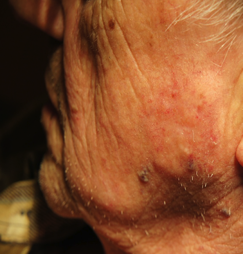
Achromic indurated mandibular plaque of 3.5 cm in diameter.

Histology (Hematoxylin and eosin staining). Dispersed spindle-shaped malignant melanocytes in a dense and diffuse fibrotic stroma in dermis; lymphoid aggregates at periphery (original magnification 200×).

Immunohistochemical examination strongly positive for S-100 protein (A) and negative for MelanA (B) (original magnification 100×).
Discussion
Desmoplasia refers to the growth of dense connective tissue of the stroma. This growth is characterized by low cellularity within a hyalinized or sclerotic stroma and by disorganized blood vessel infiltration. The diagnosis of DM is difficult because of variability in clinical appearance and a frequent absence of pigmentation. DM can mimic a scar or different tumors, such as an infundibular cyst, pilomatricoma, or dermatofibrosarcoma, as in our case. Dermoscopy is a useful aid during the evaluation of DM. The most common dermoscopic features found in DMs include atypical vascular structures, peppering, and occasional atypical globules and regression structures, such as scar-like areas and atypical crystalline structures.5,6 Histologically, most DMs are fibrosing spindle-cell tumors. This type of tumor is characterized by spindle-shaped malignant melanocytes within an abundant collagenous stroma. Lymphocytic aggregates often surround or infiltrate the tumors. The cytological atypia of DM is relatively variable, ranging from a fairly bland spindle form to marked nuclear pleomorphism. An
The application of immunohistochemistry, and particularly analysis of S-100, SOX-10 and p75, is mandatory for the diagnosis of melanoma. The distinction between paucicellular pDM and scarring may be straightforward. Immature scars may also express S-100, and pDM is mostly negative for other melanocytic markers, such as HMB45, microphthalmia transcription factor (MITF) or melan-A. Morphologic clues include nuclear atypia, poor circumscription of the lesion, lymphoid collections and strong positivity for S-100. Moreover, a diagnostic algorithm recently proposed by Weissenger
According to the different proportions of desmoplastic components in the tumors, DMs can be subdivided into pDM with more than 90% desmoplasia and mixed DM (mDM) with less than 90% desmoplasia.
2
The histological categorization is clinically relevant because pDM appears to have a distinct clinical biology (
The presence of neurotropism within melanoma, increases the risk of local recurrence.
3
The primary treatment is complete surgical excision. If nerve involvement is documented, wide excision is preferable. Due to the decreased potential for metastasis to regional lymph nodes (reported rates of 0-15%), an SLN biopsy is still controversial.3,7,9 For the current case, we preferred to perform SLN biopsy. Pawlik
Conclusions
In conclusion, DM is a rare variant of spindle-cell malignant melanoma. DM is easily misdiagnosed at an early stage because it can be confused with benign entities. A lesion biopsy and careful attention to the presence of spindle cells associated with lymphocytic aggregates in an abundant fibrotic stroma in the dermis may provide clues for diagnosis