
Abstract
The most frequent metastatic sites of the urothelial bladder cancers (UBCs) are bones, lungs, lymph nodes, liver, pleura, and brain. In the literature, skeletal muscle metastases from UBC have been rarely reported. We report a case of a 65-year-old male with metastatic myositis ossificans to obturator muscle 14 months after radical cystectomy performed for a muscle invasive transitional cell carcinoma. An abdomen computed tomography scan showed a lesion of about 8 cm in diameter in the left obturator muscle with myositis ossificans aspect. Ultrasound guided biopsy specimen of the left obturator muscle revealed poorly differentiated metastatic urothelial carcinoma with malignant myositis ossificans aspects. The patient refused additional surgery and received systemic chemotherapy and radiotherapy at the site of the lesion. The patient more than 6 months after treatment has a good performance status with a partial reduction of the mass and negative imaging for metastases in the follow-up.
Introduction
Radical cystectomy (RC) with extended pelvic lymph node dissection is the standard treatment for non-metastatic muscle-invasive transitional cell carcinoma (TCC) of the bladder. 1 However, lung metastases and secondary urothelial tumors are usually detected at routine follow-up, while pelvic metastases and distant recurrences (often bone's lesions) are predominantly symptomatic. 2 Hematogenous metastasis to skeletal muscle from TCC is extremely rare and there are few reports described in literature.3–5 We report a first case of metastatic myositis ossificans to obturator muscle 14 months after RC performed for a muscle invasive TCC.
Case Report
A 65-year-old male patient visited the Department of Urology with a history of dysuria, supra-pubic pain, urinary frequency associated with macroscopic hematuria. His past history was remarkable for gastro-esophageal reflux and hypertension. The ultrasound of the urinary tract suggested the presence of a sessile bladder wall's tumor approximately 3 cm in size engaging anterior wall and part of the bladder neck. These imaging were confirmed performing a cystoscopy. In October 2012 the patient underwent a transurethral resection of the bladder tumor (TURB). Histological examination showed a high-grade papillary TCC of the bladder with muscle invasion (pT2). The computed tomography (CT) of the abdomen-pelvis and chest X-ray was negative for metastases. A month later the patient underwent a RC and urinary diversion to an ileal conduit. The final pathology report confirmed the result of the TURB. A high-grade TCC with invasion of the bladder muscle layer was found, and TNM staging was pT2a N0M0. The prostate, the distal ureters, the proximal urethra and lymph nodes were free from tumor involvement. Patient did not receive chemotherapy in a neoadjuvant or adjuvant setting. The patient 14 months after RC presented a dull persistent pain in left groin that increases with body movements and walking. Subsequently, a full body CT and magnetic resonance imaging (MRI) of pelvis were performed and showed a lesion about 8 cm in diameter in the left obturator muscle with myositis ossificans aspect, and infiltrating branches ileum and ischio-pubic left (Figure 1). The patient did not present any other abnormalities or metastatic lesions. Transrectal ultrasonography-guided biopsy of the mass lesion in the left obturator muscle was performed. The pathological analysis of the biopsy specimen revealed poorly differentiated metastatic urothelial carcinoma with malignant myositis ossificans aspects and positive to the CK7 marker. We recommended additional surgical resection of the mass to prevent the disease development. However, the patient refused additional surgery, receiving subsequently systemic chemotherapy and radiotherapy at the site of the lesion. More than 6 months after treatment, the patient has a good performance status with a partial reduction of the mass and negative imaging for metastases in the follow-up.
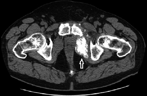
Computed tomography scan through pelvis shows mass developed within left obturator internus muscle with extensive ossification.
Discussion
Urothelial bladder cancer is histologically divided into three types: TCC (90-95% of UBC), squamous cell carcinoma (4-8% of UBC) and adenocarcinoma (1-2% of UBC). 6 Distant metastasis are detected in 33% of patients who undergo RC. 7 The most common metastatic sites are, in order of frequency, bones, lungs, and lymph nodes. The unusual metastatic sites in UBC include liver, pleura, brain and subcutaneous metastasis.6,7 The incidence of muscular metastasis is unknown, but approximately 0.16-0.3% has been reported in clinical practice, and 0.8% in an autopsy study. 4 Therefore, muscular metastasis distant from primary lesions are considered rare, even though they represent 50% of total body mass and receive a large blood flow.5,6 Several factors have been supposed to provide the resistance of skeletal muscle to metastatic disease. These factors comprise lower pH values in the muscle, muscle contractility that changes tissue pressure, and muscle capacity to remove cancer-produced lactic acid that induces tumor vascularity in tissues and facilitates tumor cell proliferation.3,8 Despite these defensive factors, metastasis to skeletal muscle have been reported in association with tumors of pancreas, kidney, colon, lung, stomach and ovary. 9 Skeletal muscle metastasis may be an incidental finding on CT of the abdomen during follow-up, because most lesions identified in these patients are neither painful nor palpable. However, the few case reports present in literature regarding skeletal muscle metastases secondary to UBCs are painful.3–5
Ossifying muscle metastases have been reported in association with primary mucinous adenocarcinomas involving the colon, stomach, small bowel, and, more rarely, breast, ovary, prostate, or skin. They are characterized by metaplastic ossification.9,10 Metastasis from a UBC have been reported in skeletal muscles,3–5,11 but we found no other report of metaplastic ossification within muscle metastases from an TCC in literature. Therefore, it is necessary to make a differential diagnosis with different types of myositis ossificans. Four types of myositis ossificans have been described by Steiner et al. 12 The most common form is myositis ossificans circumscripta which is a localized, self-limiting form secondary to penetrating, thermal or iatrogenic trauma. The key role in the pathogenesis is probably played by prolonged presence of macrophages in the muscle tissue altered by trauma and necrosis, which results in the release of mediators of osteogenesis. 13 The second variety is associated with neurological disorders such as closed head or spinal cord injury. The third type is pseudo-malignant myositis ossificans of unknown origin presenting without history of trauma. 12 This type may be often confused with malignant tumors. The fourth type is a rare genetic disease called fibrodysplasia ossificans progressive which is an unusual autosomal dominant inherited disease characterized by congenital malformations and osseous metaplasia of the muscles and connective tissue, leading to ossifications. This is fatal and, on an average, death occurs around the age of 35 years. 14 However, the cause of neoplasm-induced heterotopic ossification remains unknown. Rosenbaum et al. 15 believe that ossification occurs in direct contiguity with neoplasm, suggesting a local rather than a systemic osteogenic effect. Mesenchymal tissue with the ability to differentiate into osteoblasts must be locally available. Most skeletal muscle metastasis are detected on CT due to its follow-up use in oncologic staging, but MRI is considered superior to CT for detecting muscle abnormalities. 16 However, Pretorius et al. 11 report that 83% of metastatic lesions showed rim-enhancing intramuscular masses with hypoattenuation on enhanced CT. Ossifying muscle metastases can be seen on conventional radiographs as heterotopic ossifications in soft tissues. CT is more sensitive to calcifications and ossifications but it does not differentiate primary ossifying lesions from metastatic ones. 11 MRI findings, although not specific, report low to intermediate signal in the T1-weighted image and uniform high-signal intensity in the T2-weighted image. However, MRI with intravenous gadolinium enhancement was helpful in planning the biopsy of these lesions as it is useful to evaluate the vascularity of the lesion. 17 In our case MRI showed a 8.3×4.4 cm sized T2-weighted shows diffuse swelling and an increase in the signal of the left obturator internus muscle (Figure 2). The original arrangement of muscular fibers is not maintained. Treatment of these patients may depend on the clinical setting and the condition of the patient. Treatment options may include radiotherapy, chemotherapy and surgical excision. Excision of the painful mass may be helpful in carefully selected patients. 17 Treatments for skeletal muscle metastasis are still controversial in literature. Klune et al. 18 showed that surgical excision was selectively possible only in patients meeting various conditions and recommended radiotherapy or chemotherapy. Herring et al. 19 compared radiotherapy and surgery whit chemotherapy showing a control in terms of pain and size of metastatic lesion in 75% of patients. Nabi et al. 4 reported a mean survival rate of 8 months after receiving chemotherapy, while Pop et al. 20 reported not statistically significant survival rates among patients who underwent radiotherapy or chemotherapy compared with patients who underwent surgery. Chemotherapy frequently is the only option, due to advanced disease with multiple metastasis.

Transverse and axial T2-weighted image shows diffuse swelling and an increase in the signal of the left obturator internus muscle. The original arrangement of muscular fibers is not maintained.
Conclusions
The most frequent metastatic sites of UBC are bones, lungs, lymph nodes, liver, pleura, and brain. In the literature, skeletal muscle metastases from UBC have rarely been reported. The study case reported, to our knowledge, is the only that shows to date on ossifying skeletal muscle metastases of obturator muscle from TCC. Therefore, the incidence of this lesion in the general population should be further investigated in future case reports. Since the metastasis of tumors show to be a late event in the progression of the disease, the treatment of these patients depends on the clinical setting and their general condition.