Although many clinicopathological studies of malignant melanoma of the conjunctiva have been reported, there have been no studies of the expression and gene mutations of KIT and PDGFRA in melanoma of the conjunctiva. A 69-year-old Japanese woman consulted our hospital because of black mass (0.7 × 0.7 × 0.6 cm) in the conjunctiva. A biopsy was taken. The biopsy showed malignant epithelioid cells with melanin deposition. Immunohistochemically, the tumor was positive for S100 protein, HMB45, p53, Ki-67 (labeling=30%), KIT and PDGFRA. The tumor was negative for pancytokeratins (AE1/3 and CAM5.2). A genetic analysis using PCR-direct sequencing revealed no mutations of KIT gene (exons 9, 11, 13, and 17) and PDGFRA gene (exons 12 and 18). The pathological diagnosis was conjunctival melanoma. Despite chemotherapy, the patient developed multiple metastases of melanoma, and died of melanoma 7 years after the biopsy. In conclusion, the author reported a case of melanoma of conjunctive expressing KIT and PDGFRA proteins without gene mutations of KIT and PDGFRA.
Malignant melanoma is a highly malignant tumor, and NRAS and BRAF mutations are mainly involved in the pathogenesis of melanoma.1,2KIT gene, mapped to 4q12, encodes an oncogenic transmembranous receptor tyrosine kinase, KIT, whose ligand is stem cell factor.3 The platelet derived growth factor receptor-α (PDGFRA) gene, also mapped to 4q12, also encodes an oncogenic transmembranous receptor tyrosine kinase, PDGFRA.3 The KIT gene plays an important role in the melanocyte migration, development, differentiation and tumorigenesis.4 Previous studies have shown that activating mutations of the KIT gene may lead to tumorigenesis of cutaneous melanoma.1 Since KIT and PDGFRA genes are mapped to 4q12, it is anticipated that PDGFRA gene mutations are involved in the tumorigenesis of melanoma, as in the case of gastrointestinal stromal tumors.3 However, PDGFRA gene mutations in melanoma have rarely been examined.5–8 In addition, PDGFRA protein expression has rarely been analyzed in melanoma. These studies have been performed in Caucasians, and only two reports by Ashida et al.6 and ours7 is available in Mongoloids, including Japanese in which malignant melanoma is much more uncommon than in Caucasians.9 Ashida et al.6 reported that KIT protein expression was 48% in Japanese cutaneous melanoma and that KIT mutations were 16% in Japanese cutaneous melanoma. Our previous study7 has shown that KIT and PDGFRA expression in cutaneous melanoma was present in 92% and 100%, respectively, and that mutations of KIT and PDGFRA were recognized in 8% and 0%, respectively, in cutaneous melanoma.
Although many clinicopathological studies on melanoma of conjunctiva have been performed,10,11 there have been no studies of KIT and PDGFRA in melanoma of the conjunctiva. The author investigated the protein expression and gene mutation status of KIT and PDGFRA in a case of conjunctival melanoma of a Japanese woman.
Case Report
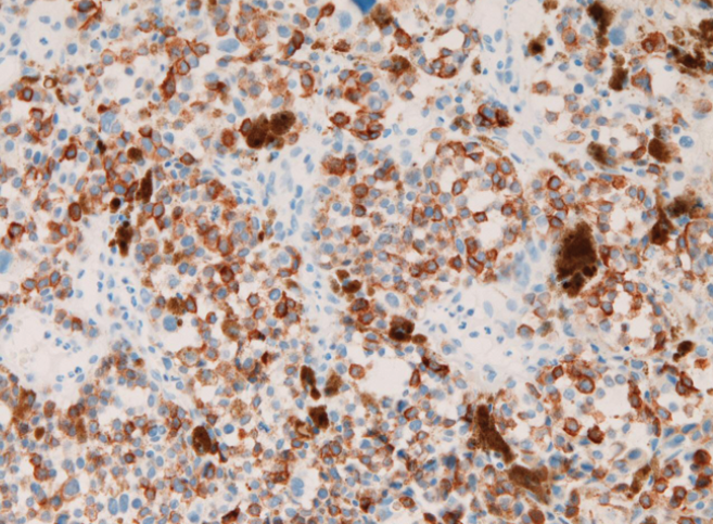
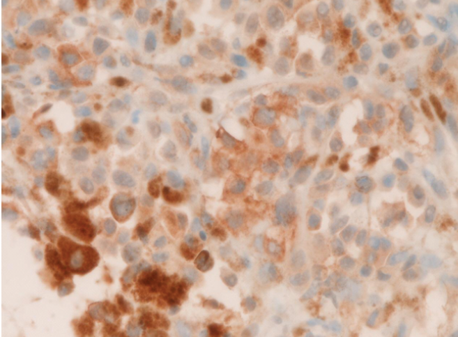
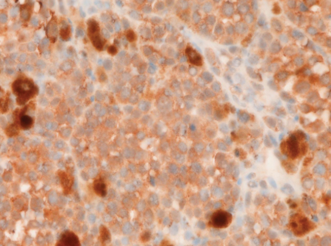
A 69-year-old Japanese woman consulted our hospital because of black mass in the conjunctiva. Physical examination revealed a black tumor measuring 0.7×0.7×0.6 cm of the right conjunctiva. A biopsy was taken, and the biopsy showed malignant epithelioid cells with brown pigment deposition (Figure 1). The brown pigment was positive with Fontana-Masson stain, and therefore was thought to be melanin. An immunohistochemical analysis was performed, using Dako's Envision method, as previously described.12–14 Immunohistochemically, the tumor cells were positive for S100 protein (Figure 2), HMB45 (Figure 3), p53, Ki-67 (labeling=30%), KIT (Figure 4) and PDGFRA (Figure 5). The tumor was negative for pancytokeratins (AE1/3 and CAM5.2).
Histology of the conjunctival tumor. Malignant epitheloid cells are seen. Brown pigment was present. These features are suspicious of conjunctival melanoma. Haematoxylin & Eosin, x200.
The tumor cells are positive for S100 protein. Immunostaining, x200.
The tumor cells are positive for HMB45. Immunostaining, x200.
The tumor cells are positive for KIT protein. Immunostaining, x300.
The tumor cells are positive for PDGFRA. Immunostaining, x300.
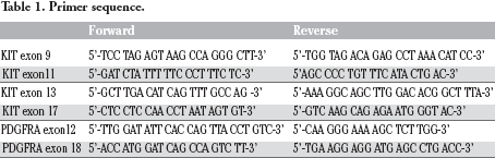
Genetic analyses of the KIT gene (exons 9, 11, 13, and 17) and PDGFRA (exons 12 and 18) gene were performed by the PCR direct sequencing method, as previously reported.15–19 The exons of both genes were selected because they are frequent mutation sites.3 The primers are shown in Table 1. In brief, genomic DNA was extracted from paraffin blocks with proteinase K digestion and phenol/chloroform extraction, and subjected to PCR for 40 cycles (94°C for one min, 52°C for one min, 72°C for one min), using a thermal cycler (GeneAmp PCR system 9700, Applied Biosystems, ABI, CA). The annealing temperature was 53°C. PCR products were extracted, and subjected to a computed automatic DNA sequencer (ABI PRISM 3100 Genetic Analyzer, Applied Biosystems, ABI, CA). These techniques revealed that there were no mutations of the KIT gene (exons 9, 11, 13, and 17) and PDGFRA gene (exons 12 and 18) in this tumor.
Primer sequence.
Forward
Reverse
KIT exon 9
5′-TCC TAG AGT AAG CCA GGG CTT-3’
5′-TGG TAG ACA GAG CCT AAA CAT CC-3’
KIT exon11
5′-GAT CTA TTT TTC CCT TTC TC-3’
5'AGC CCC TGT TTC ATA CTG AC-3’
KIT exon 13
5′-GCT TGA CAT CAG TTT GCC AG -3’
5′-AAA GGC AGC TTG GAC ACG GCT TTA-3’
KIT exon 17
5′-CTC CTC CAA CCT AAT AGT GT-3’
5′-GTC AAG CAG AGA ATG GGT AC-3’
PDGFRA exon12
5′-TTG GAT ATT CAC CAG TTA CCT GTC-3’
5′-CAA GGG AAA AGC TCT TGG-3’
PDGFRA exon 18
5′-ACC ATG GAT CAG CCA GTC TT-3’
5′-TGA AGG AGG ATG AGC CTG ACC-3’
The pathological diagnosis was conjunctival melanoma. Despite chemotherapy, the patient developed multiple metastases of melanoma, and died of melanoma 7 years after the biopsy.
Discussion
The present case is the second report of PDGFRA protein status in melanoma and is the first in conjunctival melanoma. Our previous study7 showed 100% expression of PDGFRA protein in cutaneous melanoma. The present study is the forth report of PDGFRA mutations in melanoma; the first was reported by Curtin et al.,5 who found no PDGFR mutations in 26 cutaneous melanomas. The second was reported by Sihto et al.,20 who demonstrated no PDGFRA gene mutations in 14 cutaneous melanomas. The third was reported by us; no mutations of PDGFRA gene were found in 12 cutaneous melanomas. The current case is the first report of PDGFRA gene status in the conjunctival melanoma.
The present case showed no mutations of the KIT gene. Studies of KIT mutations are scant in number in cutaneous melanoma, and are none in conjunctival melanoma. Willmore-Payne et al.21 showed only 2% of melanomas had KIT mutations. Sihto et al.20 showed no KIT mutations in 14 cutaneous melanomas. In contrast, Curtin et al.1 showed that KIT mutations were present in 39% of mucosal melanomas, in 36% of acral melanomas, 28% in melanomas of sun-damaged skin, and in 0% of melanomas of non-sun-damaged skin. Beadling et al.22 recently reported that KIT mutations were present in 23% of acral melanomas, 15.6% of mucosal melanomas, 1.7% of cutaneous melanomas, and 0% of choroidal melanomas. Handolias et al.23 reported that KIT mutations were present in 2% of melanomas and that KIT mutations were frequent in acral and sun-damaged skin melanomas and mucosal melanomas while it was very rare in non-sun-damaged skin melanoma. In the present case, no mutations were seen in the KIT gene. Since KIT mutational studies are scant in conjunctival melanoma, more studies remain to be performed.
The present case showed positive KIT protein expression in conjuctival melanoma. The percentage of KIT expression in cutaneous melanomas varies among researchers. There have been no reports of KIT expression in conjunctival melanoma, to the best of our knowledge. The percentage in the literature ranges from 21 %24 to 84%.25 Sihto et al.20 reported that KIT expression in most human solid tumors, including melanomas, were due to KIT gene amplification. More studies of the relationship between KIT gene mutations and KIT protein expression in conjunctival melanoma remain to be performed.
In conclusion, the author reported a case of melanoma of conjunctiva expressing KIT and PDGFRA proteins without gene mutations of KIT and PDGFRA. Because this is only a case report, examinations of larger number of patients are needed.
References
1.
CurtinJABusamKPinkelDBastianBCSomatic activation of KIT in distinct subtypes of melanomaJ Clin Oncol2006;24:4340–6
2.
CurtinJAFridlyandJKageshitaTDistinct sets of genetic alterations in melanomaN Engl J Med2005;353:2135–47
3.
HirotaSIsozakiKPathology of gastrointestinal stromal tumorPathol Int2006;56:1–9
4.
AlexeevVYoonKDistinctive role of c-Kit receptor tyrosine kinase signaling in mammalian melanocytesJ Invest Dermatol2006;126:1102–10
5.
CurtinJAPinkelDBastianBCAbsence of PDGFRA mutations in primary melanomaJ Invest Dermatol2008;128:488–9
6.
AshidaATakataMMurataHPathological activation of KIT in metastatic tumors of acral and mucosal melanomasInt J Cancer2009;124:862–8
7.
TeradaTLow incidence of KIT gene mutations and no PDGFRA gene mutations in primary cutaneous melanoma: an immunohistochemical and molecular genetic study of Japanese casesInt J Clin Oncol2010;15:453–6
8.
TeradaTAmelanotic malignant melanoma of the esophagus: report of two cases with immunohistocheimcal and molecular genetic study of KIT and PDGFRAWorld J Gastroenterol2009;15:2679–83
9.
SoberAJLewRAKohHKBarnhillRLEpidemiology of cutaneous melanoma: an updateDermatol Clin1991;9:617–29
10.
FolbergRMcLeanIWZimmermanLEMalignant melanoma of the conjunctivaHum Pathol1985;16:136–43
11.
De PotterPShieldsCLShieldsJAMendukeHClinical predictive factors for the development of recurrence and metastasis of conjunctival melanoma: a review of 68 casesBr J Ophthalmol1993;77:624–30
12.
TeradaTKawaguchiMFurukawaKMinute mixed ductal-endocrine carcinoma of the pancreas with predominant intraductal growthPathol Int2002;52:740–6
13.
TeradaTTaniguchiMIntraductal oncocytic papillary neoplasm of the liverPathol Int2004;54:116–23
14.
TeradaTKawaguchiMPrimary clear cell adenocarcinoma of the peritoneumTohoku J Exp Med2005;206:271–5
15.
TeradaTGastrointestinal stromal tumor of the uterus: a case report with genetic analyses of c-kit and PDGFRA genesInt J Gynecol Oncol2009;28:29–34
16.
TeradaTMediastinal seminoma with multiple KIT gene mutationsPathology2009;41:695–7
17.
TeradaTMutations and protein expression of KIT and PDGFRA genes in ipsilateral testicular seminomas: an immunohistochemical and molecular genetic studyAppl Immunohistochem Mol Morphol2011;19:450–3
18.
TeradaTPrimary extragastrointestinal stromal tumors of the transverse mesocolon without c-kit mutations but with PDGFRA mutationsMed Oncol2009;26:233–7
19.
TeradaTPrimary multiple extragastroin-testinal stromal tumors of the omentum with different mutations of c-kit geneWorld J Gastroenterol2008;14:7256–9
20.
SihtoHSarlomo-RikaraMTynnienenOKIT and platelet-derived growth factor receptor alpha tyrosine kinase gene mutations and KIT amplifications in human solid tumorsJ Clin Oncol2005;23:49–57
21.
Willmore-PayneCHoldenJATrippsSLayfieldLJHuman malignant melanoma: detection of BRAF- and c-kit-activating mutations by high-resolution amplicon melting analysisHum Pathol2005;36:486–93
22.
BeadlingCJacobson-DunlopEHodiFSKIT gene mutation and copy number in melanoma subtypesClin Cancer Res2008;14:6821–8
23.
HandliasDSalemiRMurrayWMutations in KIT occur at low frequency in melanomas arising from anatomical sites associated with chronic and intermittent sun exposurePigment Cell Melanoma Res2010Epub ahead of print
24.
ArberDATamayaRWeissLMParaffin section detection of the c-kit gene product (CD117) in human tissues: value in the diagnosis of mast cell disordersHum Pathol1998;29:498–504
25.
MontoneKTvan BellePElenitsasRElderDEProto-oncogene c-kit in malignant melanoma: protein loss with tumor progressionMod Pathol1997;10:939–44