
Abstract
We report a case of a giant melanoma on the back with a very extreme Breslow thickness. On physical examination a large odorous and ulcerating tumour was seen adjacent to two large crusted lesions, probably in transit metastases. In the right and left axilla enlarged lymph nodes were palpated. The patient underwent salvage surgery consisting of a complete wide excision of the tumors on the back as well as axillary lymph node dissection on both sides. Histopathology showed a malignant melanoma with a Breslow thickness of 48 mm. Four of fifteen nodes in the right axilla and one of nine nodes in the left axilla, were positive for metastatic disease. Also various in transit and subcutaneous metastases were found in the wide excision specimen. The interest of our observation relies in the rarity of a melanoma with such an extreme Breslow thickness and the difficulty in performing adequate palliative therapy that offers quality of life by means of tumor control.
Keywords
Introduction
Today, most melanoma patients are diagnosed early with a low Breslow thickness. 1 However, patient delay can be an important factor for melanoma to grow rapidly.
The incidence of cutaneous melanoma has risen rapidly in the last decades and estimates predict a further increase in the next years. In the Netherlands for example, the number of melanoma patients increased from 2400 patients in 2000 to 3500 in 2005, suggesting that the predicted number of 4800 patients in 2015 is an estimation that is too low. 2
The incidence of thin melanomas (Breslow thickness ≤1.0 mm) has increased as was shown in a recent Dutch study over the period 1980–2002. 3 These observations reflect a greater patient and physician awareness to recognize early melanoma with consequently a higher cure rate. Among the well known prognostic factors, such as ulceration, mitotic rate and sentinel lymph node status, Breslow thickness remains the most important predictor of long term survival.4,5 Patients diagnosed with thick melanomas (Breslow thickness ≥4 mm) are at bad risk.
Case Report
A 56-year old woman without a medical history was referred by her general physician to our outpatient department because of three large lesions on the back which had grown significantly in the last weeks. On physical examination a large ulcerating odorous skin tumour was found on the back measuring 8 by 6 cm. Adjacent to it two additional crusted lesions were noted measuring 4 and 1.5 cm respectively (Figures 1 and 2). Enlarged lymph nodes were palpated in the right and left axilla and on the back two subcutaneous nodules were appreciated. Biopsy of the largest tumor showed a malignant melanoma. Although a chest X ray was negative for metastases as well as a negative abdominal ultrasound, the serum LDH was increased to 1140 U/L which was very suspect for distant metastasis. In order to perform good palliative care it was decided to treat the local tumor aggressively with surgery. The three tumors were excised en block in one prep rate and a combined lymph node dissection of both axillae was performed. A split skin graft (SSG) was used to cover the wound on the back. Histopathology revealed a radically removed ulcerating malignant melanoma with a Breslow thickness of 48 mm. Four of fifteen nodes in the right axilla and one of nine nodes in the left axilla were tumour positive. In addition various in transit metastases were found in the wide excision specimen of which the largest was 3.5 cm in diameter. Ten weeks later after recovering from surgery before being able to commence with systemic chemotherapy patient developed multiple and painful nodes in the lower right neck region. Because she became critically ill local radiotherapy was omitted and she died two weeks later, unexpectedly.
A giant melanoma on the back.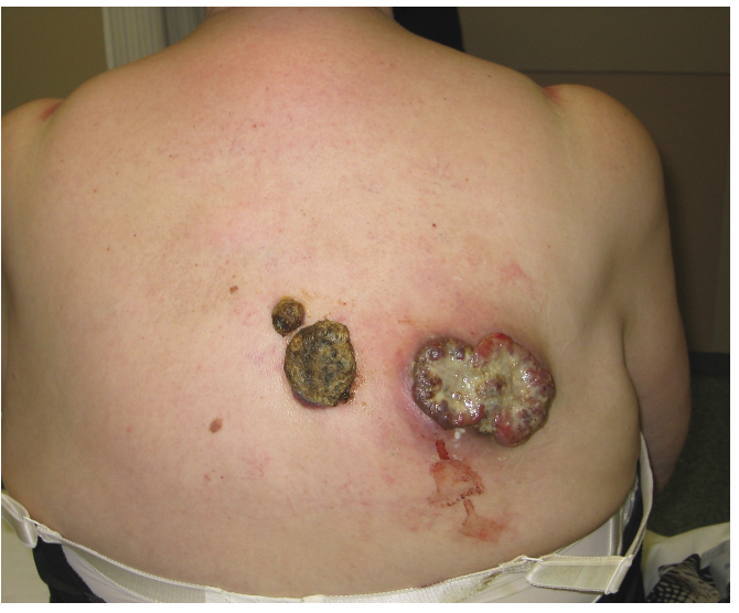
A ulcerating tumor on the back.
Discussion
The interest of this case relies in the rarity of the extreme Breslow thickness and the difficulty to perform appropriate salvage treatment.
In the database of the Netherlands Cancer Registry only one other melanoma with this magnitude of thickness (70 mm) is documented in the period 1994–2005. 6 Thick melanomas (>4 mm) are relatively common. In the final version of the AJCC (American Joint Commitee on Cancer) melanoma staging 14% of the included 17.600 melanoma patients had a melanoma with a Breslow thickness >4 mm.4 However, giant melanomas of 48 mm are rarely encountered. To our knowledge, this is one of the largest melanoma's described in literature. 7
Patients who present with thicker melanomas are generally of later age.8,9 One of the reasons for this could be a decreased awareness in elderly patients combined with more difficulties to recognize or detect changes of their skin. Also self negligence and fear of visiting a doctor may be reasons for delay in actually visiting a physician. 8 Our patient, for example, had never visited a general practitioner before.
Despite a normal chest X-ray and a negative abdominal ultrasound most probably our patient had disseminated disease when she first presented. The serum LDH at first consultation was increased to 1140 U/L which is very suspect for distant metastasis.
Notwithstanding this, surgery was chosen in order to achieve palliative local tumour control. This local intervention resulted in 3 months of relatively acceptable quality of life.
When melanoma patients present with an advanced stage of local disease as in the above described case, the consulting physician has to think twice before starting therapy with a curative intent. The answer for curative therapy in melanoma in general has to be sought in early recognition and early presentation as opposed to the situation of our patient.
After having performed the biopsy together with the elevated LDH and palpable axillary lymph nodes on both sides the patient could already be staged as T4N2M1c. These findings, bringing her in a palliative situation, made a PET-CT scan, with the aim to prove distant metastases, a needless investigation.
Giant melanomas like these are rarely encountered. With the aim of tumor control, principles of wide surgical excision and therapeutic lymph node dissection can still be applied for giant melanomas. 10 Despite the size of a giant melanoma, the work up and principles of wide surgical excision remain the same. There is no benefit with a greater than two-centimeter margin in melanomas with thickness greater than four millimeters, as the risk of dying from metastatic disease is greater than the risk of recurrence. 11
Other therapy options instead of salvage surgery could be radiation therapy or palliative chemotherapy. For radiation therapy in this case the tumor surface would have been too large with a very low response rate. According to the Dutch melanoma guideline the standard palliative chemotherapy should be DTIC with a very low response rate. 12
Treatment results for stage IV melanoma remain unsatisfactory with a one year survival of 33%. 4 No systemic therapy has demonstrated to affect overall survival, although the recent immunotherapy with Ipilimumab and the introduction of the BRAF pathway inhibitors have shown promising results.13,14
In conclusion, despite advances in adjuvant therapies, the primary treatment for a giant melanoma can remain surgical even when there is no curative intent.
In this case, treatment was performed with palliative intent as time was precious and unnecessary diagnostic research was to be avoided. Consequently, since the described melanoma tumour was too large for radiotherapy, salvage surgery was chosen as the best palliation treatment and for local control of the primary tumour and the axilary lymph node metastases. Adjuvant palliative systemic treatment was planned afterwards but unfortunately this was cancelled when the patient presented with locoregional recurrent disease and became too weak for further palliative therapy.