
Abstract
Anal cancer usually presents with a visible or palpable tumour. In this case we describe a 54-year old man diagnosed with Cancer of Unknown Primary (CUP) with a single inguinal node as the only finding. Thorough examination failed to identify any primary tumour. The patient was treated with lymph node dissection and not until nearly two years after initial diagnosis, was the primary tumour found, and the patient was diagnosed with anal cancer. The patient was treated with chemoradiotherapy and 45 months after initial diagnosis there is still no sign of relapse. This case illustrates, that anal cancer can metastasise before the primary tumour is detectable. Furthermore, it demonstrates the necessity of thorough clinical follow-up after treatment of CUP since the primary tumour was found later. Finally this is a case of a long-term survivor following treatment for metastatic inguinal lymph nodes from an initially unknown primary cancer.
Introduction
Anal cancer is a rare disease accounting for only about 1.5% of all of the gastrointestinal cancers. 1 Symptoms are often few and include rectal bleeding, pain and the sensation of a mass in the anal region, and as many as 20% of the patients have no symptoms at all. 2
Metastatic Cancer of Unknown Primary (CUP) constitutes a heterogonous group of metastatic tumours where the primary tumour cannot be identified despite thorough clinical examinations. CUP may account for as many as 3–5% of all cancers. 3
We present a case with anal cancer debuting as CUP with a single lymph node in the right inguinal region as the only finding.
Case Report
54-year old man presents with an enlarged lymph node in the right inguinal region. The patient is formerly healthy and is in a good clinical condition. There are no other symptoms. Examination reveals a 2×4 cm large lymph node in the right inguinal region but no other palpable lymph nodes in any other region. Rectal digital examination is normal. A biopsy is taken and the histology shows highly differentiated carcinoma cells. The tumour tissue is CK7+ and CK20−, most likely representing a squamous cell carcinoma, and the pathologists conclude that it could be either a malignant transformation of an epithelial cyst or metastasis from lung or elsewhere. Cystoscopy, urine cytology, ultrasound of the abdomen, gastroscopy and anoscopy are performed and do not reveal any primary tumour. A colonoscopy has been performed in November 2005 and was normal. CT of the thorax and abdomen reveals a 7.5 mm process in the left lower lobe near the hilus, and a few enlarged lymph nodes in the mediastinum. Bronchoscopy is macroscopically normal and biopsies are without malignancy. The CT-scan is re-evaluated and the small process in the left lung is not believed to be a primary lung tumour. Thus, at this point there is still no evidence of a primary tumour.
The patient is referred to Department of Oncology with the diagnosis of CUP. The biopsy is revised and the possible options are still malignant transformation of epithelial inclusion cyst or cystic metastasis from squamous cell carcinoma. The patient remains in good clinical condition. A whole-body FDG-PET/CT is performed and shows a malignantly-looking lymph node in the right inguinal region as the only finding (Figure 1).
PET/CT-scan showing a single malignantly-looking lymph node in the right inguinal region as the only finding.
Lymph node dissection is performed with the removal of seven lymph nodes from the right inguinal region and, histologically, only one of the lymph nodes is malignant. Histology shows that a metastasis to lymph node seems more likely than primary cystic carcinoma but no further diagnosis can be established. Due to the fact that the operation might not have been radical the patient is advised to adjuvant radiotherapy with 2 Gy × 23. The patient, however, does not wish radiotherapy towards the right inguinal region in fear of side effects, and the patient is referred back to Department of Surgery for clinical follow-up.
Twenty-three months after initial diagnosis, a control PET/CT reveals a suspicious lesion in the rectum/anal canal. A biopsy is taken and the histology shows squamous cell carcinoma – the same cells as were found in the right inguinal region.
The patient is referred back to Department of Oncology and 3D-transanal ultrasound and PET/CT (Figure 2) reveals an anal cancer T2N1M0 (though formerly N2 because of the right inguinal lymph node metastasis). The patient is treated with radiotherapy concomitant with cisplatin and 5-FU. The patient is advised to elective inguinal irradiation since the disease debuted in the right inguinal region, but the patient still refuses inguinal irradiation because of possible side-effects.
PET/CT-scan revealing malignant-looking uptake in the anal canal.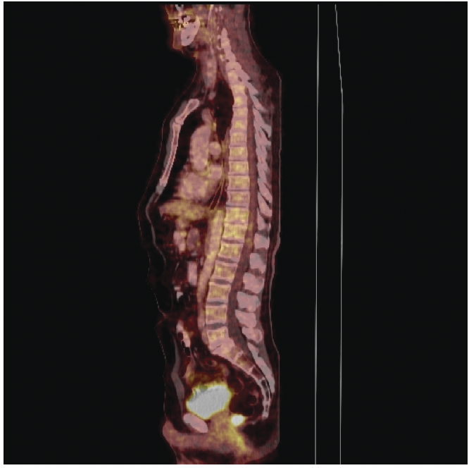
Eighteen months after completion of chemoradiotherapy for anal cancer there is still no sign of relapse. So far the patient has lived 45 months since initial cancer diagnosis.
Discussion
Patients with CUP pose a challenge to clinical oncologists. Since the primary tumour is not known, the treatment cannot be directed towards one specific cancer disease, and the prognosis is often poor. The group of patients with isolated metastatic inguinal lymph nodes (squamous carcinoma) constitutes a small but favourable sub-set of the patients with CUP. In these patients examination towards identifying the primary tumour should include anorectal examination, gynaecological examination and probably also a cystoscopy. 3 The recommended treatment for isolated metastatic inguinal lymph nodes continues to be inguinal lymph node dissection with or without adjuvant radiotherapy, and long-term survivors have been reported. 4
We have presented a case in which a patient presented with a single lymph node metastasis in the right inguinal region as the only finding. Despite thorough clinical examination including whole-body PET/CT the primary tumour was not found. Not until nearly two years later did the primary tumour appear on a control PET/CT. Normally, at the time of diagnosis, patients with anal cancer present with a palpable or visible tumour in the anal canal, 5 but our case illustrates that anal cancer can metastasise before the primary tumour becomes large enough to be identified.
The case also demonstrates the necessity of thorough clinical follow-up after completion of therapy for lymph node metastases of unknown origin, since the primary tumour was found in the clinical follow-up period. The extent of this clinical follow-up is debatable but should probably include a CT of the abdomen and thorax every six months. Furthermore, anorectal and gynaecological examinations should be included in the clinical examination in addition to the CT. These examinations are inexpensive and easy to perform, and inguinal lymph nodes are a known predilection site for the spread of both anal cancer and some gynaecological cancers.
As mentioned earlier in the discussion, adjuvant radiation therapy following inguinal lymph node dissection is optional in patients with isolated metastatic inguinal lymph nodes. The patient in this report declined adjuvant inguinal irradiation following lymph node dissection, and later, when treated for anal cancer, the patient also declined prophylactic inguinal irradiation.
According to a multivariate analysis in the Phase III EORTC-trial
6
inguinal node metastasis is an independent prognostic factor for treatment failure and overall survival in anal cancer. In most centres patients with anal cancer are treated with prophylactic inguinal irradiation but the optimal dose of this radiation has yet to be established. In a large study on 243 patients with anal cancer Papillon
In conclusion, this case illustrates, that anal cancer can metastasise before the primary tumour is detectable. Furthermore it demonstrates the necessity of thorough clinical follow-up after treatment of CUP since the primary tumour was found later. Finally, this is a case of a long-term survivor following treatment for isolated metastatic inguinal lymph nodes.