
Abstract
Background
Developmental dental anomalies are seen as abnormalities in tooth size, shape, position, and structure due to multiple reasons during various stages of tooth development. These anomalies can create disturbances in dental arch lengths and occlusions. Hence, it is very important to treat, recognise and perform proper treatment. The purpose of this study was to find out the prevalence and distribution of selected developmental anomalies in shape, size and position of teeth in the Saudi population of Taif Region.
Design and methods
The study was based on the clinical examination of 2481 adults who are Saudi nationals came for dental treatment from September 2019 to February 2020, at Taif University Dental Hospital, Saudi Arabia. These patients were examined clinically for developmental dental anomalies affecting shape, size and position.
Results
We found that a total of 512 individuals (20.63%) had developmental anomalies and out of which 386 persons (15.56%) had at least one developmental dental anomaly. The frequency and distribution of anomalies of shape and size, number and position were 46.8%, 26.9% and 42.9% respectively. In the present study, 15.56% individuals exhibited at least one anomaly, 8.54% subjects had more than one anomaly and 79.36%.did not any developmental anomaly. On comparison, statistically significant results were seen between different groups of anomalies.
Conclusions
The present study had varying results for the prevalence rate of selected dental anomalies. This variation in results might be due racial differences or discrepancy in sample size or/and diagnostic or inclusion criteria. Treatment of developmental anomalies necessitates a multidisciplinary approach and mostly may comprise of orthodontic correction or prosthetic replacement.
Introduction
Morphological variations of tooth are common developmental anomalies due to disturbance during morpho-differentiation of odontogenesis. 1 The relevant information about prevalence and distribution of such dental anomalies may go unnoticed during routine clinical examination other than chief complain of patient, such teeth are asymptomatic and are discovered on careful clinical and radiographic examination of the oral cavity. The most common complication of dental anomalies is malocclusion, change in eruption sequence and path (ectopic eruption), difficult in restorative, endodontic or surgical procedures. Identification and timely intervention at an early stage is needed to minimize or prevent complications. 2 The aim of this study is to identify, analyze few selected of developmental dental anomalies in the Taif region of Kingdom of Saudi Arabia.
Design and Methods
The present study was conducted on 2481 patients who attended the outpatient clinic, Faculty of Dentistry, Taif University Dental Hospital, at Taif, Saudi Arabia, from September 2019 to February 2020. The ethical approval for the study was obtained from the Institutional Ethics committee, prior to the start of the study. This cross-sectional study is based on clinical examination and panoramic radiographs. Only Saudi nationals aged more than 18 years were included after taking their informed consent. Individuals having history of any systematic diseases, any common syndrome, cleft lip and palate, any tooth extraction due to caries or trauma or for orthodontic reasons and others were excluded from this study. Demographic details including age and gender of patients along with detailed medical, dental and family histories were recorded. A detailed clinical examination was done along with panoramic radiographs for presence of any dental anomalies and the following dental anomalies were assessed.
Anomalies in tooth number: congenital missing (Hypodontia) and supernumerary tooth (Hyperdontia)
Anomalies in tooth size: Macrodontia and Microdontia
Anomalies in tooth shape: Talons cusp, Fusion, Taurodontism, Dens Evaginatus and others
Anomalies in tooth position: Ectopic eruption and Rotation.
A comprehensive analysis was done on the data collected using spreadsheet (Excel 2000; Microsoft Office, Microsoft Corporation, USA) and analyzed using the Statistical Package for Social Sciences version 20 (SPSS Inc., Chicago, USA). All the results were formulated and presented as Tables 1–4. Comparative analysis was done between different study groups of anomalies using Spearman's rank Correlation test and the correlation was significant at the P<0.01 level for all groups as presented in Table 4.
Significance for public health
The assessment of prevalence and distribution of dental anomalies in the Kingdom of Saudi Arabia is very much required. These dental anomalies are significant and show no symptoms, seen on careful examination. These mostly result in malocclusion, change in eruption, increased vulnerability to caries, compromised esthetics and problems in permanent teeth, difficulties in endodontic or surgical procedure of the affected tooth, so on. It is very essential to take care of these anomalies, if not can produce disturbances in maxillary and mandibular dental arch and occlusions. Further, these might pose complication in planning the orthodontic treatment. Hence to know the prevalence rate and distribution of such anomalies will help various dental faculties/specialists to identify and recognize prevailing dental anomalies early and perform proper treatment planning. There are no such studies in adult populations of Saudi Arabia. This study has indicated that about 20% of the participants had dental anomalies and was observed more in males of about 63.3%.
Results
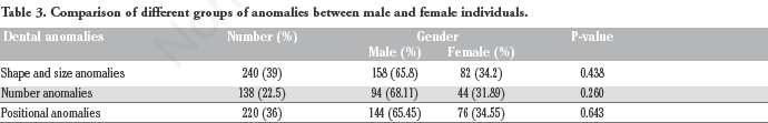
Out of 2481 subjects [1444 (26.86%) males and 1037 (41.79%) females], a total of 512 individuals (20.63%) had developmental dental anomalies. The distribution by gender was 324 males (63.4%) and 188 females (36.6%). The frequency and distribution of the developmental dental anomalies are shown in Table 1. Out of the total 512 individuals, 386 (15.56%) exhibited at least one anomaly and 212 (8.54%) subjects displayed more than one anomaly (Table 2). Large number of individuals (1969) did not have any anomaly which was about 79.36%. There were no statistically significant differences among males and females, when compared for different groups of anomalies such as shape, size and position (Table 3). On comparison between groups, all the anomalies were significantly most prevalent (P<0.05) as shown in Table 4.
Distribution and prevalence of developmental dental anomalies.

Frequencies of dental anomalies in the total subjects.

Comparison of different groups of anomalies between male and female individuals.
Comparative analysis between different study groups.

*Indicates statistically significant (P<0.01).
Among the anomalies in the present study, the most frequent developmental dental anomaly was rotation (24.6%), followed by ectopic eruption (18.3%), belong to group of positional anomalies. The second most common group was size and shape anomalies which included microdontia (15.2%). Among the number anomaly group was Hyperdontia or supernumerary teeth (15.6%), which had a higher frequency than hypodontia (11.3%). Anomalies related to shape such as Dens Evaginatus (2.1%), Talon cusp (5.4%), fusion (3.3%) were in smaller numbers. All these data are presented in Table 1.
Discussion
Developmental anomalies of permanent dentition cause complications such as malocclusion, increased susceptibility to caries, esthetics, and problems in eruption of succedaneous teeth, difficulties in endodontic or surgical procedure of the affected teeth. These may pose problem subsequently causing irreversible damages. In general, these anomalies remain overlooked/ignored/unnoticed in routine clinical practice and are rare chief complaints of the patients. 3 The prevalence rate and distribution of such anomalies will help various dental faculties/specialists to identify and recognize prevailing dental anomalies early as well as in comprehensive treatment.
Although there are studies related to the prevalence of individual dental anomalies either in shape or size, no studies on prevalence and distribution of the developmental dental anomalies in adult populations of Saudi Arabia were found. However, only one similar study among Saudi children has been published. 4 Previous studies indicated that the frequencies of many dental anomalies are varied in different populations. Thisvariation in results were mainly due to racial differences, variable sampling techniques and inconsistent or dissimilar diagnostic criteria.2,5,6
In the current study 24.67% of the sample had anomalies, out of which <10% had only one dental anomaly and around 15.11% showed more than one type of anomaly. These findings were similar to the study conducted by Sogra et al. 7 and reported that 12% had at least one dental anomaly, and 5% having more than one anomaly. However, results of other studies have noted higher than the present rate of prevalence for several dental anomalies.8,9
The most common dental anomalies belonged to shape, followed by position and number anomalies. This group of anomalies was more prevalent than shape, structural and number anomalies. The frequently occurring anomaly of position was rotation of teeth (20.58%), followed by ectopic eruption (18.3%) (Table 1). The prevalence of ectopic eruption has been reported to be in the range of 0.7% to 7.9%.10,11 However, a study by Afify et al., repor ted as low as 0.7% in Saudi population. 11
In our study, the prevalence of Hypodontia found to be affecting 58 (11.3%) individuals which was higher compared to previously reported studies.10,12 A study in Indian population found to be affecting 10.9% of pretreatment orthodontic patients 13 and found to be 8.1% and 8.5% in Australian and Japanese orthodontic patients respectively.2,14 However, in another study it was found to be 2.7% in Mexican orthodontic patients. 15
The prevalence of talon cusp ranged between 1-8% of the population and this anomaly found to be affecting 28 (5.4%) individuals in our study. However, the previous study in Saudi children reported to be 1.4% for talon cusp.4,16 In the present study, fusion was the rarest anomaly compared to other shape anomalies, with a prevalence of 3.3% which is similar to previously reported studies. 17 The prevalence of fusion ranged from 0.5% to 5% depending on the geographic area, race and genetic factors. 18
In the current study, we noted 10 (1.9%) of the patients having taurodontism and found to be almost similar to previous studies.4,19 The prevalence was found to be higher by 8.0% as reported by Darwazeh et al. in Jordanian patients. 20 It was reported to be higher in the French population with a prevalence of around 15%. 21 The prevalence of dens evaginatus in the present study was 2.1% and previous studies have shown a range of 1.01% in the Thai population and 3% in Hong Kong Chinese patients.22,23
Dilaceration was noted in about 2.5% of the individuals in our study and Hamasha et al. 24 the occurrence of root dilacerations was 3.7% of the dentition. However, dilacerations occurring in mesial and distal directions are clearly visible with radiographs for diagnosis of root dilaceration. The prevalence of dilaceration reported to be very least of about 0.39 in one study and 16.48% in another study.12,25 Among various studies, it was found to be 5.29% in Iranian population, 26 and 0.18% in French population. 21 Very few or limited studies are available on the prevalence of supernumerary roots. The prevalence is very much varying from 0.034% to 5.9% as reported previously.12,27 We found it to be 6.4% among 33 persons with male predilection as shown in Table 1. The variations in the prevalence rates of the developmental anomalies are mainly due to sampling technique, ethnic diversity, and different inclusion criteria.
Conclusions
The present study showed that 20.63% of the participants had dental anomalies. Most anomalies were observed in males about 63.3%. Timely identification and intervention will minimize the complications in the permanent dentition. Rarely, developmental anomalies of teeth are associated with many syndromes and genetic diseases and hence it is very important and helpful in precise and timely diagnosis of complex genetic abnormalities of the craniofacial region and in the treatment for better esthetic and function in the future.
Footnotes
The authors declare no potential conflict of interest.