
Abstract
Human amniotic fluid-derived stem cells (AFSCs) represent a novel class of broadly multipotent stem cells sharing characteristics of both embryonic and adult stem cells. However, both the origin of these cells and their actual properties in terms of pluripotent differentiation potential are still debated. In order to verify the presence of features of pluripotency in human second trimester AFSCs, we have investigated the ability of these cells to form in vitro three-dimensional aggregates, known as embryoid bodies (EBs), and to express specific genes of embryonic stem cells (ESCs) and primordial germ cells (PGCs). EBs were obtained after 5 days of AFSC culture in suspension and showed positivity for alkaline phosphatase (AP) staining and for specific markers of pluripotency (OCT4 and SOX2). Moreover, EB-derived cells showed the expression of specific transcripts of the three germ layers. RT-PCR analysis, carried out at different culture times (second, third, fourth, fifth, and eighth passages), revealed the presence of specific markers of ESCs (such as FGF4 and DAPPA4), as well as of markers typical of PGCs and, in particular, genes involved in early stages of germ cell development (Fragilis, Stella, Vasa, c-Kit, Rnf17). Finally, the expression of genes related to the control of DNA methylation (DNMT3A, DNMT3b1, DNMT1, DNMT3L, MBD1, MBD2, MBD3, MDB4, MeCP2), as well as the lack of inactivation of the X-chromosome in female samples, was also demonstrated. Taken together, these data provide further evidence for the presence of common features among human AFSCs, PGCs, and ESCs.
Introduction
In recent years, great interest has been devoted to human amniotic fluid-derived stem cells (AFSCs), a novel class of stem cells characterized by expression of specific markers of pluripotency even after several passage numbers, high proliferation rate, ability to differentiate into cells of all the three embryonic germ layers, and absence of tumor formation after transplant in nude mice (1–4,8–10, 17,20,29). Owing to these features, AFSCs are considered a peculiar class of stem cells with intermediate properties between embryonic stem cells (ESCs) and lineage-restricted adult progenitor cells. As a consequence, several studies have focused their attention on the study of AFSCs and their possible application in regenerative medicine (1–3, 8,17,20). Although the majority of published reports used v-Kit Hardy-Zuckerman 4 Feline Sarcoma Viral Oncogene Homolog positive [cluster of differentiation 117+ (CD117+); c-KIT+] selected AFSCs for their purposes (5,7,10,11,15), other studies have investigated the features of unselected, directly cultured, AFSCs (1,17,31). The main debated topics concerning AFSCs are related to their origin and their actual properties in terms of pluripotent differentiation potential (19). In fact, several studies have demonstrated the presence of a wide spectrum of cells of different origins within human amniotic fluid (AF), derived from the skin and the digestive tract of the developing embryo/fetus, as well as from the amniotic membranes (2,3). However, different authors have suggested that AFSCs may have characteristics similar to pluripotent cells of the epiblast, leading to a hypothesis that during embryogenesis some cells migrate from the epiblast to the AF (12,26,30). Nevertheless, so far no definitive experimental support for this hypothesis has been provided, and the debate on the origin of these cells is still open. To better characterize the features of pluripotency in human second trimester AFSCs and their possible origin from epiblast, we investigated AFSC transcriptional profiles, particularly genes specifically expressed in ESCs and primordial germ cells (PGCs), and tested AFSCs' ability to form in vitro three-dimensional aggregates, known as embryoid bodies (EBs) (35). These analyses revealed that human second trimester AFSCs are able to express markers typical of PGCs, in particular genes involved in early stages of germ cell development, and show several features of pluripotency.
Materials and Methods
AF samples were obtained from 12 women undergoing amniocentesis for prenatal diagnosis at 16 weeks of pregnancy after written informed consent, out of a total of 314 amniocenteses carried out during the study period (January 2011–January 2013). The major indications for amniocentesis were positive maternal serum triple test, advanced maternal age with positive maternal serum triple test, and advanced maternal age solely. The mean (±SD) maternal age at amniocentesis was 37.66 ± 3.75 years (Table 1). The study has been approved by the ethics committee of the “G. d'Annunzio” University of Chieti-Pescara, ASL Lanciano-Chieti-Vasto, Italy.
Summary of Data of 12 Samples of Amniotic Fluid From Which Amniotic Fluid-Derived Stem Cells (AFSCs) Were Isolated

Isolation and Culture of Human AFSCs
AFSCs were cultured in Iscove's modified Dulbecco's medium (IMDM), supplemented with 20% fetal bovine serum (FBS), 100 U/ml penicillin, 100 μg/ml streptomycin, 2 mM L-glutamine, 5 ng/ml basic fibroblast growth factor (FGF2) (all Sigma-Aldrich, Milan, Italy) and incubated at 37°C with 5% humidified CO2. This culture medium, characterized by high serum content in conjunction with IMDM, was chosen due to the reported ability to select a homogeneous cell population (1). At 80% confluence, cells were treated with 0.05% trypsin-ethylenediaminetetraacetic acid (EDTA) (Sigma-Aldrich) and passaged every 2 days with a 1:3 split beyond the 10th passage.
Endothelial Differentiation of AFSCs
For endothelial differentiation, carried out in order to provide differentiated cells to be used for comparison with undifferentiated cells in expression studies, AFSCs at passage 6 were transferred into an appropriate medium [IMDM supplemented with 50 ng/ml vascular endothelial growth factor (VEGF; Sigma-Aldrich)] for 15 days. AFSCs under endothelial differentiation conditions have developed an epithelial-like morphology and have presented functional characteristics of endothelial cells by forming vascular tube-like structures in Matrigel. AFSCs (3 × 10⁵ cells/ ml) were seeded into a six-well plate precoated with Matrigel (10 mg/ml; BD Pharmingen, San Jose, CA, USA) and cultivated for 15 days. Moreover, endothelial differentiation was assessed by studying expression of endothelial markers [VEGF, von Willebrand Factor (vWF), vascular endothelial growth factor receptor 2 (VEGFR-2), and vascular endothelial cadherin (VE-cadherin)] by RT-PCR (Table 2).
List of Human-Specific Primer Sequences Used in RT-PCR to Confirm Endothelial Differentiation
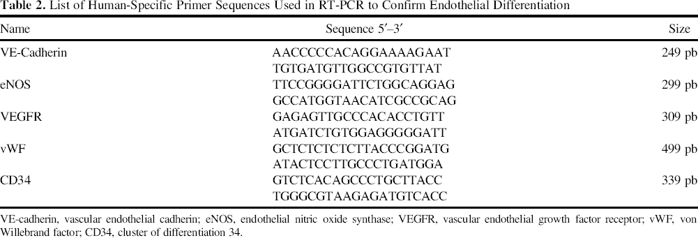
VE-cadherin, vascular endothelial cadherin; eNOS, endothelial nitric oxide synthase; VEGFR, vascular endothelial growth factor receptor; vWF, von Willebrand factor; CD34, cluster of differentiation 34.
Embryoid Body Formation Induced by the “Hanging Drop” Method
To form EBs (see Fig. 1 for schematic), human AFSCs at the eighth passage were harvested with 0.05% trypsin and cultured in suspension in IMDM containing 15% FBS, 1 mM glutamine, 0.1 mM β-mercaptoethanol (Sigma-Aldrich), and 1% penicillin–streptomycin. The 25-μl drops (~1,000–10,000 cells) were plated on the lid of Petri dishes (M-Medical, Milan, Italy), which were inverted and covered onto a dish filled with 8 ml phosphate-buffered saline (PBS; M-Medical) to maintain the humidity (hanging drop method). After 4 days of incubation, EBs were resuspended in the same medium and continued to grow in suspension for 2 days. After a total of 6 days in suspension, the EBs were plated onto gelatin-coated (0.1%; Sigma-Aldrich) tissue culture plates for morphological analysis.

Schematic diagram of sequential steps performed for obtaining and analyzing EB-like structures. AFSCs, amniotic fluid-derived stem cells; EBs, embryoid bodies; AP, alkaline phosphatase; Sox2, sex-determining region Y box 2; OCT4, octamer-binding transcription factor 4.
Alkaline Phosphatase (AP) Staining
Both AFSC cultures and EBs were fixed with 4% paraformaldehyde (Sigma-Aldrich) for 10 min at room temperature and incubated for 2 h with BCIP/NBT liquid substrate system (Sigma-Aldrich) at room temperature. The reaction was stopped by removing the substrate solution and washing with distilled water.
Expression Studies by RT-PCR
Total RNA was extracted using Eurogold Total RNA Mini Kit (Euroclone, Milan, Italy) from different cell lines, namely: i) undifferentiated AFSCs at the third, fourth, fifth, and eighth passage; ii) AF-derived endothelial progenitor cells (AF-EPS), and iii) cells from EBs. cDNA was synthesized from total RNA using RevertAid First Strand cDNA Synthesis Kit (Fermentas, M-Medical) and oligo (dT) primers. The cDNA was amplified according to the protocol of fast Kapa2G Hot Start: 95°C for 1 min for predenaturation, followed by 35 cycles of 95°C for 10 s, 45–58°C for 30 s, and 72°C for 15 s, with a final extension of 30 s at 72°C. RNA samples incubated without reverse transcriptase served as negative controls to verify the absence of DNA. PCR products were resolved by electrophoresis (Euroclone) on 3% agarose gels (Euroclone) and visualized by ethidium bromide (Euroclone) staining. RT-PCR analysis was carried out in order to investigate the expression of i) typical markers of ESCs and PGCs, ii) alternatively spliced genes involved in development and differentiation, iii) genes involved in DNA methylation, and iv) genes related to X chromosome inactivation (Table 3). Relative quantification of the expression levels of each gene was normalized with housekeeping gene glyceraldehyde 3-phosphate dehydrogenase (GAPDH). The primers used for expression studies are listed in Tables 4 and 5.
List of Investigated Genes (See Text/Figure Legends for Definitions)

List of Specific Primer Sequences of Human Embryonic Stem Cells (hESCs) and Primordial Germ Cells (PGCs) Used in RT-PCR

List of Specific Primer Sequences for Alternative Splice Variants of DNA Methyltransferases (DNMTs), Methyl-CpG-Binding Domain Proteins (MBDs), X Inactive Specific Transcript Sense (Xist), and Antisense (Tsix) Used in RT-PCR

Cardiogenic Differentiation of EBs
At the end of incubation day 4, EBs were grown in suspension for 2 days in IMDM containing 20% FBS, 100 U/ml penicillin, 100 μg/ml streptomycin, 2 mM L-glutamine, 10 μM 5-aza-2′-deoxycytidine (Sigma-Aldrich), and 0.1 μM ascorbic acid (Sigma-Aldrich). After a total of 6 days in suspension, cells were transferred into gelatincoated tissue culture dishes and grown for 10 days in medium without ascorbic acid and 5-aza-2′-deoxycytidine.
Morphological Assays in Light and Electron Microscopy
EBs at different periods of culture were fixed with a 3% paraformaldehyde solution for 15 min at room temperature in PBS 1×, stained with hematoxylin eosin solution (Bio-Optica, Milano SpA, Milan, Italy) at room temperature, and observed under a ZEISS Axioskop light microscope (Carl Zeiss AG, Milan, Italy) equipped with a Coolsnap video camera (Photometrics, Tucson, AZ, USA) For electron microscopy analyses, EB samples were prefixed with 2.5% glutaraldehyde (Electron Microscopy Sciences, Rome, Italy) in 0.1 M cacodylate buffer (Electron Microscopy Sciences), pH 7.3, for 40 min at 4°C and postfixed with 1% osmium tetroxide (Electron Microscopy Sciences) for 40 min at 4°C. After being dehydrated in alcohol at progressively higher concentrations, samples were embedded in Spurr resin (Electron Microscopy Sciences). Semithin sections were stained with 1% toluidine blue (Sigma-Aldrich) and analyzed with a ZEISS Axioskop light microscope equipped with a Coolsnap digital camera (Carl Zeiss AG). Ultrathin sections were cut with a Reichert ultramicrotome (Reichert, Inc., Teramo, Italy), mounted on 200 mesh cupper grids (Electron Microscopy Sciences, Fort Washington, WA, USA), and photographed by means of a ZEISS-109 electron microscope equipped with a Gatan 830Z00W44 digital camera (Pleasanton, CA, USA).
Immunostaining of Cultured EBs
EBs were fixed for 15 min with a 3% paraformaldehyde solution at room temperature in 1× Dulbecco's phosphate-buffered saline (PBS), pH 7.6, supplemented with 2% sucrose (Sigma-Aldrich,). After cell membrane permeabilization with 0.5% Triton X-100 (MP Biomedicals Europe, Milan, Italy), EBs were incubated with 10% bovine serum albumin (BSA; Sigma-Aldrich) in 1× Dulbecco's phosphate-buffered saline for 30 min at room temperature, followed by a 45-min incubation at room temperature with 10 μl of a phycoerythrin (PE)-conjugated mouse anti-human octamer-binding transcription factor 3/4 (OCT3/4) antibody (1:10,000; BD Pharmingen, San Jose, CA, USA), diluted in 1% BSA/PBS. For sex-determining region Y box 2 (SOX2) or cardiac myosin heavy chain immune labeling, samples were first incubated for 60 min at room temperature with a mouse anti-human SOX2 antibody (1:2,000: Euroclone) or a mouse anti-human cardiac myosin heavy chain antibody (1:3,000; Abcam, Cambridge, UK) diluted in 1% BSA/PBS. After rinsing three times with 1× PBS, samples SOX2 labeled were incubated for 45 min at 37°C with a goat anti-mouse IgG-fluorescein isothiocyanate (FITC) (Jackson Immuno Research, West Grove PA, USA) diluted 1:100 in 1% BSA/PBS, while samples cardiac myosin heavy chain labeled were incubated for 45 min at 37°C with a goat anti-mouse IgG-Alexa Fluor® 488-conjugated secondary antibody (Invitrogen, Molecular Probes, Eugene, OR, USA) diluted 1:100 in 1% BSA/PBS. After labeling, nuclei were counterstained with 4′,6-diamidino-2-phenylindole (DAPI)-mounting medium (Vector Laboratories, Inc., Burlingame, CA, USA). The observations were carried out with a ZEISS AXIOSKOPE light microscope equipped with a Coolsnap Videocamera and with the MetaMorph® 6.1 Software System to analyze images (Universal Imaging Corp., Downingtown, PA, USA).
Results
AFSC Selection and Morphology
In the present study, we at first investigated in AFSCs the transcriptional profiles of genes known to be abundantly and uniquely expressed in human ESCs and PGCs (26), as well as the expression of alternatively spliced genes involved in development and differentiation (14,27,38) and in epigenetic mechanisms (16,33). To this aim, we cultured AF samples obtained from women undergoing amniocentesis for prenatal diagnosis at 16–19 weeks of pregnancy without c-KIT+ selection. During the first 5 days of culture, AFSCs adhered in plastic culture dishes, and both fibroblast-like cells and epithelioid cells appeared. After the third passage, most cells exhibited a fibroblast-like phenotype, with disappearance of the marked morphological differences (Fig. 2a). Between the third and the fourth passage, AFSCs frequently formed spherical colonies, which were positive for AP staining (Fig. 2b). The cells were cultured beyond the 10th passage without any evident alteration of their proliferation ability, morphology, and karyotype. Directed endothelial differentiation to AF-EPS was confirmed by RT-PCR for endothelial markers and vascular tube-like formation on growing the cells in Matrigel (data not shown).

Isolation of hAFSCs, expression of the typical markers of hESCs and PGCs, and formation of EBs. (a) Fibroblast-like morphology of AFSCs; (b) AP-positive cell colonies; (c) expression of specific markers of pluripotency and embryonic stem cells (ESCs) detected by RT-PCR in human AFSCs at the second, third, fourth, fifth, and seventh passages; (d) expression of premeiotic, meiotic, and postmeiotic markers in AFSCs at the second, third, fourth, fifth, and seventh passages; (e) EB-like structures on day 8 and on day 15; (f) expression of three germ layers markers detected by RT-PCR in the cells from EB-like structures; (g) AP staining of EB-like structures 2 days after attachment in cell culture. Klf4, Kruppel-like factor 4; hTERT, human telomerase reverse transcriptase; FGF4, fibroblast growth factor 4; DPPA2, developmental pluripotency associated 2; ESG1 DPPA5, embryonal stem cell specific gene 1, developmental pluripotency associated 5; GDF3, growth differentiation factor 3; GAPDH, glyceraldehyde 3-phosphate dehydrogenase; Stra 8, stimulated by retinoic acid 8; Piwil 2, piwi-like RNA-mediated gene silencing 2; DAZL, deleted in azoospermia-like DAZL; Rnh2, ribonuclease inhibitor 2; Ovol1, ovo-like zinc finger 1; Boll, boule-like RNA-binding protein; Creb 3/4;, cAMP-responsive element-binding protein 3/4; cycl 1, cylicin 1; GATA4, guanine–adenine–thymine–adenine-binding protein 4; AFP, α fetoprotein; Nkx2.5, NK2 homeobox 5.
Expression Studies
For expression studies, RT-PCR analyses were carried out on AFSCs at the second, third, fourth, fifth, and seventh passages, always showing the expression of the classical markers of pluripotency: OCT4, NANOG, c-MYC, and SOX2 (Fig. 2c). Furthermore, the expression at all passages of other specific markers of ESCs [Kruppel-like factor 4 (KLF4), human telomerase reverse transcriptase (hTERT), c-KIT, fibroblast growth factor 4 (FGF4), developmental pluripotency associated 2 (DPPA2), DPPA4, DPPA5 (embryonal stem cell-specific gene 1; ESG1), growth differentiation factor 3 (GDF3)] was also observed (Fig. 2c). To verify the possible origin of AFSCs from epiblast, we studied the expression of premeiotic, meiotic, and postmeiotic markers of PGCs, evidencing at all passages the presence of markers typical of the early stages of germ cell development [Fragilis, Stella (DPPA3) and Vasa]. Interestingly, Fragilis is also expressed in ESCs and embryonic germ (EG) cells, indicating a potential role of this gene in the pluripotency status (36). Other premeiotic markers [deleted in azoospermia-like (DAZL), piwi-like RNA-mediated gene silencing 2 (Piwil 2), stimulated by retinoic acid gene 8 (Stra8)] were expressed by AFSCs. Among these, only DAZL had been previously reported as being expressed also in c-KIT+ AFSCs (32). In addition, we demonstrated expression in AFSCs of all the investigated meiotic [ovo-like zinc finger 1 (OVOL1), boule-like RNA-binding protein (BOLL)] and postmeiotic [Haprin, Acrosin, cAMP-responsive element-binding protein 3/4 (Creb3/4), cylicin 1 (Cycl1)] markers. Among these, a peculiar feature was shown by the Cycl1 gene; it was expressed only starting from the seventh passage (Fig. 2d). These results indicate the presence among AFSCs of a cell population sharing molecular features common to pluripotent PGCs.
EB Formation
In a second approach aimed to the study of the features of pluripotency of AFSCs, we investigated the ability of these cells to form in vitro three-dimensional aggregates (EBs), which are widely considered an optimal starting point for the differentiation of pluripotent stem cells into three germ layers and thus as an appropriate way to test pluripotency (37). AFSCs were grown in suspension in uncoated Petri dishes, and after 8 days of culture (4 days in hanging drop, 2 days in suspension, and 2 days after attachment in cell culture) (Fig. 1), spherical aggregates appeared (Fig. 2e). At day 15 (7 days after attachment in cell culture), the detachment of cells from the periphery of the spheres was observed (Fig. 2e). In each experiment, starting from 1 million cultured AFSCs, we obtained approximately 80–85 EB structures. The sizes of EBs formed at day 8 were approximately 56,497.6 ± 2,388.0 μm2 area, 618.9 ± 23.4 μm perimeter, 193.4 ± 3.72 μm width, and the EB sizes sharply increased at day 14, respectively, 64,709.1 ± 5,345.8 area, 787.5 ± 199.6 perimeter, 246.0 ± 67.6 width. Expression studies carried out on cells from EB-like structures after their treatment with trypsin showed the presence of specific transcripts of the three embryonic germ layers, such as guanine–adenine–thymine–adenine-binding protein 4 (GATA4; endoderm), GATA6 and α fetoprotein (AFP; mesoderm), Nestin (ectoderm), and the early cardiac transcription factor NK2 homeobox 5 (Nkx2.5) (13,18) (Fig. 2f). This suggests that the EB-forming cells have the potential to differentiate in vitro into derivatives of all three primary germ layers. EBs were positive for AP staining, which represents another typical feature of pluripotent cells (Fig. 2g). Routine hematoxylin–eosin staining procedure allowed us to detect the presence of two main EB cell populations: the core cell population, characterized by a smaller size and heterochromatic nuclei, and the peripheral cell population, consisting of cells characterized by higher cellular and nuclear dimensions, starting to detach from the EB mass and suggestive of differentiating cells (Fig. 3a). To better evaluate the ultrastructural characteristics and organization of EB cell compartments, transmission electron microscopy procedures and analyses were carried out. Observations performed on semithin sections obtained from EBs at day 14 of culture revealed the presence in the EB mass of a hollow cavity surrounded by a crown of cells more packed together toward the inner cavity and more spaced at the EB periphery (Fig. 3b4). Interestingly, at a higher magnification, the presence of a high number of electron-dense granules resembling myelinic figures, as well as the budding and extracellular accumulation of vesicles with an electron-transparent homogeneous content was displayed (Fig. 3b1–3). Immunofluorescence analysis revealed that the EBs express the markers of pluripotency OCT4 and SOX2 (Fig. 3c). Under the treatment with ascorbic acid and 5-aza-2′-deoxycytidine, the cell mass derived from EBs started to differentiate into cardiomyocytes, as demonstrated by the cytoplasmic presence of α-myosin heavy chain (α-MHC) organized in parallel-oriented filaments evidenced by immunocytochemistry after 10 days of differentiation (Fig. 3d). These data demonstrate that human AFSC-derived EB-like structures, when cultured in appropriate conditions, can give rise to terminally differentiated cardiomyocytes. However, the efficiency of this process appears to be quite low, since beating cells were only sometimes observed, mainly at the periphery of the EB.

Morphological patterns of EBs. (a) Hematoxylin eosin staining of EBs demonstrating the presence of different cell populations with different morphological and staining characteristics; (b1) semithin section of a 14-day cultured EB stained with toluidine blue solution; the inset at a lower magnification shows the whole embryoid body containing a hollow cavity and a crown of peripheral cells and resembling a blastocyst structure; (b2) cell cytoplasm displaying different types of granules; (b3) extracellular release of vesicles with an electron-transparent homogeneous content; (b4) electron-dense cytoplasmic granules resembling myelinic figures; (c) specific markers of pluripotency detected in cells from EB-like structures with immunofluorescence (OCT4: 2D deconvolution; SOX2: 2D Photoshop reconstruction). (d) Cells derived from EB-like structures positive for myosin marker after the differentiation into cardiomyocytes. DAPI, 4′,6-diamidino-2-phenylindole; MHC, cardiac myosin heavy chain.
Alternative Splice Variants Study
A further approach used to investigate the features of pluripotency in AFSCs was based on the study of alternative splice variants, which represent novel biomarkers of pluripotent stem cells, playing several splice isoform key roles in lineage commitment and self-renewal (6). For this reason, we examined the expression of alternative splicing of genes involved in cell proliferation and differentiation in three cell types: undifferentiated AFSCs at the seventh passage, EBs, and amniotic fluid-derived endothelial precursors (AF-EPS), obtained by cultivating AFSCs in an appropriate medium supplemented with VEGF for 15 days. Most of the alternative splice variants were expressed in all the three classes of cells analyzed (Fig. 4). However, DNA methyltransferase 3 β (DNMT3B) exon 10 was uniquely expressed in EBs and completely absent in the other two types of cells. DNMT3B is a member of the DNA methyltransferase family, encoding for 40 different isoforms and involved in de novo methylation after embryonic implantation. DNMT3B exon 10 containing mRNA transcripts are present in ESCs but not in somatic cells. Thus, our results suggest that EB-forming cells are able to reacquire characteristics similar to pluripotent stem cells. On the other hand, analysis of DNMT3B catalytic domain encoded by exons 20–22 showed the presence of the full-length isoform in all of the three cell types (Fig. 4a). In addition, we studied alternative splice variants of spalt-like transcription factor 4 (Sall4) gene, a transcription factor essential for pluripotency, present with two isoforms (Sall4A and Sall4B) differing by the inclusion or exclusion of exon 2 (21). Only Sall4B is able to maintain the pluripotent state by binding preferentially to highly expressed loci in ESCs. The expression of Sall4A isoform was detected in all three types of cells analyzed, while Sall4B isoform was only detected in cells derived from EBs and in undifferentiated AFSCs (Fig. 4b). We next monitored the expression of genes encoding for DNA methyltransferase (DNMTs) (22,28) and methyl-CpG-binding domain proteins (MBDs) in AFSCs, EBs, and AF-EPSs. Transcripts of four classes of DNMTs (DNMT1, DNMT2, DNMT3A, and DNMT3B) were widely expressed in AFSCs and AF-EPSs. The same result was obtained for the expression profiles of the various MBD genes [MBD1, MBD2, MBD3, MBD4, and methyl CpG-binding protein 2 (MeCP2)] (Fig. 5a). In contrast, EBs showed a different expression profile. In fact, only the expression of DNMT1, DNMT2, and DNMT3B was detected, while DNMT3A was not expressed (Fig. 5a). Another marker of pluripotency in stem cells is represented by the presence of two active X chromosomes, occurring during the embryonic development and the formation of germ cell precursors. To verify the status of X chromosome inactivation, we analyzed the expression of X inactive specific transcript (XIST) and X inactive specific transcript, antisense (TSIX) genes in the undifferentiated AFSCs, EBs, and AF-EPSs. Transcripts of the XIST gene were detected in female AFSCs and AF-EPS lines, but not in male samples (Fig. 5b). Conversely, these cells do not show TSIX gene expression, indicating the presence of an inactive X chromosome. On the other hand, XIST was not expressed in female EBs, while the expression of the antisense repressor TSIX was observed (Fig. 5b). These data suggest a reactivation of the X chromosome in EBs, demonstrating that three-dimensional spheres obtained in vitro from AFSCs are capable of acquiring some pluripotent abilities. Based on these results, human female AFSCs can be categorized into two epigenetic classes: i) undifferentiated and differentiated AFSCs, that consistently exhibit XIST+/TSIX- cells, with consequent X chromosome inactivation, and ii) EBs, composed by XIST-/TSIX+ cells, in which X chromosome reactivation takes place.

Expression of alternative splice variants in cells derived from EB-like structures. (a) RT-PCR analysis performed on undifferentiated AFSCs, amniotic fluid-derived endothelial precursors (AF-EPSs), and cells from EB-like structures; (b) alternative splicing patterns of spalt-like transcription factor 4 (SALL4) analyzed by RT-PCR. Expression of isoforms SALL4A and SALL4B were present in AFSCs and cells from EB-like structures. P2RX5, purinergic receptor P2X, ligand-gated ion channel, 5; NDKA, nucleoside diphosphate kinase A; STAT3, signal transducer and activator of transcription 3; NUBP2, nucleotide-binding protein 2; SHC1, Src homology 2 domain-containing transforming protein 1; CDC25A, cell division cycle 25A; TBC1D3P2, Tre-2, Bub2p, and Cdc16p domain (TBC1) family, member 3 pseudogene 2; DNMT3B, DNA methyltransferase 3 β.

Expression of DNMTs, MBDs, XIST, and TSIX genes in AFSCs, AF-EPSs, and cells from EB-like structures. (a) AFSCs and AF-EPSs showed the expression of DNMTs type 1, 2, 3A, 3B, methyl-CpG-binding domain protein 1 (MBD1), MBD2, MBD3, MBD4, and methyl CpG-binding protein 2 (MeCP2). Cells from EB-like structures expressed the transcripts of DNMT type 1, 2, and 3B; (b) RT-PCR for X inactive specific transcript (XIST) and X inactive specific transcript antisense (TSIX) expression performed on female AFMSCs, EBs, and AF-EPS. XIST transcripts are detected only in female AFSCs and AF-EPS, while the expression of the gene TSIX was present in female EBs and absent in female undifferentiated and differentiated AFMSCs.
Discussion
Taken together, all the obtained results are in agreement with the hypothesis that AFSCs have similar properties to PGCs. This hypothesis is very interesting because PGCs originate from the proximal epiblast adjacent to the extraembryonic ectoderm, then passing through the primitive streak, and giving rise to several extraembryonic mesodermal lineages and to germ cells (39). Until a short time ago, no experimental support to this hypothesis had been provided. However, more recently, the group of Moschidou et al. (23) reported that human first-trimester AF contains cells of germ origin that can be reverted to pluripotency when supplemented with valproic acid. In fact, first-trimester c-Kit+-selected AFSCs, when reprogrammed to pluripotency, are able to express ESC-specific markers, maintain repression of X-inactivation, are positive for AP staining and are capable of forming EBs. The same group suggested that also mid-trimester c-KIT+ AFSCs show features of pluripotency when cultured in appropriate media (24). In the present study, we confirmed these findings also in unselected AF cells from the second trimester of pregnancy, since in our experience, these cells share common characteristics with ESCs, including expression of the key genes involved in the process of cellular reprogramming (OCT4, SOX2, c-MYC, and KLF4) and with PGCs, as evidenced by the expression of genes involved in the early stages of germ cell development (Fragilis, Stella, Vasa, and c-Kit). On the basis of this evidence, and according to the suggestions of Moschidou et al. (23) and Dobreva et al. (12), a small percentage of PGCs are likely lost into the AF during migration in the genital ridge, and this could explain the presence of early markers of germ cells in the unselected AFSCs. Additionally, in the present study, we have demonstrated that second trimester AFSCs are able to form in vitro three-dimensional aggregates, similar to EBs. Interestingly, when AFSCs are aggregated to form EBs, they reacquire pluripotency potential and features of early stage embryogenesis that are otherwise lost. In fact, cells from AFSC-derived EBs, in addition to the expression of pluripotency markers (OCT4, SOX2, NANOG, and KLF4), which are present also in unselected AFSC cultures, display a number of specific features such as i) expression of alternatively spliced exons specific of pluripotent stem cells, such as the exon 10 of DNMT3B and the b isoform of Sall4; ii) expression of markers representative of the three embryonic germ layers (such as GATA4, GATA6, AFP, and Nestin), and iii) absence of the X chromosome inactivation. This latter aspect is certainly very interesting since the reactivation of the inactive X chromosome appears to be tightly correlated with genomic reprogramming events and occurs at least twice during mammalian development: once in the ESCs prior to blastocyst implantation, and thereafter in the epiblast, in particular in the PGCs or PGC precursor cells. The expression of several specific markers of ESCs and especially typical markers of PGCs appears to be a specific feature of second trimester-unselected AFSCs, since c-Kit+-selected cells obtained from the second trimester of pregnancy do not demonstrate these properties (24). As a consequence, we hypothesize that the protocol based on the direct culture of AF cells without any selection could allow them to retain a small percentage of PGCs or PGC progenitors that are likely discarded or lost during the procedure of c-KIT selection. In conclusion, and although at present AFSCs are classified into an intermediate stage between ESCs and adult stem cells, their molecular and biological characteristics are closer to the PGCs and are increasingly distant from multipotent stem cells. When considering that amniocentesis is a widely accepted standard procedure of prenatal diagnosis, the banking of AFS cell lines could have a strong impact in public health, thus opening the way both for their possible clinical application in humans, and for their use for the modeling of human genetic disease, which could be easily and frequently detected during prenatal diagnosis. Given these considerations, AF cells could represent an interesting alternative to induced pluripotent stem (iPS) cells for the study of human diseases. In fact, embryoid body-like structures arising from AFSCs are able to reacquire pluripotency potential and features of early stage embryogenesis and may represent a promising solution for cell-based therapy and regenerative medicine. So far, AFSCs have been used by our group in preclinical studies to treat pathological conditions such as ischemic brain injury with encouraging results (34). The outcome of these studies supported the idea that immunomodulatory and paracrine effects are predominant in the therapeutic properties of these cells (34). This notion is important since it is emerging in the scientific world that postengraftment differentiation and proliferation are probably not the primary mechanism by which cell therapy confers significant benefits. Despite the increased knowledge about the mechanisms involved in the therapeutic effects of AFSCs, further research focusing on this theme is required to design strategies aimed at increasing the efficiency of therapies for the treatment of various human diseases.
Footnotes
Acknowledgments
This manuscript is in memory of Stefano Tetč, a colleague and a friend, who prematurely passed away on March the 14th, 2014. This study was supported by grants from the Italian Minister of University and Research (MIUR) to A.P. and from the Cari-Chieti Foundation (Italy) to I.A., R.D.P., S.T., and L.S. Author contributions: Conceived and designed the experiments: I.A., G.P., A.P., S.T., L.S. Performed the experiments: I.A., R.D.P., M.A., M.A.C., L.C., S.S., M.A.D.'A., A.D.B., V.M., F.P. Analyzed the data: I.A., L.S., R.D.P. Contributed reagents/materials/analysis tools: L.S., S.T., R.D.P., A.D.B. Wrote the article: I.A., L.S., R.D.P., C.V.B. The authors declare no competing financial interests.