
Abstract
The pancreatic and duodenal homeobox gene 1 (Pdx-1) plays a key role in normal pancreas development and is required for maintaining the normal function of islets. In this study, we examined whether human adipose tissue-derived stem cells (hASCs) could differentiate into insulin-producing cells by exogenously expressed Pdx-1. hASCs were infected with recombinant adenovirus encoding the mouse Pdx-1 gene and differentiated under high-glucose conditions. Insulin transcript levels and the expression of key transcription factors required for pancreatic development including FoxA2, Nkx2.2, and NeuroD were significantly increased by exogenous Pdx-1 overexpression. The expression of Nkx6.1 was found only in Pdx-1-induced hASCs. In addition to transcripts for transcription factors involved in pancreatic development, transcripts for the GLP-1 receptor, glucokinase, and glucose transporter, which are required for maintaining the function of pancreatic β-cells, were observed only in Pdx-1-induced hASCs. Pdx-1-induced hASCs exhibited insulin secretion in response to glucose challenge in vitro. When Pdx-1-induced hASCs were transplanted into streptozotocin (STZ)-induced diabetic mice, they reduced blood glucose levels, although they did not restore normoglycemia. These results demonstrate that the expression of exogenous Pdx-1 is sufficient to induce pancreatic differentiation in vitro but does not induce the fully functional, mature insulin-producing cells that are required for restoring normoglycemia in vivo.
Keywords
Introduction
Islet transplantation is a promising cell-based therapy for diabetes. However, the unavailability of sufficient numbers of pancreas donors makes islet transplantation applicable only in a limited number of cases. Therefore, studies have increasingly focused on producing transplantable pancreatic β-cell substitutes. One of the most studied approaches is genetic reprogramming of stem cells using the pancreatic and duodenal homeobox gene 1 (Pdx-1).
Pdx-1 plays a key role in normal pancreas development and is required for maintaining the normal function of islets (22). Pdx-1 binds to and transactivates an insulin promoter (17); it is involved not only in regulating insulin gene expression but also in regulating the expression of many other β-cell-specific genes (14, 15, 24, 25).
Genetic ablation of Pdx-1 in mice leads to the loss of the entire pancreas (6) and results in the development of diabetes (1). In humans, patients bearing a mutant insulin promoter factor 1 (Ipf-1; the human form of Pdx-1) gene exhibit agenesis of the pancreas at birth (23).
Several research groups have reported that ectopic expression of Pdx-1 induces the formation of insulin-producing cells from embryonic stem (ES) cells, adult bone marrow, or intestinal stem cells (9, 11, 16). Furthermore, ectopic expression of Pdx-1 converts adult liver cells into functional insulin-producing cells, reversing hyperglycemia (5, 10, 21). This finding suggests that Pdx-1 is a master regulatory factor capable of inducing transdifferentiation from differentiated cells whose fates are already determined.
We have previously demonstrated that human adipose tissue-derived stem cells (hASCs) have the potential to differentiate into pancreatic lineage cells (12). In this study, we examined whether the exogenous expression of Pdx-1 was capable of inducing pancreatic differentiation from hASCs. We found that overexpression of Pdx-1 induced the differentiation of hASCs into aggregates of insulin-producing cells and showed that these cells exhibited insulin secretion in response to glucose stimulation in vitro. Moreover, when transplanted into streptozotocin (STZ)-induced diabetic mice, they reduced blood glucose levels. However, they did not fully reverse hyperglycemia and restore normoglycemia in vivo.
Materials and Methods
Culture of hASCs
Human adipose tissue was obtained from three male donors (age range: 54–70 years) who were supposed to get abdominal surgery at the Asan Medical Center (Seoul, Korea). The study was approved by the Asan Medical Center Institutional Review Board, and all donors provided informed consent. hASCs isolated from adipose tissue were cultured in low-glucose Dulbecco's modified Eagle's medium (DMEM; Gibco, Life Technologies, Long Island, NY, USA) as previously described by Zuk et al. (26). Briefly, the stromal vascular fraction (SVF) was isolated by extensively washing adipose tissue with phosphate-buffered saline (PBS; Gibco) followed by digestion of the extracellular matrix by incubating with 0.075% type I collagenase (Sigma, St Louis, MO, USA) at 37°C for 30 min. After neutralizing enzyme activity with DMEM containing 10% fetal bovine serum (FBS; Gibco), the solution was centrifuged at 1,200 rpm for 10 min to obtain the SVF pellet. The pellet was resuspended in 0.16 M NH4Cl for 5 min to lyse red blood cells and then centrifuged at 1,200 rpm for 10 min. The collected SVF pellet was filtered through a 100-μm nylon mesh (BD Falcon, Bedford, MA, USA) to remove cellular debris and incubated overnight at 37°C/5% CO2 in control medium (DMEM containing 10% FBS, 1% antibiotic/antimycotic solution; Gibco). Following incubation, the plate was washed with PBS to remove nonadherent cells and adhered cells were maintained in control medium at 37°C/5% CO2. The medium was replaced every third day thereafter.
Adenovirus Production and Induction of Differentiation by Pdx-1 Infection
Full-length mouse Pdx-1 cDNA was purchased from Open Biosystems (Huntsville, AL, USA). An adenoviral construct expressing Pdx-1 gene was produced using the Adeno-X ViraTrack Expression System 2 (Clontech, Mountain View, CA, USA) following the manufacturer's protocols. Briefly, the Pdx-1 gene was cloned into the pDNR-CMV donor vector, and the resulting pDNR-CMV-Pdx-1 construct was used to transfer the Pdx-1 gene into the pLP-Adeno-X-ViraTrak-ZsGreen acceptor vector through Cre-LoxP recombination. After Cre-loxP recombination was completed, the recombinant adenoviral vector was introduced into Supercharge EZ10 ElectroCompetent cells (Clontech) using electroporation. The entire transformants were inoculated on Luria Bertani Agar (LB; Becton, Dickinson & Company, Sparks Glencoe, MD, USA)/chloramphenicol (Sigma)/sucrose (Sigma) plates to recover the colonies originated from the competent cells harboring the recombinant adenoviral vectors. After 24–30 h, about 50 colonies of various sizes were obtained and small- to-medium-sized colonies were exclusively picked. PCR colony screening analysis using Adeno-X LP primer Mix was carried out to further confirm the presence of the recombinant adenoviral vector. Once confirmed, recombinant pAd-Pdx-1-ZsGreen was digested with PacI to expose the inverted terminal repeats, and PacI-digested pAd- Pdx-1-ZsGreen was transfected into HEK293 cells (Prof. Heuiran Lee, Department of Microbiology, University of Ulsan, College of Medicine, Seoul, Korea). One day after seeding, passage-five hASCs were infected with adenovirus expressing Pdx-1 at a multiplicity of infection (MOI) of 300. Forty-eight hours later, Pdx-1-induced cells were switched to DMEM containing high glucose and maintained for up to 4 weeks. hASCs without Pdx-1 infection, but exposed to the same differentiating protocol as Pdx-1-induced hASCs, were used as a control.
Immunocytochemistry
Cells were fixed in 4% paraformaldehyde (Merck, Hohenbrunn, Germany) in PBS for 20 min at room temperature and then washed three times with PBS containing 0.1% bovine serum albumin (BSA; Sigma). Cells were permeabilized using 0.3% Triton X-100 (Sigma) in PBS containing 0.1% BSA and 10% normal goat or donkey serum (Vector Laboratories, Inc., Burlingame, CA, USA). Cells were incubated overnight at 4°C with primary antibody. After washing, cells were incubated with fluorescence-labeled secondary antibodies for 2 h at room temperature. Cells were then mounted in Vectashield mounting medium containing 4′,6-diamidino-2-phenylindole (DAPI; Vector Laboratories) and examined using a fluorescence microscope (Olympus, Tokyo, Japan).
The following antibodies were used at the indicated concentrations: goat anti-Pdx-1 (Santa Cruz Biotechnology, Inc., Santa Cruz, CA, USA; 1:1,000), mouse anti-insulin (Sigma; 1:500), rabbit anti-human C-peptide (Linco Research, St. Charles, MO, USA; 1:100), Alexa Fluor 555-conjugated donkey anti-goat IgG (Invitrogen Molecular Probes, Eugene, OR, USA; 1:1,000), Cy3-conjugated goat anti-mouse secondary antibody (Jackson Immuno Research Laboratories, West Grove, PA; 1:400), and Cy3-conjugated goat anti-rabbit secondary antibody (Jackson ImmunoResearch Laboratories; 1:400).
Quantification of Insulin-Reactive Cells
Quantification of insulin-reactive cells (aggregates of insulin-producing cells) was analyzed using an image analyzer. For image analysis, three randomly selected fields containing aggregates of insulin-producing cells were photographed using an Olympus DP71 digital camera system and Olympus Ix 71 software and analyzed using ImagePro-Plus 5.1 software (Media Cybernetics, Silver Spring, MD, USA).
Reverse Transcription
Primer Sequences for RT-PCR
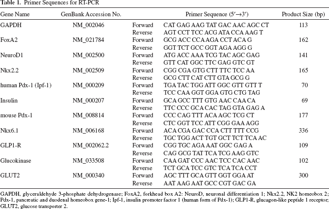
GAPDH, glyceraldehyde 3-phosphate dehydrogenase; FoxA2, forkhead box A2: NeuroD, neuronal differentiation 1; Nkx2.2, NK2 homeobox 2; Pdx-1, pancreatic and duodenal homeobox gene-1; Ipf-1, insulin promoter factor 1 (human form of Pdx-1); GLP1-R, glucagon-like peptide 1 receptor; GLUT2, glucose transporter 2.
Measurement of Insulin Secretion
Cells were washed five times with PBS and incubated for 1 h in Krebs–Ringer bicarbonate (KRB) buffer (120 mM NaCl, 2.5 mM CaCl2, 1.1 mM MgCl2, 25 mM NaHCO3, 0.1% BSA) containing 5.6 mM glucose. Cells were then washed again and incubated in KRB buffer containing 23 mM glucose for another 1 h. Supernatants from cells stimulated with 5.5 or 23 mM glucose were collected and analyzed using a human insulin ELISA kit (Mercodia, Sylveniusgatan, Sweden) according to the manufacturer's instructions.
Transplantation of Pdx-1-Induced Aggregate of Insulin-Producing Cells
Eight-week-old, male BALB/c nude mice (OrientBio, Kyunggi, Korea) were made hyperglycemic with a single, 250 mg/kg intraperitoneal injection of streptozotocin (STZ, Sigma). Three days after STZ injection, diabetic mice with blood glucose levels greater than 400 mg/dl were used for transplantation. After anesthetizing, the left kidney was exposed and a small nick was made using a 23-gauge needle (Korea Vaccine Co. Ltd., Seoul, Korea). Control or Pdx-1-induced hASCs (2 × 10⁶ cells) were then transplanted under the left kidney capsule using a PE50 tube (Becton Dickinson & Company). Blood glucose levels in blood samples obtained from the tail vein were monitored using a Lifescan OneTouch SureStep (Johnson & Johnson Co., Inc., Milpitas, CA, USA). All experimental procedures involving animals were approved by the Institutional Animal Care and Use Committee of the Asan Medical Center.
Immunohistochemistry
Kidneys that received Pdx-1-induced hASCs were removed after 30 days. Removed kidneys were fixed, embedded in paraffin, and stained with hematoxylin and eosin (Sigma) for general kidney histology and analyzed by immunohistochemistry for insulin, as described above.
Statistical Analysis
Statistical analyses were performed using Student's t tests.
Results
Pdx-1-Induced Pancreatic Differentiation in Human Adipose Tissue-Derived Stem Cells
To induce pancreatic differentiation, we infected fifth passage hASCs with recombinant adenovirus (MOI, 300). Two days after infection, over 60% of cells were immunoreactive for Pdx-1 in the nucleus of infected hASCs, whereas no Pdx-1-immunoreactive cells were found among control hASCs (Fig. 1). At this point, Pdx-1-induced hASCs were switched to DMEM with high glucose and maintained for up to 4 weeks. During this period, morphological changes occurred in Pdx-1-induced hASCs, which gradually aggregated and formed clusters; the morphology of control cells remained unchanged. Immunocytochemical analyses showed that insulin and C-peptide positivity was associated with clustered Pdx-1-induced hASCs (Fig. 2). The yield of the aggregate insulin-producing cells was obtained by quantitative analysis and calculated to be approximately 11%. In agreement with immunocytochemical results, insulin transcript levels were substantially increased in Pdx-1-induced hASCs compared with controls (Fig. 3A).

Expression of Pdx-1 in hASCs. Pancreatic and duodenal homeobox gene-1 (Pdx-1) expression after adenoviral infection [multiplicity of infection (MOI), 300] was assessed by immunocytochemistry. Pdx-1 protein was detected in the nucleus of Pdx-1-induced human adipose-derived stem cells (hASCs), but no Pdx-1 expression was found in control cultures. Nuclei were stained with DAPI.

Immunocytochemical analysis for insulin and C-peptide. After differentiation for 4 weeks, insulin- and C-peptide-positive cells were detected only among Pdx-1-induced hASCs. Nuclei were stained with DAPI.

Expression of pancreas-related genes in ectopic Pdx-1-induced hASCs. (A) An RT-PCR analysis showed that genes involved in pancreas development [Neuronal differentiation 1 (NeuroD), forkhead box A2 (FoxA2), and NK2 homeobox 2 (Nkx2.2)] were upregulated in Pdx-1-induced hASCs. Expression of endogenous Ipf-1 (insulin promoter factor 1; human Pdx-1) was observed only in hASCs induced by exogenously added mouse Pdx-1. Endogenous Ipf-1 and exogenously expressed mouse Pdx-1 were distinguished using species-specific primers. Genes involved in maintaining pancreatic β-cell function were expressed only in Pdx-1-induced cultures. (B) Quantitative real-time PCR confirmed the changes in gene expression observed in conventional RT-PCR analyses. hASCs without Pdx-1 infection, but exposed to the same differentiation protocol as Pdx-1-induced hASCs, were used as a control. Total RNA from a human pancreas (20-year-old Caucasian male) was used as a positive control. Data are presented as means ± SEMs of three independent experiments (*p < 0.01 and **p < 0.001 vs. respective controls). GAPDH, glyceraldehyde 3-phosphate dehydrogenase; GLP1r, glucagon-like peptide 1 receptor; GLUT2, glucose transporter 2.
In order to dissect the mechanisms underlying the Pdx-1-induced pancreatic differentiation of hASCs, we performed RT-PCR and assessed the expression of genes involved in pancreatic development. RT-PCR analyses using species-specific primer pairs showed that the endogenous human Ipf-1 was activated in exogenous mouse Pdx-1-induced hASCs (Fig. 3A). The expression of key transcription factors required for pancreatic development, including forkhead box A2 (FoxA2), NK2 homeobox 2 (Nkx2.2), and Neuronal differentiation 1 (NeuroD) were present at basal levels in the control group and were upregulated by Pdx-1-induced differentiation (Fig. 3A). Nkx6.1 expression was detected only in Pdx-1-induced hASCs. In addition to transcripts for transcription factors related to pancreatic development, transcripts for glucagon-like peptide 1 (GLP-1) receptor, glucokinase (GK), and glucose transporter 2 (GLUT2), which are required for maintaining the function of pancreatic β-cells, were observed only in Pdx-1-induced hASCs. The changes in gene expression observed by conventional RT-PCR analyses were further confirmed by quantitative real-time RT-PCR (Fig. 3B).
Insulin Secretion in PDX-1-Induced hASCs by Glucose Stimulation
To determine whether Pdx-1-induced hASCs were capable of insulin secretion, we incubated Pdx-1-induced hASCs with KRB buffer containing low (5.6 mM) or high (23 mM) concentrations of glucose and assayed culture supernatants for human insulin, as described earlier. As shown in Figure 4, Pdx-1-induced hASCs secreted insulin in response to glucose stimulation.

Assay of in vitro insulin secretion by Pdx-1-induced hASCs. Insulin released in response to 5.6 and 23 mM (high glucose) was measured. Data are presented as means ± SEMs of three independent experiments (*p < 0.001 vs. 5.6 mM glucose).
Transplantation of Pdx-1-Induced hASCs Into Diabetic Mice
To determine whether the Pdx-1-induced hASCs were capable of regulating blood glucose in vivo, we transplanted Pdx-1-induced hASCs into the left kidney capsule of diabetic nude mice. Mice in the control group were transplanted with hASCs without Pdx-1 but differentiated in the same manner as Pdx-1-induced hASCs. Although transplantation of Pdx-1-induced hASCs did not reverse hyperglycemia and restore normoglycemia (Fig. 5A), it did reduce glucose levels compared to mice that received control hASCs, preventing the development of a severely hyperglycemic state. After 4 weeks of transplantation, the kidney was recovered and histological assessment was performed. In the histological kidney examination, we did not find islet like structures (Fig. 5B). Immunohistochemical analyses showed that Pdx-1-induced hASCs transplanted under the kidney capsule stained positively for insulin. In contrast, no insulin-positive cells were found in sections from the control mice (Fig. 5B).
Assessment of in vivo function of transplanted cells. (A) Changes in blood glucose levels in diabetic mice were measured. After differentiation, 2 × 10⁶ control or Pdx-1-induced hASCs were transplanted under the left renal capsule of diabetic mice (n = 10 mice/group) Pdx-1-induced hASCs reduced blood glucose levels to 300 mg/dl, a level that was maintained in the absence of severe status for 1 month. Mice transplanted with control hASCs showed no decrease in blood glucose levels and died within 1 week. Data are presented as means + SEMs (*p < 0.05, **p < 0.01, and ***p < 0.001 vs. respective controls). STZ, streptozotocin. (B) Immunohistochemical analysis of transplanted cells. Thirty days after transplantation, mice were sacrificed and kidneys were removed for histological examination. Paraffin-embedded kidney blocks were sectioned (thickness, 5 μm), and sections were stained with hematoxylin and eosin (H&E staining) (a) and analyzed for insulin by immunohistochemistry. (b) Control hASCs; (c) Pdx-1-induced hASCs. Arrow indicates positive staining. Cell nuclei were stained with DAPI.
Discussion
The efficient generation of insulin-producing cells from adult stem cells is an important clinical goal because of the potential of such cells to substitute for islet transplantations in the treatment of diabetes. It has been reported that human adipose tissue, which is available in large quantities as a waste product of cosmetic liposuction, contains multipotent stem cells (26), and have been shown to possess immunosuppressive properties and to survive in xenotransplantation (8, 18, 20). In our previous work, we showed that hASCs exhibit some characteristics of pluripotent embryonic stem cells and have the potential to differentiate into pancreatic lineage cells (12). Thus, human adipose tissue represents a source of autologous stem cells sufficient to meet the potential clinical demand for engineered insulin-producing cells.
Several studies have attempted to induce insulin-producing cells using differentiating agents (2, 3) and through overexpression of the transcription factor Pdx-1 (5, 9–11, 16, 21), which is critical in pancreatic development and maintenance of normal pancreatic function (22).
In this study, we evaluated the ability of Pdx-1 to induce insulin-producing cells by infecting hASCs with adenovirus expressing Pdx-1.
Initially, we investigated whether genes involved in pancreatic development and function were expressed in Pdx-1-induced hASCs and compared the results with human pancreas. Overexpression of exogenous mouse Pdx-1 induced the expression of endogenous human Ipf-1 after differentiation. After differentiation for 4 weeks, Pdx-1-induced differentiated cultures exhibited increased expression levels of transcription factors involved in β-cell development (i.e., FoxA2, NeuroD, Nkx2.2). Expression of GLUT2 and GK, which are required for the maintenance of pancreatic β-cell function, was observed only in Pdx-1-induced hASCs, as was expression of the GLP-1 receptor. The gene expression pattern of Pdx-1-induced hASCs was similar to that of human pancreas. These results indicate that Pdx-1 alone was capable of inducing the expression of other genes involved in pancreatic differentiation and suggest that these differentiated cells might have the ability to secrete insulin in response to glucose. We tested this latter supposition by analyzing the secretion of insulin in response to glucose stimulation, which is an important indicator of functional pancreatic β-cells. Our results confirmed that glucose did indeed stimulate insulin secretion in these Pdx1-induced hASCs.
To extend these in vitro results to an in vivo setting, we transplanted Pdx-1-induced hASCs into STZ-induced diabetic mice. The severity of hyperglycemia was reduced in these mice; however, blood glucose levels remained high, indicating that, although Pdx1-induced hASCs were functional, their activity in vivo was weak. Our results are in accord with two recent papers by Lin et al. (13) and Kajiyama et al. (7), who reported limited success using Pdx-1-induced hASCs. In vivo data from both studies demonstrated that Pdx-1-induced hASCs did not convert hyperglycemia to normoglycemia, and only modestly improved blood glucose levels.
One possible explanation for this limited in vivo efficacy is that the number of transplanted cells is insufficient to normalize hyperglycemia. Alternatively, Pdx-1-induced hASCs may be less mature, despite the ability of these differentiated cells to secrete insulin in response to glucose stimulation in vitro. A third possibility is that, although high glucose is one factor needed for pancreatic differentiation, maintaining cells under high-glucose conditions during differentiation may have paradoxically severely reduced insulin expression in β-cells and promoted β-cell apoptosis (4, 19).
In summary, our findings suggest that hASCs differentiated into aggregates of insulin-producing cells in vitro after being infected with an adenovirus expressing Pdx-1 without any supplementation of the specific factors known to induce pancreatic differentiation. However, Pdx-1 alone is not sufficient to induce functionally mature insulin-producing cells from hASCs. Despite the inability of Pdx-1-induced hASCs to restore normoglycemia in diabetic animals, our findings present the possibility of gene modification-based cell therapy using hASCs. For application to human patients, further studies are needed to establish the requirements for inducing functional maturation of differentiated cells and more safe and effective methods to overcome limitation caused by genetic manipulation using a viral vector.
Footnotes
Acknowledgments
This work was supported by National Research Foundation of Korea Grant funded by the Korean Government (313-2008-2-E00299). The authors declare no conflicts of interest.