
Abstract
Negative cosignaling molecules play an important role in regulating T-cell responses to alloantigen stimulation. We recently reported that adenoviral-mediated transduction of islet allografts with B7-H4 inhibits allograft rejection. In this study, we investigate the mechanism for B7-H4-induced prolongation of mouse islet allograft survival. Streptozotocin-induced diabetic C57BL/6 mice were rendered normoglycemic by renal subcapsular implants of B7-H4-transduced BALB/c islets. Grafts and spleens were removed after days 2, 10, and 60 (n = 8 each) for characterization of kinetics of Foxp3 and interleukin 10 (IL-10) expression. Mixed lymphocyte reaction (MLR) was done at day 60. Ten mice were subjected to nephrectomy at 60 days and then five were implanted with secondary BALB/c islets and five were given third-party CBA/J islets. An increase in Foxp3 and IL-10 mRNA expression was detected in recipients' spleens at day 60 and this was associated with increased quantities of Foxp3+ cells. Splenocytes at day 60 showed hyporesponsiveness during MLR to alloantigen stimulation. Proliferation was partially restored after CD25+ T-cell depletion. Secondary BALB/c islets survived for 79 ± 29 days compared with 21 ± 3.6 days for CBA/J islets (p < 0.001). Local expression of B7-H4 induces long-term unresponsiveness to donor-specific alloantigens, and is associated with T regulatory cells, suggesting the development of tolerance.
Introduction
Transplantation of islets to restore pancreatic β-cell function is a promising means for treating type 1 diabetes (T1D) in the clinical setting. The success of islet transplantation is largely reliant on effective immunosuppressive therapy. Current immunosuppressive drugs are able to control acute rejection efficiently. Changes to the immunosuppressive regimen in the Edmonton Protocol improved graft survival at 1 year, but toxic effects of these drugs on β-cells have been implicated as a major factor in the long-term dysfunction of islet allografts (14,29,30,40,41). In order to improve β-cell function, rationalization of immunosuppressive therapy is needed using a new generation of drugs with minimal side effects. Greater immunoselectivity is desirable with the overall objective of tolerance induction.
Immune-mediated rejection is mainly controlled by an alloreactive T-cell response to transplanted grafts. The specificity for alloreactivity is determined by the recognition of the peptide presented on the major histocompatibility complex (MHC) and T-cell receptor (TCR). However, the antigen-specific signal generated by the TCR alone is insufficient for optimal T-cell activation (19). Full T-cell activation requires a second positive cosignal, such as is provided by CD28 stimulation (15,19). In the presence of a negative cosignaling molecule, T-cell proliferation is terminated and likely results in a state of anergy, which is defined as inability to respond when reexposed to the same antigen (15,19). The ability of negative cosignals to generate anergy provides us with an attractive strategy to inhibit allograft rejection and to induce tolerance. Generation of tolerance with cosignaling blockade has been reported (5,13,23,35,38) to occur through various mechanisms including anergy, clonal deletion, and immune regulation/suppression. For example, blocking both CD28-B7 and CD40-CD40 ligand binding results in apoptosis of proliferating T cells and permanent engraftment of cardiac allografts (20). On the other hand, combined cytotoxic T lymphocyte antigen-4 (CTLA-4). Ig and anti-CD40L treatment results in tolerance to alloantigen in vivo through induction of T regulatory cells (Tregs) within the local graft environment and/or peripheral tissues (23,35). Combined anti-BTLA (B and T lymphocyte attenuator) and CTLA-4.Ig can also induce local accumulation of forkhead box P3+ (Foxp3+) T cells and tolerance in mouse islet allografts (37). Several studies have demonstrated that it is feasible to withdraw continuing immunosuppressive therapy after blockade of cosignaling molecules (5,13,23,35,38).
B7-H4 is a newly identified negative cosignaling molecule in the CD28-B7 family (24,26,31,39,44). We recently reported that local expression of B7-H4 by a recombinant adenovirus (Ad-B7-H4) prolongs murine islet allograft survival in a strongly immunogenic combination, from BALB/c to C57BL/6 (39). Islets expressing B7-H4 survived significant longer, with half of the recipients surviving beyond 60 days (the experimental end point). These promising data prompted us to explore whether alloantigen tolerance arises in mice receiving B7-H4-transduced islets. In the present study, we sought evidence that long-term survival of B7-H4-transduced islets is associated with donor-specific tolerance rather than with immune ignorance.
Materials and Methods
Mice
C57BL/6 (H-2b), BALB/c (H-2d), and CBA/J (H-2K) mice were purchased from Jackson Laboratory and housed in the Jack Bell Research Centre under conventional conditions. All mice were cared for according to the guidelines of the Canadian Council on Animal Care and regulations of the University of British Columbia.
Primary and Secondary Transplantations
BALB/c mice were used as both primary and secondary transplant donors into C57BL/6 recipients. CBA/J mice were used as third-party control donors for secondary transplants. C57BL/6 mice were also used as syngeneic donors for C57BL/6 recipient controls in some experiments. In the primary transplant, a group of 400 islets was isolated and transplanted into the left renal capsule of recipients rendered diabetic by a single intraperitoneal (IP) injection of streptozotocin (STZ, Sigma, Oakville, Canada) at the dose of 200 mg/kg 3–4 days before transplantation, as described previously (39). The BALB/c donor islets used for primary transplant were transduced with 5 plaque-forming units (pfu) of Ad-B7-H4 or Ad-LacZ, as described previously (39). To test for immunologic tolerance, the kidneys bearing the primary islet grafts were removed after long-term function (over 60 days). These nephrectomized mice were retransplanted with the same donor strain (BALB/c) or third-party (CBA/J) islets into the right kidney capsule. No immunosuppressive treatment was provided.
Mixed Lymphocyte Reaction (MLR) Assay
Irradiated (3000 rads) splenic leukocytes (SLC) (3 times; 105) from donor strain BALB/c or CBA/J mice as stimulators were cultured in triplicate with 1 times; 105 SLCs as responder cells (at a stimulator/responder ratio of 3:1) obtained from nontransplanted wild-type C57BL/6 or long-term surviving recipients transplanted with Ad-B7-H4-transduced islets (39). [3H]Methylthymidine was added 18 h before harvesting. Recombinant mouse interleukin-2 (IL-2) at 100 U/ml was added in some MLR assays.
Detection of IL-2 Secretion by ELISPOT
Irradiated (3000 rads) antigen presenting cells (APCs; StemCell Inc., Vancouver, Canada) from BALB/c mice were cultured in triplicate with 1 times; 106 purified CD3+ T cells (StemCell Inc.) from C57BL/6 or long-term surviving recipients transplanted with Ad-B7-H4-transduced islets in a anti-IL-2 precoated plate. Detection of IL-2 was performed according to the manufacturer's instructions (eBioscience, San Diego, CA).
Depletion of CD25+ T Cells
CD4+CD25+ T cells were depleted from SLCs by microbeads coated with anti-CD25 monoclonal antibody (mAb) (Miltenyi Biotec Inc.). The negatively selected CD25- cells were collected. Purity and viability, as determined by fluorescence activated cell sorting (FACS) analysis and trypan blue staining, respectively, exceeded 95% in all cases.
Flow Cytometric Analysis of Lymphocyte Phenotypes in Lymph Nodes and Spleens
Single-cell suspensions were stained with fluorochrome-conjugated mAbs. MAbs against CD4, CD25, and Foxp3 were purchased from eBioscience. Intracellular staining of Foxp3 was performed using a commercially available kit (eBioscience). Cells were analyzed on FACSCanto (BD Bioscience, Mississauga, Canada) using De Novo software (De Novo Software, Los Angeles, CA).
Real-Time PCR Quantification of Genes Associated with T Cell in Grafts and Spleens of Transplant Recipients
Long-term surviving primary allografts were removed with the aid of a dissection microscope. The graft site was subjected to RNA extraction. Real-time PCR was performed as previously described (39). Relative expression level was expressed as 2–(CTubiquitin–CTgene) (where CT is cycling threshold), with ubiquitin RNA as the endogenous control. The primer pairs are listed in Table 1. Mouse glyceraldehyde 3-phosphate dehydrogenase (GAPDH) was used as an internal control for normalization.
PCR Primer Pairs
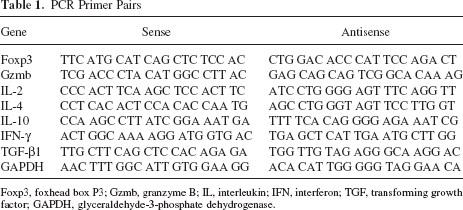
Foxp3, foxhead box P3; Gzmb, granzyme B; IL, interleukin; IFN, interferon; TGF, transforming growth factor; GAPDH, glyceraldehyde-3-phosphate dehydrogenase.
Histological Analysis
The islet graft-bearing kidney was fixed in 4% paraformaldehyde and paraffin embedded. For CD3 (Abcam, Cambridge, USA) and Foxp3 (eBioscience) staining, endogenous peroxidase activity was blocked with 3% hydrogen peroxide for 15 min at room temperature. Horseradish peroxidase (HRP)-conjugated anti-rabbit or anti-rat antibody was used for secondary antibody. Brown color was developed by 3,3′-diaminobenzidine substrate (Sigma). Double staining of insulin (DAKO, Mississauga, Canada) and CD45 (BD Bioscience) was performed using secondary Texas Red- or fluorescein isothiocyanate (FITC)-conjugated antibodies. Isletitis grade was assessed by Yoon's method (grade: 0, normal islets; 1, mononuclear infiltration, largely in the periphery, in less than 25% of the islet; 2, 25–50% of islet showing mononuclear infiltration; 3, over 50% of islet showing mononuclear infiltration; and 4, small, retracted islet with few mononuclear cells).
Statistics
Graft survival curves were analyzed using Kaplan-Meier life-table analysis (Prism 5.0, La Jolla, CA) and the two sets of survival curves were compared by the log-rank test. Student's t-test (two-tailed and paired analysis) or one-way analysis of variance (ANOVA) (Fisher's PLSD post hoc test) was used for comparison of means between experimental groups examined by MLR, real-time PCR, and FACS assays. Differences were considered significant if p < 0.05, unless stated otherwise.
Results
A Regulatory Phenotype Develops in Lymphocytes of B7-H4-Transduced Islet Grafts and Periphery
Expression of genes associated with regulatory T cells was sought in both the graft and the periphery. Foxp3, IL-10, and transforming growth factor-β1 (TGF-β1) mRNA expression in islet grafts and spleens of long-term surviving and syngeneic recipients were analyzed by quantitative real-time PCR (Q-PCR) on days 2, 10, and 60 (n = 8 in each group). Tissues from syngeneic (C57BL/6 → C57BL/6) recipients were used for comparison. Figure 1A shows that Foxp3 expression in both islet graft and spleens from D60_Tx recipients was higher than that of the syngeneic transplant controls, but statistical significance was achieved only on the day 60 after first transplant (Fig. 1A). TGF-β1 was expressed at a similar level to syngeneic controls in all tissue analyzed (data not shown). In contrast, IL-10 expression was significantly increased in the spleen at day 60 (p = 0.005, n = 8) (Fig. 1B).

High levels of Foxhead box P3 (Foxp3), regulatory T cells (Tregs), and interleukin (IL)-10 were found in the grafts and periphery. Results from syngeneic islet transplant recipients and D60_Tx recipients are shown in white and gray bars, respectively. 18s mRNA was used as loading control. (A) mRNA expression level in the grafts and spleens. There was a significantly increased expression of Foxp3 in grafts and spleens of D60_Tx recipients, compared with that of the syngeneic recipients (p = 0.03 and 0.05). (B) IL-10 mRNA expression level in the grafts and spleens. IL-10 expression level was significantly higher in the D60_Tx mice, compared with syngeneic recipients at day 60 in the spleen (p = 0.005). Kinetics of Foxp3+ cells in the local renal lymph nodes (C) and spleens (D). Percentages of renal lymph node Foxp3+ cells in the D60_Tx mice were significantly higher than that in the syngeneic mice at day 2 posttransplant (p = 0.004). Frequencies of spleen Foxp3+ Tregs in the D60_Tx mice were significantly higher than that in the syngeneic mice at day 60 posttransplant (p = 0.04). Each group consisted of 8 recipients.
Foxp3+, CD4+CD25+Foxp3+ Tregs, and CD4+CD25- Foxp3- Teffs (T effector cells) in the draining renal lymph nodes and spleens of long-term surviving graft recipients were quantified by FACS. Figure 1C shows that increased Foxp3+ Tregs were detected in the renal lymph nodes at day 2 posttransplant (p = 0.004, n = 8). However, there was no significant difference in absolute number (6.05 ± 1.84 times; 104 vs. 9.03 ± 2.97 times; 104, p = 0.06). There was no difference in the quantity of CD4+CD25- Foxp3- Teffs in either renal lymph nodes or spleens of the two groups of recipients (data not shown). At day 60, a significantly higher percentage of Foxp3+ Tregs was detected in the spleen of the mice that had B7-H4-transduced islets compared with the syngeneic recipients (3.40 ± 0.64 vs. 4.54 ± 1.01, respectively, p = 0.04, n = 8) (Fig. 1D). There was also shown a significant increase in absolute number (7.57 ± 2.91 times; 105 vs. 1.06 ± 1.71 times; 106, respectively, p = 0.05).
MLR Demonstrates Attenuated Proliferation of Splenic Lymphocytes for Long-Term Surviving Recipients with B7-H4-Transduced Islets
We examined the proliferation of SLCs from either naive C57BL/6 mice (B6 mice) or C57BL/6 mice bearing 60-day surviving B7-H4-transduced BALB/c islet grafts (D60_Tx mice) in response to BALB/c stimulator cells. Figure 2A shows that SLCs from D60_Tx mice demonstrated attenuated proliferation (5.3 ± 1.2 × 10³ cpm) compared with cells from naive B6 mice (10.2 ± 1.3 × 10³ cpm) (p = 0.003). The response of D60_Tx SLCs to other alloantigen was evaluated by challenging them with third-party CBA/J (H-2k) stimulator cells. SLCs from both naive B6 and transplanted D60_Tx mice demonstrated similar responses to CBA/J cells (p = 0.14 for D60_Tx cells stimulated with BALB/c vs. CBA/J, n = 8) (Fig. 2A). Hence, SLCs from D60_Tx recipients are anergic to primary donor alloantigen but mount a normal proliferative response to other alloantigens.

Mixed lymphocyte reaction (MLR) for lymphocytes from long-term surviving recipients with Ad-B7-H4-transduced islets showed hyporesponsiveness. (A) Splenic lymphocytes (SLCs) harvested from spleens of naive C57BL/6 (B6) mice or mice transplanted with Ad-B7-H4-treated BALB/c islets at 60 days (D60) were cocultured with γ-irradiated (at 3000 rad) stimulators from either donor-specific BALB/c (BAL) (H-2d) or third-party CBA/J at a responder/stimulator (R/S) ratio of 1:3 for 3 days in a MLR assay. Cell proliferation was calculated as mean counts per minute (cpm ± SE) of triplicate culture wells. Response of SLCs from D60_Tx recipients was significantly lower than those from naive B6 mice (p = 0.003). Proliferation of D60_Tx cells to donor-specific and third-party controls was significantly different (p = 0.03). There was no significant difference between naive B6 mice and D60_Tx mice in response to CBA/J stimulation (p = 0.14). (B) SLCs from naive B6 mice showed hyporesponsiveness in the presence of D60_Tx SLCs. A 1:1 mixture of naive B6 and D60_Tx responder cells was cocultured with γ-irradiated BALB/c. Proliferation in this MLR assay was significantly lower than that of the control B6 without responder from D60_Tx mice (p = 0.04). (C) The presence of CD4+CD25+ is required for the anergic state of SLCs from D60_Tx mice. CD4+CD25+ were depleted from D60_Tx SLCs (Dep_D60) mice and were cocultured with irradiated alloantigen BALB/c SLCs. MLR cultures of responders from D60_Tx with or without depletion of CD4+CD25+ population, or nontransplanted wild-type C57BL/6 responding to BALB/c alloantigen stimulation, were done in parallel. The ability of D60_Tx cells to respond to antigen stimulation was significantly increased after CD25 depletion (p = 0.03). (D) IL-2 addition could reverse the suppressive state in D60_Tx cells. Proliferation was significantly higher after exogenous IL-2 (100 U/ml) was added to the MLR culture (p = 0.001). Each group consisted of 8 recipients. (E). Relative expression level of IL-2 in the coculture of B6 or D60_Tx plus BALB/c was measured by real-time PCR. (F) Frequency of IL-2 secretion in the MLR culture was detected by ELISPOT. Purified CD3+ T cells (106) from B6 or D60_Tx were cocultured with irradiated BALB/c antigen presenting cells (APCs) from splenocytes. The number of spots in the coculture of D60+BAL was significantly lower than that of B6+BAL coculture (p = 0.05). Results are generated from three independent experiments. Each group consisted of 5–8 recipients.
Figure 2B demonstrates an experiment with a 1:1 mixture of SLCs from naive B6 and D60_Tx transplanted mice. Cell proliferation was (6.2 ± 2.4 × 10³ cpm) in response to BALB/c (H-2d) donor antigen stimulation compared with (10.2 ± 1.3 × 10³ cpm) for naive B6 cells (p = 0.04), demonstrating that D60_Tx SLCs caused suppression of naive SLC proliferation in response to donor alloantigen stimulation.
The Presence of CD4+CD25+ T Cells Is Required for Hyporesponsiveness to Alloantigen Stimulation
To investigate the mechanism of donor-specific hyporesponsiveness observed in vitro, the role of CD4+CD25+ T cells was examined. CD4+CD25+ Tregs were depleted from SLCs isolated from D60_Tx mice and were subjected to MLR with BALB/c stimulators. As shown in Figure 2C, depletion of CD4+CD25+ T cells restored the response of D60_Tx cells to alloantigen stimulation, suggesting that CD4+CD25+ T cells plays a role in B7-H4-induced islet allograft tolerance.
We next tested whether or not exogenously added IL-2 could reverse the donor-specific alloantigen hyporesponsiveness. The addition of IL-2 to the MLR coculture resulted in increased proliferation compared with cocultures without IL-2 (11.9 ± 2.1 × 10³ vs. 5.3 ± 1.2 × 10³ cpm) (p = 0.001) (Fig. 2D). The results revealed that exogenous provision of IL-2 to the MLR coculture ablated the suppressive response mediated by CD4+ CD25+. Therefore, the hyporesponsive state in the D60_Tx cells is IL-2 dependent.
Production of IL-2 in the MLR cocultures was also detected. At both mRNA and protein levels, transcription (Fig. 2E) and secretion (data not shown) were lower in the coculture of D60-Tx than in that of B6, in response to BALB/c stimulation. The reduced amount of IL-2 in the former coculture could be due to either a decline in the number of cells able to secrete IL-2, or to impaired function of IL-2 secretion from each cell. To test these two possibilities, ELISPOT was performed to test IL-2 secretion at a single-cell level. This showed a significant decrease in the number of spots in the former co-culture, suggesting that the low production of IL-2 was in part through the reduced numbers of cells able to secrete the proliferative cytokine IL-2 (p = 0.05, n = 5) (Fig. 2F).
B7-H4 Inhibits Proinflammatory Cytokines in the Allografts
It has been shown previously that polarization of the T helper 1 (Th1) response results in graft failure, whereas a Th2 response is associated with graft acceptance (32,45). In order to test whether or not the protective role of B7-H4 in the alloimmune response is due to the shift of Th1/Th2 balance, IL-2 (for monitoring T cell proliferation), interferon-γ (IFN-γ; Th1), IL-4 (Th2), and granzyme B [Gzmb, for monitoring destructive cytotoxic T lymphocyte (CTL) response] was quantitated by real-time PCR 10 days after the first transplantation. Transcription of IL-2 was lower in Ad-B7-H4-treated mice compared with Ad-Laz-treated mice, but this was not significant (Fig. 3). In contrast, both IFN-γ and Gzmb were significantly decreased in Ad-B7-H4-treated mice (p = 0.007, p = 0.04 for IFN-γ and Gzmb, respectively) (Fig. 3). Expression of IL-4 was similar in the two groups, suggesting that B7-H4 preferentially inhibits Th1 and CTL responses locally. There was no significant change of these cytokines in the serum or spleen, suggesting a local effect of B7-H4 in the islet allograft at an early stage.

Cytokine RNA expression in the grafts. Relative mRNA expression of IL-2, interferon-γ (IFN-γ, IL-4), and granzyme B (Gzmb) was measured by real-time PCR at 10 days post-first transplant in Ad-Lacz (shown in squares, labeled as “control”) or Ad-B7-H4 (shown in triangles, labeled as “B7-H4”) recipients. The syngeneic transplantation of B6 to B6 was used for comparison and considered as an expression level of 1. A significant reduction of both IFN-γ and Gzmb was detected (p = 0.007 and p = 0.04, respectively, n = 5).
Second Set Islet Allografts Survive After Removal of Long-Term Surviving Ad-B7-H4-Transduced Islet Grafts
Figure 4A demonstrates the results of random blood glucose levels following transplantation of B7-H4-transduced islets, subsequent graft nephrectomy at day 60, then secondary transplantation from the same donor haplotype (BALB/c) without any further immunosuppressive treatment. Figure 4B compares the survival of secondary transplants implanted with BALB/c donor-specific islets versus third-party CBA/J islets. Three of five BALB/c donor-specific allografts survived indefinitely (>100 days), and the other two failed with delayed kinetics on days 38 and 58, respectively (p = 0.001 compared with third-party control). CBA/J islet grafts failed at 21 ± 4 days.

B7-H4 induced unresponsiveness to second set donor islet allografts, but not third-party islets. (A) Blood glucose levels in the long-term surviving recipients with Ad-B7-H4-transduced BALB/c islets. After removal of the primary islet-engrafted left kidney, D60_Tx mice became hyperglycemic, then were retransplanted with a second donor-specific BALB/c islet allograft (T2x; n = 5) without any immune treatment. (B) Comparison of survival of islet grafts after removal of the primary islet-engrafted left kidney, D60_Tx mice were retransplantation with a second donor-specific BALB/c islet allograft (black line, n = 5) or with third-party control CBA/J islets (gray line, n = 5) without any further treatment. Donor-specific, second transplanted recipients survived significantly longer than that of third-party control (p = 0.001, n = 5, mean 79 vs. 21 days, respectively).
Inflammation, β-Cell Function, and Immune Infiltrates Are Distinct in the Secondary Transplantation Recipients
In order to analyze the immune responses of accepted or rejected recipients in second set transplantation, grafts were harvested from secondary transplanted mice and stained for hematoxylin and eosin (H&E), insulin, CD45, CD3, and Foxp3. Infiltrates of CD45 leukocytes and residues of insulin-positive cells were observed in two failed grafts that rejected at day 38 and 58 after secondary transplant. In contrast, three long-term survivors showed well-preserved islets with a minimal amount of infiltration (Fig. 5A, B). Further identification of the infiltration in failed grafts revealed that majority were CD3+ (330 ± 29 per field) and very few Foxp3+ T cells (17 ± 8 per field) (Fig. 5C, D), suggesting that alloreactive T cells were responsible for the failure of two allografts upon secondary transplantation. Moreover, the percentage of Foxp3+ versus CD3+ cells in the surviving allografts exceeded that in the failed allografts (39.8% vs. 5.1%) (Fig. 5E).

Histology of rejected and surviving allografts after secondary transplant showed distinct patterns. (A) Rejected and surviving grafts were stained with H&E, anti-insulin (red), and CD45 (green). (B) Grafts from two failed and three surviving recipients were harvested and stained with H&E. The slides were scored according to Yoon's methods. (C) Rejected and surviving grafts were stained with CD3 and Foxp3 (brown). (D) CD3+ and Foxp3+ cells were quantified and shown as numbers per field. (E). Ratio of Foxp3+ cells in the CD3+ population. Data represent two rejected and three long-term surviving recipients after secondary transplantation with donor-specific BALB/c islets. Scale bars: 50 μm.
Figure 6 shows the percentage of Foxp3+, Treg, and Teff cells in the renal lymph node and spleen of failed or surviving recipients after secondary transplant. No significant differences were observed among the different subsets, suggesting that Tregs do not play a dominant role in maintaining second set transplantation in the periphery.

Fluorescence-activated cell sorting (FACS) analysis of Foxp3+, Tregs, and T effector cells (Teffs) in the spleens and renal lymph nodes in two failed and three surviving recipients after secondary transplantation. (A) Representative plots of forward scatter (FSC) versus side scatter (SSC), CD4+ versus CD25+, CD25+ versus Foxp3+, and histogram of Foxp3+ T cells. (B) Percentage of Foxp3+ Tregs, and Teffs in the spleen and lymph node (LN) after secondary transplant. There are no significant differences between the two groups. There were 100,000 events collected.
Discussion
B7-H4 has been recognized as a potent negative costimulatory regulator of T-cell responses and function (24,26,31,44). Our previous study indicated that B7-H4. Ig protein arrests cell cycle progression of activated CD4+ T cells in G0/G1 phase and induces apoptosis of both activated CD4+ and CD8+ T cells from patients with T1D (24). Cell-associated B7-H4 in transfectants of human β-cells clearly inhibits the cytotoxicity of β-cell antigen-specific T-cell clones derived from patients with T1D to targeted human β-cells (24). We previously reported that B7-H4 expressed on donor islets improved islet transplantation outcomes (39). Reduced numbers of alloreactive CD8+ cells were detected in the allografts transduced with Ad-B7-H4 compared with untreated allografts at day 10 posttransplant (39). This observation suggests that long-term survival of tolerant mice could be due to reduced immune responses controlled by local B7-H4 expression. Therefore, B7-H4-treated recipients are unable to destroy a well-established allograft that recruits only a small number of effector cells to the graft site by B7-H4-mediated negative cosignaling. In the present study, we showed that T cells isolated from recipient mice with long-term surviving BALB/c allografts responded to donor-specific alloantigen (BALB/c) significantly less than to third-party alloantigen (CBA/J) in MLR coculture. This result was further confirmed in vivo by the enhanced survival of BALB/c over CBA/J secondary allografts transplanted into mice with long-term surviving BALB/c primary allografts. Three out of five recipients had islet survival times of >100 days upon retransplantation with the first-donor (BALB/c) islets without any further immune treatment. More importantly, these mice rejected islets from CBA/J mice shortly after second transplant, excluding the possibility of nonspecific immune suppression or immune ignorance. This result is in line with other studies, which confirm that donor-specific tolerance, rather than ignorance, is achievable by cosignaling blockade (5,13,23,35,38).
Several mechanisms have been proposed for tolerance induction. The role of CD4+CD25+ T cells as suppressive cells is well established in allograft protection and tolerance induction. Some argue that the presence of CD4+CD25+ T cells is not a unique feature of allograft acceptance (1). In our model, we established that the presence of CD4+CD25+ T cells is required for hyporesponsiveness to alloantigen stimulation in recipients. As well, we detected a significant increase in Foxp3 expression in the spleens of the B7-H4-transduced islet recipient mice compared with syngeneic islet recipient mice. This finding is consistent with the notion that the level of CD4+CD25+Foxp3+ Tregs is directly associated with allograft acceptance (1). Tregs can be divided into two distinct groups based on their origins, specificity, and mechanism of action (25). CD4+CD25+ naturally occurring Tregs (nTregs) are generated in thymus during normal T-cell development and enter the periphery as functionally mature T cells. nTregs suppress effector T-cell proliferation in vitro through a cell contact-dependent manner (11). nTregs depend on IL-2 for survival in combination with TCR engagement (9,36). Consistent with this, the present study has established that the hyporesponsiveness- mediated by CD4+CD25+ T cells in the long-term surviving mice is IL-2 dependent. The contribution of nTregs in inducing tolerance is best illustrated by the spontaneous development of autoimmune disease in normal mice when CD4+CD25+ T cells are depleted (4). nTregs were first identified by their ability to suppress murine allogeneic bone marrow transplantation. Cotransfer of purified CD4+CD25+ Tregs with naive T cells significantly delayed graft versus host disease. Depletion of CD4+CD25+ T cells completely abrogated tolerance generated by CD40/CD40L or CTLA-4 in vitro and in vivo (34). Similar protection was reported in solid transplantation (12). The data presented in our current study support the idea that tolerance induction by B7-H4 is associated with Treg-mediated suppression.
Despite the ability of nTreg to maintain tolerance, they cannot fully protect grafts from destruction (4,12, 25). A second group of Tregs called inducible Tregs (iTregs) can be induced in the periphery and include both Foxp3+ Th3 and Foxp3- Tregs (Tr1) cells. Th3 cells have been shown to elicit immune tolerance through the secretion of TGF-β1 (7). Tr1 cells, on the other hand, act through and secrete large amounts of IL-10 (10). Tr1 cells tend to migrate toward sites of inflammation, while nTregs are predominately found in lymphoid organs (28). The relative role of each of the different types of Tregs in allograft survival is not well characterized and may depend on many factors, including the type of and site of the allograft. Some studies have demonstrated that high levels of Tregs in the graft and/or periphery are associated with graft survival (2,23,28,35). We have detected a significant increase in IL-10 expression at the same time point when Tregs were examined, implying that IL-10-induced iTregs might play a role in tolerance induction. Furthermore, although TGF-β plays a critical role in the induction of Foxp3+ Treg cells (3,33), and exerts suppressive actions on many immune cell types, its importance is not observed in all systems. Consistent with other studies showing that production of TGF-β by Tregs is not essential for suppression in vivo (18,21), our data showed that TGF-β mRNA expression did not change significantly in the local graft or the spleen of the recipients with long-term surviving allografts, compared with mice receiving syngeneic grafts, indicating that it might play a minimal role in B7-H4-mediated tolerance induction. The contribution of TGF-β to the induction of iTregs from several studies remains unclear, but it may relate to its ability to induce IL-10 expression (22,25). Kryczek et al. demonstrated that one mechanism by which Treg cells activate APCs is through IL-10-induced B7-H4 expression on these cells (16). The importance of IL-10 is also suggested since in the tumor environment IL-10 is responsible for upregulation of APC B7-H4 expression, resulting in B7-H4-dependent suppressive APCs in the tumor microenvironment (17). Our current data also suggest a role for IL-10-dependent mechanisms in B7-H4-mediated inhibition of allotransplant rejection in our model.
An important finding in this study is that protection of islet allografts by B7-H4 is moderated through inhibition of the Th1 response. Loss of function of B7-H4 in vivo results in a mounted Th1 response (33), suggesting B7-H4 preferentially inhibits Th1 polarization. This may be due to a high level of expression of B7-H4 receptors on the Th1 subset (42). We did not observe an influence on the Th2 response after B7-H4 treatment, as was shown in Yuan et al.'s study (43). The reason for this may be due to the different system we used. In their study, diabetic C57BL/6 mice were injected intraperitoneally with B7-H4-transfected transgenic pancreatic β (NIT) cells. In our study, diabetic C57BL/6 mice were transplanted with Ad-B7-H4-transduced BALB/c islets under the kidney capsule. In fact, we observed local effects in the early stage, whereas Yuan et al. detected systemic changes in general. Although different systems were used in our study and theirs, both groups observed a systemic effect on Treg involvement. We did detect a high expression of Foxp3 in local draining lymph nodes. This result may reflect a robust alloreactive response in the renal lymph node, when immature APCs encounter alloantigen from the graft and migrate to the lymph node and proliferate there. Alternatively, B7-H4 limited priming T cells inefficiently at early time point, as transcription of IL-2 in the allograft was lower, but not significantly reduced. Consistent with this notion, we also stained a massive infiltrate in the early stage (39), suggesting inefficient suppression of alloreactive T-cell priming by B7-H4. At an early stage, B7-H4 was able to control local inflammatory cytokine production, such as IFN-γ and granzyme B. At a later stage, B7-H4-mediated induction of Tregs in the periphery facilitated a reduced response to alloantigen stimulation.
The action of Tregs is reversible. B7-H4 is able to induce Tregs in the local intragraft and periphery. However, induction of Tregs seems to be independent on B7-H4 expression. We tested a similar low expression of B7-H4 in five second set transplant recipients. We showed previously that expression of B7-H4 remained about only 5% at day 60 posttransplant due to the replication-defective adenoviral vector used in our model. The reason, in part, may be due to the Treg-mediated hyporesponsiveness in the spleen since hyporesponsiveness is restored upon depletion of CD4+CD25+. In the absence of B7-H4, three second set transplant recipients without any immune treatment survived more than 100 days, but the other two failed at 38 and 58 days, respectively. The average surviving time of two failed recipients is longer than recipients transplanted with donor nonspecific third-party CBA/J islets (48 ± 10 vs. 21 ± 4 days). The failure is associated with massive alloreactive CD3+ T-cell infiltrates into the graft. A similar expression of Tregs in the periphery of two failed and three surviving recipients after second set transplants demonstrated that factors other than Tregs may contribute to tolerance maintenance, such as inefficient inhibition of memory T cells generated in second set transplants. Because of the small size of second set transplants, we cannot conclude on the relative contribution of Tregs and Teffs in determining the fate of secondary transplant. However, our data suggest that suppressive Tregs and cytopathogenic Teffs can be reprogrammed upon secondary donor-specific antigen stimulation. The importance of Tregs and Teffs in generating and maintaining transplantation tolerance has been described in many other studies (6,8,27,46). Our data suggest that Tregs are involved in tolerance induction but play a minimal role in tolerance maintenance.
Taken together, this study indicates that acceptance of a second graft without ongoing immune treatment is not a consequence of reduced immunogenicity against donor antigen. Instead, regulatory T cells may participate in this tolerance induction by B7-H4-mediated alterations of this graft and/or peripheral microenvironment. The relative contribution of Tregs in the local graft versus peripheral tissues can be further determined by transplantation of islet-engrafted kidneys and by adoptive transfer of splenocytes (or Tregs) from long-term surviving recipients to naive recipients. The current data support the hypothesis that tolerance induction mediated by B7-H4 may be mediated through both nTregs in the graft and IL-10-expressing iTreg modulation in the periphery. Further studies to characterize these hypotheses are under way. The mechanism of tolerance induction by B7-H4 through modulating Tregs will be further investigated. For example, the interaction of Th1, Th2, Th17, and Treg and the homing of specific APCs from grafts to local draining lymph nodes can be further investigated. Regardless of the precise mechanism involved, our findings suggest that alloantigen-specific tolerance can be elicited through the action of B7-H4 and that we can take advantage of its action to design a new generation of β-cell compatible immunosuppressive regimens.
Footnotes
Acknowledgments
This project was funded by the Juvenile Diabetes Research Foundation, the Canadian Institutes of Health Research (MOP-79414), and the Michael Smith Foundation for Health Research. X.W. is recipient of fellowships from the CIHR/Michael Smith Foundation for Health Research Strategic Training Program in Transplantation Research and University of British Columbia Graduate program. C.B.V. is a Michael Smith Foundation for Health Research Senior Scholar. The authors thank Drs. James D. Johnson, Annette Plesner and Janet K. Chantler for technical support on mouse islet isolation, transplantation, and immunostaining, respectively. The authors provided the following: participated in research design (X.W., J.H., C.B.V., A.M., D.O., G.L.W.); participated in the writing of the manuscript (X.W., A.M., D.L.M., D.O., G.L.W.); participated in the performance of the research (X.W., J.H.); contributed new reagents or analytic tools (L.C.); participated in data analysis (X.W., J.H., A.M., Z.A., D.O., G.L.W.). The authors declare no conflict of interest.