
Abstract
Cultured human melanocytes are increasingly being used in the treatment of vitiligo. The growth media contain various types of mitogenic factors, both recombinant human (e.g., rhbFGF and rhSCF) and synthetic (e.g., TPA). High concentrations of mitogenic factors accelerate the cell cycle, and consequently may increase the risk of carcinogenesis of transplanted cells. Mutations of genes of the RAS/RAF/MEK/ERK signaling pathway are very often found in the early stages of the development of melanoma. TPA is considered to be an oncogenic factor, but so far there is no evidence to show that it is responsible for damage to the genetic material of cultured melanocytes. The aim of our study was to assess the risk of the development of mutations in selected genes of the RAS/RAF/MEK/ERK signaling pathway during the culturing of melanocytes in various growth media. Based on the results obtained, it can be concluded that TPA and high concentrations of other growth factors intensify the proliferation of melanocytes, without the risk of damage to the HRAS (exon 1 and 2), KRAS (exon 1 and 2), NRAS (exon 1 and 2), and BRAF (exon 11 and 15) genes. In order to assess the total safety of the transplantation of cultured melanocytes, it is necessary to carry out further studies on other signaling pathways as well as carry out biological tests on an animal model.
Introduction
Vitiligo is a common dermatosis characterized by the presence of depigmented patches, as a result of the destruction of pigment cells (melanocytes). The localization of the vitiligo is diverse, and in extreme cases the depigmentation may affect almost the whole body surface. One of the methods for the treatment of slow repigmenting and stable vitiligo patches is the transplantation of autologous melanocytes cultured with different techniques (4, 6). The growth media that allow for faster proliferation of melanocytes than in normal epidermis contain many mitogens that accelerate the mitosis of melanocytes. TPA (12-O-tetradecanoylphorbol-13-acetate) is a potent mitogen and a potential oncogenic factor that, according to some authors, may lead to the cancer transformation of melanocytes under cell culture conditions. High concentrations of mitogenic factors and the rapid cell cycle increase risk of mutagenesis and can lead to initiation of carcinogenesis in cell culture conditions (11).
Melanoma originates from the pigment cells. The increasing incidence of melanoma of the skin has lead researchers to look for mechanisms that lead to the cancer transformation of normal melanocytes. Sunburn in childhood is the most dangerous risk factor increasing the likelihood of melanomagenesis. Risk factors also include PUVA therapy, exposure to ionizing radiation, skin phototypes I and II, the presence of giant congenital nevi, atypical moles and FAMMM syndrome, immune-suppression, advanced age, and some genetic disorders, such as xeroderma pigmentosum. Familial melanoma is found in about 10% of cases (5, 20).
Environmental risk factors (sunburn, PUVA therapy, the exposure to ionizing radiation, vinyl chloride, certain types of HPV infections) result in the damage to genes controlling, among others, the cell cycle, the process of apoptosis, and intercellular interaction. DNA damage predisposing to the development of melanoma often involves such genes as NRAS, BRAF, PTEN, p16INK4A, APAF1, MITF, BCL-2, cKIT, TP53, AKT3, and mTOR (14).
The RAS/RAF/MEK/ERK signaling pathway occurs in eukariotic cells and controls proliferation, migration, differentiation, and apoptosis processes. Growth factors, hormones, and various potentially oncogenic agents activate this pathway (Fig. 1). Mutations of the genes encoding protein of the RAS/RAF/MEK/ERK signaling pathway were observed in primary melanoma and melanoma cell lines. BRAF mutations were also observed in benign cells and dysplastic moles, suggesting the participation of the RAS/RAF/MEK/ERK signaling pathway in the initiation of melanomagenesis (3, 7, 16).

RAS/RAF/MEK/ERK signaling pathway. In the G0 phase of the cell cycle, RAS is bound to GDP (guanosine diposphate). Upon binding of growth factors to RTKs (receptor tyrosine kinases), GDP is exchanged for GTP (guanosine triphosphate). This process require of GDP/GTP change factors transmission to the cell membrane where RAS resides. The SOS (Son of Sevenless) exchange factor is towed to the cell membrane by the Grb2 (growth factor receptor-bound protein-2) adapter protein. GTP-bound RAS activates RAF, which initiates a cascade of protein phosphorylation. Phosphorylated MEK activates next protein ERK. Phosphorylated ERK is transmitted into the nucleus where it activates number of transcription factors responsible for cell cycle, apoptosis and differentiation process regulation.
The aim of our study was to assess the risk of the development of mutations in selected genes of the RAS/RAF/MEK/ERK signaling pathway during the culturing of melanocytes in various growth media.
Material and Methods
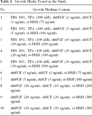
The source of melanocytes for the cell culture was the roofs of suction blisters that were formed on the inner side of the forearm of patients suffering from the acrofacial form of vitiligo. Fifteen patients with the stable form of the disease and with skin phototypes II and III qualified for the study. The epidermis was treated with an enzymatic bath in 0.25% solution of trypsin/EDTA, and then the cell suspension was transferred to culture vessels containing the basic growth medium DMEM/Ham's F-12, supplemented with mitogenes and melanogenesis-enhancing factors of various concentrations. The composition of the 10 tested media is shown in Table 1. For the first 3 days of culture, geneticin was added to all the media in order to eliminate fibroblasts and keratinocytes. The media were changed two to three times weekly. The presence of melanocytes in the cultures was confirmed by means of immunocytochemical assay (anti-HMB-45).
Growth Media Tested in the Study
The population doubling time (PDT) of melanocytes was calculated for all the tested growth media. After 15 passages, the melanocytes underwent cytogenetic tests using the PCR-SSCP method. Cytogenetic analysis was carried out on the HRAS (exon 1 and 2), KRAS (exon 1 and 2), NRAS (exon 1 and 2), and BRAF (exon 11 and 15) genes. In order to exclude germline mutations in the tested genes, blood leukocytes obtained from the peripheral blood of the patients also underwent cytogenetic analysis.
The study was approved by the Ethics Committee of the Ludwik Rydygier Medical College and appropriate informed consent was obtained from all the human subjects involved.
Results and Discussion
Melanocytes from all 15 patients were cultured (Fig. 2). There was no statistically significant difference in melanocyte population doubling time (PDT) for various media. The shortest PDT (about 24 h) was observed in the medium number 4, containing FBS 10%, TPA (100 nM), rhbFGF (20 ng/ml), rhSCF (20 ng/ml), and α-MSH (300 ng/ml). Cytogenetic tests carried out after 15 passages did not reveal any mutations in genes HRAS (exon 1 and 2), KRAS (exon 1 and 2), NRAS (exon 1 and 2), and BRAF (exon 11 and 15). No mutation of the tested genes was observed in the peripheral blood leukocytes of the patients.

Melanocytes after 4 weeks in culture conditions (medium number 4). Original magnification: 100x. Scale bar: 20 μm.
Eisinger and Marko were the first to use TPA and cholera toxin in the culture of melanocytes in 1982 (9). During the years that followed, the culture techniques were improved by adding or replacing different growth factors in the melanocyte growth media. In 1987, Lerner et al. performed the first transplantation of autologous cultured melanocytes in a patient with vitiligo, obtaining a good cosmetic effect. A suitable number of melanocytes for the transplantation were obtained using the medium elaborated by Eisinger and Marko, supplemented with 3-isobutyl-1-methylxanthine (IBMX) (13). Since the first transplantation of cultured melanocytes, there have been several articles on the possible mutagenic properties of TPA. The phenomenon of the expression of antigens characteristic for melanoma, on the surface of rapidly proliferating melanocytes in vitro, is also known. Among the most frequently detected antigens are melanotransferrin p97, integrine β3 subunit for the vitronectin receptor, gangliosides GD3 and 9-O-acetyl GD3, chondroitin sulphate proteoglycan, and MelCAM/MUC18/CD146. Cultured melanocytes with altered surface antigens have a normal diploid karyotype and do not have the biological characteristics of melanoma (10). Cells growing in culture divide quickly and, during a short time, lose their mitotic potential, which results in cellular senescence. Cellular senescence is characterized by many features, but the most prominent is accumulation of errors within the DNA strands.
High proliferation rate of cultured melanocytes potentially increases risk of the proto-oncogenes and the suppressor genes damage, which lead to the malfunction of the signaling pathways controlling the processes of growth, differentiation, and apoptosis. In melanoma, mutations affect the genes encoding protein of the RAS/RAF/MEK/ERK signaling pathway, among others. The most common genetic change in melanoma cells resulting in abnormalities in the Ras/Raf/MEK/ERK signaling pathway is a mutation in the BRAF gene, which is found in 60–70% of melanoma cases (14). Mutations in the BRAF gene occur with higher frequency in melanomas during the vertical growth phase and in metastatic melanomas (62–80%), compared to melanomas during the phase of superficial growth (10–33%) (8). High frequency of BRAF gene mutation (70–88%) is found in pigmented nevi, which may indicate the participation of the BRAF oncogene in the initiation of melanoma. Mutations in the BRAF gene have a substitution character and occur in exon 11 or 15. The most common defect of the BRAF gene is mutation in exon 15 (the exchange of va-line for glutamic acid, lysine, arginine, or aspartic acid at codon 599 or 600) (16).
The RAS family genes play a significant role in the cancer transformation of melanoma. Mutations in the NRAS genes affect codons 11, 12, 13, and 18 in exon 1 and codons 59 and 61 in exon 2. The most frequently observed mutation is the substitution of glutamine by lysine or arginine in codon 61. The mutation of the NRAS gene is observed in 5–37% cases of sporadic melanoma (2, 7). It has been demonstrated that mutations in the NRAS gene occur more often in familial rather than in sporadic melanomas. Mutations in codon 61 of the NRAS gene were detected in 95% of cases of melanoma with germinal mutation of gene CDKN2A (15). A defective NRAS gene was identified at various clinical stages of melanoma: in preinvasive melanoma (melanoma in situ), superficial spreading melanoma (SSM), and in the metastatic phase of melanoma. It was observed that mutations in exon 1 of the NRAS gene occur more frequently during cancer initiation, while mutations in exon 2 occur at later phases of melanoma development (7). Ugurel et al. identified mutations of the NRAS gene in 26 of 105 (24.8%) whereas Reinfenberger et al. found 2 of 6 (33%) analyzed melanoma cell lines (17, 21).
Mutations in the HRAS and KRAS genes in melanoma are observed very rarely. Demunter et al. assessed the incidence of KRAS gene mutations in melanoma. The study involved 69 cases of primary melanomas, 35 metastatic melanomas, and 7 benign pigmented nevi. Mutations in the KRAS gene did not occur in any of the 111 cases analyzed (7). In 1 of the 37 analyzed cases of melanoma, Reifenberger and associates identified a mutation in exon 1 of the KRAS gene, in the form of the substitution of glycine by aspartic acid in codon 12. Mutation of the KRAS gene was discovered in a patient with metastatic melanoma to the brain. Mutation of the HRAS gene was not observed in any of the 37 cases analyzed (17). Shukla et al. identified a mutation in exon 1 of the KRAS gene in 3 of the 22 analyzed cases of primary melanoma. In one case, mutation in exon 1 of the HRAS gene was also identified (18). A significantly higher frequency of the mutation of the HRAS gene, equal to 29%, was observed in Spitz nevi compared to melanoma (22).
Damage to the BRAF and MC1R genes was mostly observed in cases of melanoma occurring in areas of the skin not exposed to intense sun radiation (12). Changes in the BRAF gene must therefore be induced by mutagens other than ultraviolet radiation. One of the potential factors initiating mutations may be TPA as well as high concentrations of other growth factors. It has not yet been clearly established in what way TPA could transform a normal pigment cell into a malignant one. One of the possible mechanisms is apoptosis suppression in culture conditions (1). Recent studies have revealed that TPA causes the resistance of melanocytes to the transforming growth factor-β (TGF-β), which is a natural inhibitor of the proliferation of the pigment cells (19). The growth media for melanocytes, which are to be transplanted to patients with vitiligo, only contain factors that promote cell proliferation of melanin synthesis of the pigment cells and therefore the relationship described above does not explain the mechanism of a possible melanomagenesis in the growth media examined by us. Based on the results of our study, it can be concluded that high concentrations of TPA, as well as other growth factors, do not damage the genes of the RAS/RAF/MEK/ERK signaling pathway in cell culture conditions. It is necessary to conduct further studies evaluating the safety of carrying out the transplantation of autologous cultured melanocytes, especially in cases of using media containing high concentrations of growth factors. Besides genetic studies, it is advisable to carry out biological tests on a properly selected animal model.
Footnotes
Acknowledgment
This study was supported by the State Committee for Scientific Research in Poland (No. N402 11431/3433).