
Abstract
BACKGROUND:
Slow kVp switching technique is an important approach to realize dual-energy CT (DECT) imaging, but its performance has not been thoroughly investigated yet.
OBJECTIVE:
This study aims at comparing and evaluating the DECT imaging performance of different slow kVp switching protocols, and thus helps determining the optimal system settings.
METHODS:
To investigate the impact of energy separation, two different beam filtration schemes are compared: the stationary beam filtration and dynamic beam filtration. Moreover, uniform tube voltage modulation and weighted tube voltage modulation are compared along with various modulation frequencies. A model-based direct decomposition algorithm is employed to generate the water and iodine material bases. Both numerical and physical experiments are conducted to verify the slow kVp switching DECT imaging performance.
RESULTS:
Numerical and experimental results demonstrate that the material decomposition is less sensitive to beam filtration, voltage modulation type and modulation frequency. As a result, robust material-specific quantitative decomposition can be achieved in slow kVp switching DECT imaging.
CONCLUSIONS:
Quantitative DECT imaging can be implemented with slow kVp switching under a variety of system settings.
Introduction
After several decades’ development, dual-energy CT (DECT) imaging [1] has become one of the most advanced CT imaging techniques with the capability of quantitatively distinguishing materials having different compositions. For some specific medical applications, besides, the total radiation dose of the DECT imaging technique might also be lower [2, 3] than the conventional CT imaging technique. The foundation of DECT imaging relies on the non-linear attenuation responses of materials to X-ray photons carrying different energies [4]. As a result, DECT imaging needs to measure the object using at least two distinct X-ray beam energies, namely, one has low-energy, and the other has high-energy. For medical imaging applications, usually, the emitted X-ray photons from the X-ray tube owns a very wide energy distribution. Therefore, certain X-ray beam energy modulation strategies are needed to ease the distinction of materials [5].
By far, several X-ray beam energy modulation techniques have been developed. In general, they can be categorized into two groups: one group relies on modulating the voltage of X-ray tube, and the other group relies on manipulating the responses of X-ray detector. Among the first type of solutions, the tube potential switching is a very promising technique to enrich the variations of X-ray beam spectra. For instance, the well-known ultra-fast kVp-switching technique [6] developed by the GE Healthcare alternates the X-ray tube potential between 80 kVp and 140 kVp during the acquisitions of the DECT projections. Differently, the dual-source DECT imaging technique [7, 8] developed by the SIEMENS Healthcare collects the low-energy 80 kVp and the high-energy 140 kVp DECT projections from two independent tube-detector sub-systems that are orthogonally positioned in the same gantry. Additionally, the low-energy and the high-energy DECT projections can also be acquired from two successive CT scans: one at 80 kVp tube potential, and the other at 140 kVp tube potential. Regarding to the aforementioned second type of DECT imaging solutions, novel X-ray detectors are utilized to acquire the DECT projections. For example, the dual-layer X-ray detector [9, 10] is developed by the Philips Healthcare to acquire the low-energy and high-energy X-ray photons at the same time from the two detector layers. Most recently, the energy-resolving single photon counting detector (PCD) has been incorporated to detect the polychromatic X-ray photons with certain energy thresholds [11, 12]. In principle, spectral CT imaging with more than two X-ray beam energies can also be achieved with PCDs.
Until now, such advanced DECT imaging are merely available on the high-end commercial diagnostic CT scanners. One major cause is the high expense of the hardware, either the high-end X-ray tube or the cutting-edge X-ray detector. Over the past decade, many efforts have been made to reduce the cost of DECT modalities. For example, the twin-beam DECT technique was proposed by adding a certain beam filter [13, 14], e.g., a gold-and-tin filter, in front of the X-ray tube to generate low-energy and high-energy spectra. Moreover, Petrongolo et al. [15] proposed to insert a primary beam modulator between the X-ray source and the object to generate angularly varied beam spectra on the detector surface. Similarly, Tivnan et al. [16, 17] proposed to use a spatial-spectral filter consisting of an array of K-edge materials to modulate the X-ray beam spectra. On the other hand, the feasibility of DECT imaging using slow kVp modulation is proposed [18] as well. In it, the X-ray tube voltage is slowly varied as the gantry rotates around the object. Compared to the ultra-fast kVp switching technique, the spectral information acquired from the slow kVp switching method is angularly sparse, and thus partially breaks the spectral completeness. To compensate, many novel material-specific CT image reconstruction algorithms are developed for low kVp switching DECT imaging. For example, Szczykutowicz et al. proposed a prior image constrained decomposition algorithm (PICCS) [19]. In it, a prior CT image is generated at first from all projection (mixed spectral information) using the conventional filtered-backprojection (FBP) algorithm. Afterwards, the low-energy and high-energy CT images are reconstructed from the acquired spectral projections using the PICCS algorithm [20] to allow the image-domain material decomposition. Moreover, the one-step material decomposition method can also be used to generate the material-specific CT images as long as the kVp modulated beam spectra are well incorporated in the forward imaging model. For example, the joint statistical iterative material image reconstruction algorithm [21] proposed by Mechlem et al and the non-convex primal-dual reconstruction algorithm [22] proposed by Chen et al. In addition, the convolutional neural network (CNN) based deep learning technique can also be used to reconstruct the basis images for the slow kVp switching DECT imaging [23, 24].
In this study, the DECT material-specific imaging performance of a variety of slow kVp modulation settings are evaluated. This is important in determining the key parameters such as the beam filtration [25] and the voltage switching frequency [19] when designing a real DECT imaging system work with slow kVp switching technique. In particular, this study assumes that the X-ray beam is filtered by two different methods: one with fixed filtration for all tube potentials, and the other varies the beam filtration along with the tube potential. Namely, higher tube voltage corresponds to thicker beam filtration. Studies [26, 27] have shown that the beam filtration is important in separating the beam energy and thus improving the decomposition accuracy as well. In addition, two distinct tube voltage variation modes are assumed in this study: the first mode assigns equal duration for each tube voltage within a complete modulation period, and the second mode assigns longer duration for the lowest and highest tube voltages within a complete modulation period. Similarly, such tube voltage variations are important in enhancing the beam energy separation and thus improving the decomposition accuracy [25]. Moreover, the modulation frequency (denoted as ω), which is important for investigating the impact of angular sparsity [28] of the beam spectra, are also compared in this study. A model-based one-step material decomposition algorithm based on the work of Mechlem et al [21] that directly reconstructs the basis images from the acquired projections is adopted. DECT imaging with slow kVp switching is performed with numerical phantom, physical phantom and a biological specimen in this evaluation study.
The remainder of this article is organized as follows: Section II presents the slow kVp switching based sparse-spectra CT imaging model, the model-based bases decomposition algorithm, the numerical and experimental settings. Section III presents the numerical and experimental results, Section IV presents the discussions and a brief conclusion is drawn in Section V.
Method
Imaging model
In the following discussions, the X-ray tube voltage is assumed to vary slowly between Umin and Umax during one gantry rotation, see the illustration in Fig. 1. Since the kVp switching frequency is quite low, therefore, the tube potential could be kept for a certain duration over a small gantry rotation span before switching into the next tube potential (lower kVp or higher kVp). In total, ω periods of kVp switches are assumed during one gantry rotation.
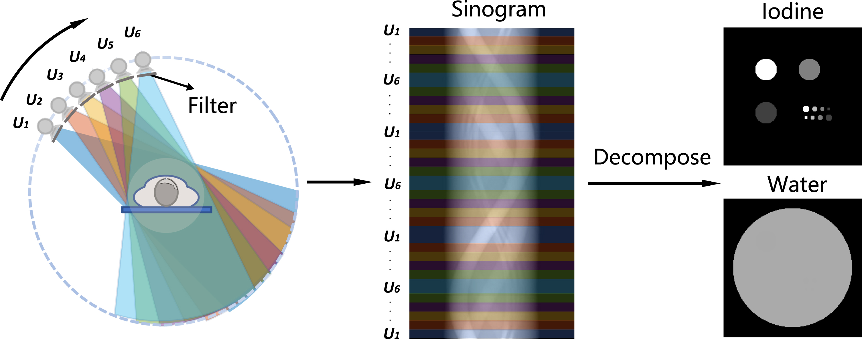
Illustration of DECT imaging based upon slow kVp switching technique.
Herein, the acquired projections are expressed as a linear combination of several pre-selected basis materials b, namely:
In the next numerical experiments, two sets of X-ray beam filtration were simulated: the first set used stationary beam filtration of 1.5 mm Aluminium (Al) and 0.5 mm Copper (Cu) for all of the varied tube potentials, and the second set employed dynamic filtration, i.e., varied Cu plate thickness for each tube voltage, as listed in Table 1. The generated X-ray beam spectra under the above two types of filtration are plotted in Fig. 2(a) and (b), separately. Those X-ray spectra were calculated in SpekCalc [32]. Additionally, the tube potentials were assumed to vary consecutively under two different kVp switching modes. In the first kVp switching mode, the tube voltage varied uniformly, i.e., each voltage level endures the identical period, see Fig. 2(c). In the second kVp switching mode, the lowest 75 kVp tube potential and highest 125 kVp tube potential were set to more occupation of the entire period while the other tube potentials in between equally share the rest occupation, see Fig. 2(d). Moreover, six different voltage modulation frequencies (the number of voltage variation periods per gantry rotation), i.e., ω = 0.5, 1, 3, 5, 15 and 25, were also investigated.
Key parameters used for experimental data acquisition
Key parameters used for experimental data acquisition
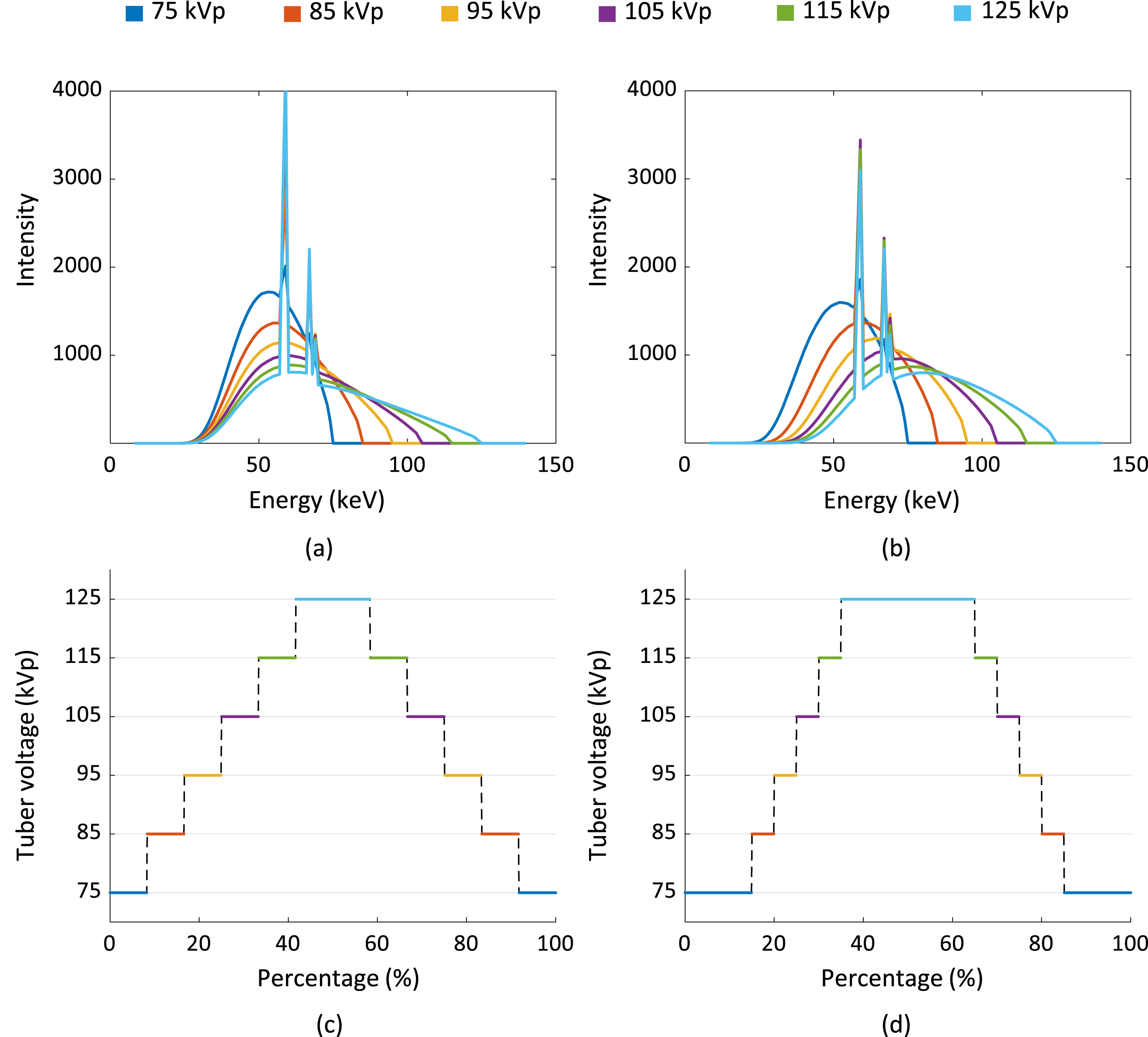
The X-ray spectra obtained (a) with stationary beam filtration and (b) dynamic beam filtration. The tube potential modulation mode having (c) uniform distribution and (d) weighted distribution.
In addition, a digital phantom containing different iodine solution inserts (5 mg/ml - 20 mg/ml) was assumed, see Fig. 3(a) for more details. The imaging geometry of the numerical experiment was set identical to the experimental setup. In particular, the source to rotation distance (SOD) was set to 1156.3 mm, and the source to detector distance (SDD) was set to 1560.3 mm. The incident number of photons was set to 5 × 10⁴ for all X-ray beam spectra. Under this condition, the image noise levels of the numerical experiments look similar as of the physical experiments.
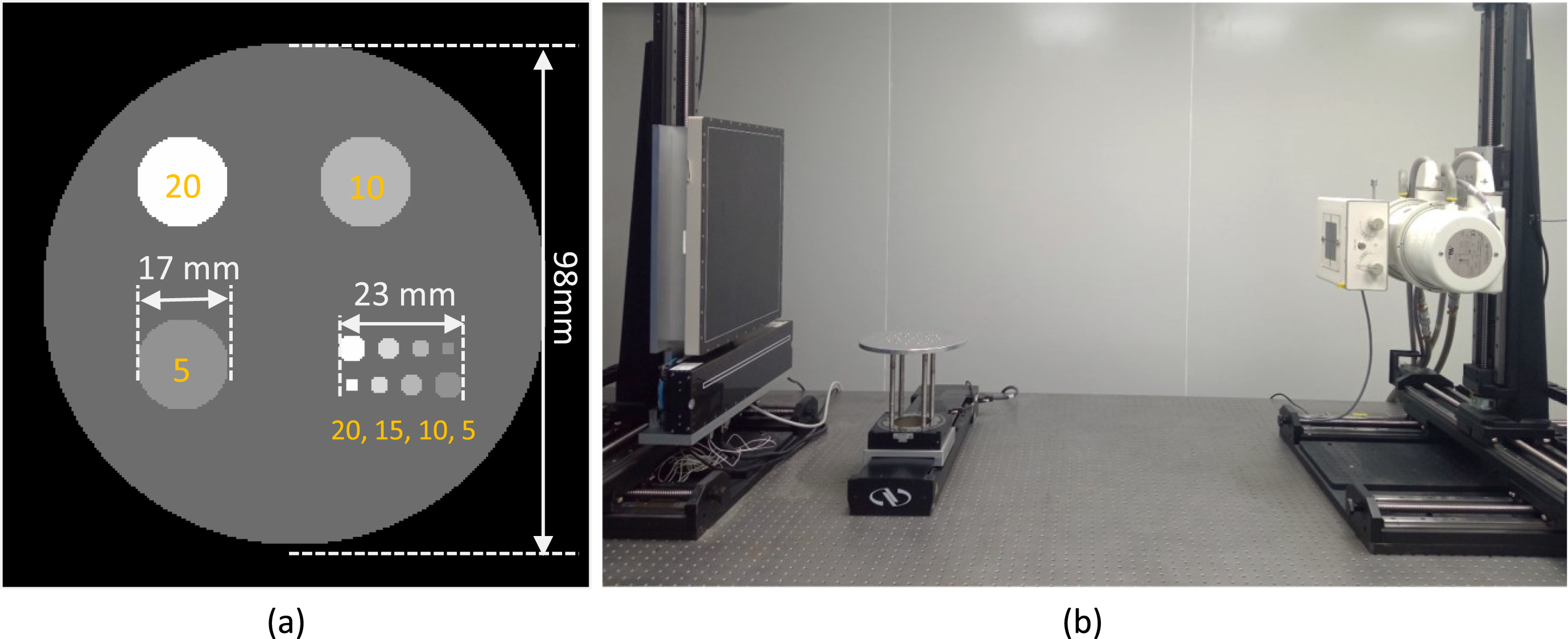
(a) Illustration of the numerical phantom used in this study, the densities (unit: mg/ml) of iodine inserts and phantom dimensions are marked, (b) photograph of the experimental benchtop system.
Experiments were performed on our benchtop DECT imaging system equipped with a medical-grade X-ray tube (G-242, Varex, UT, USA) and a flat-panel detector (4343CB, Varex, UT, USA), see Fig. 3(b). The tube voltage varied from 75 kVp up to 125 kVp with a 10 kVp interval. The effective detector pixel size was 0.417 mm × 0.417 mm. For each tube voltage, the object was scanned over 360 degree with 0.4 degree sampling interval with a certain beam filtration, see Table 1. Afterwards, the needed slow kVp switching CT imaging data were manually synthesized from these acquired six full-rotation projections with respect to certain kVp switching mode and ω value. An iodine phantom and a pork specimen were scanned separately. The iodine solutions (5 mg/ml - 20 mg/ml) were sealed in the Eppendorf tubes that having 20 mm diameter. To reduce the Compton scatters, the X-ray beam was collimated into a narrow width (less than 20 mm on the detector surface).
Parameter selection
Empirically, the size of neighboring pixels in
Iterative parameters used for image reconstruction
Iterative parameters used for image reconstruction
Simulation results
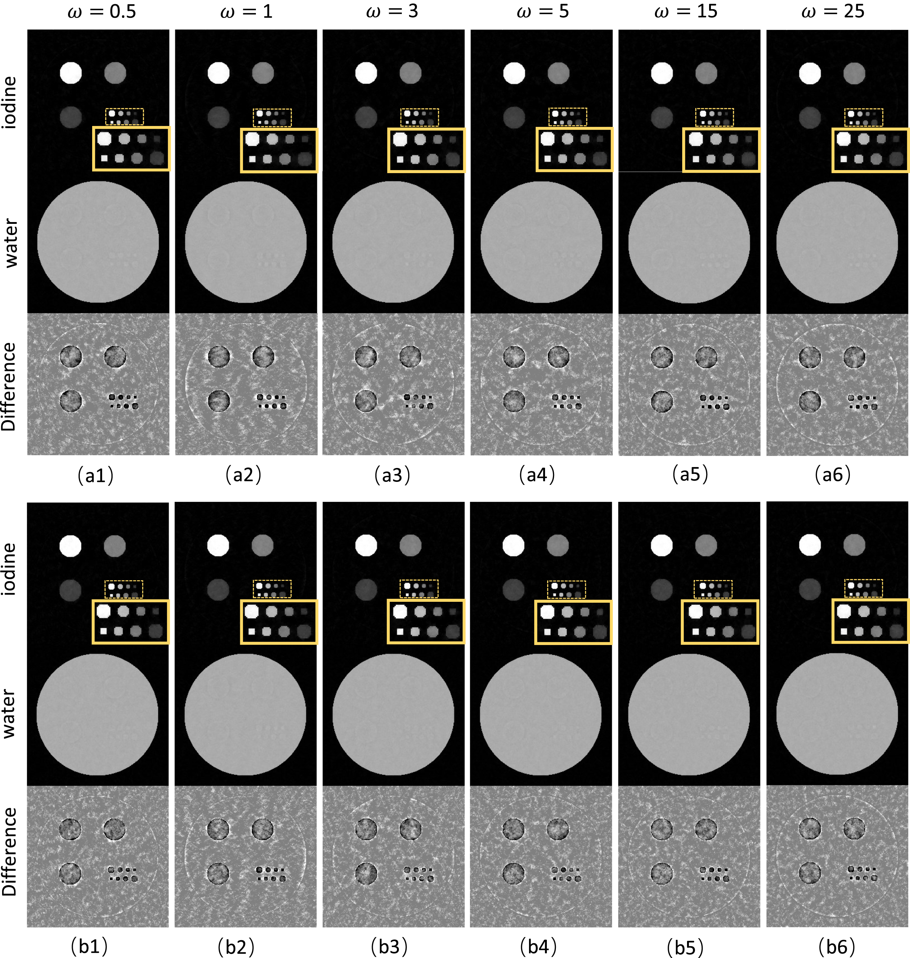
The material decomposition results obtained with the stationary filtration are shown in Fig. 4. From left to right, the basis images are generated under increased kVp modulation frequency ω. The decomposed basis images in the top three rows are obtained with uniform kVp modulation, and the ones in the bottom three rows are obtained with weighted kVp modulation. As observed, the water bases and iodine bases can be well separated under all system settings. Namely, the beam filtration and the kVp modulation frequency barely impact the DECT decomposition accuracy.
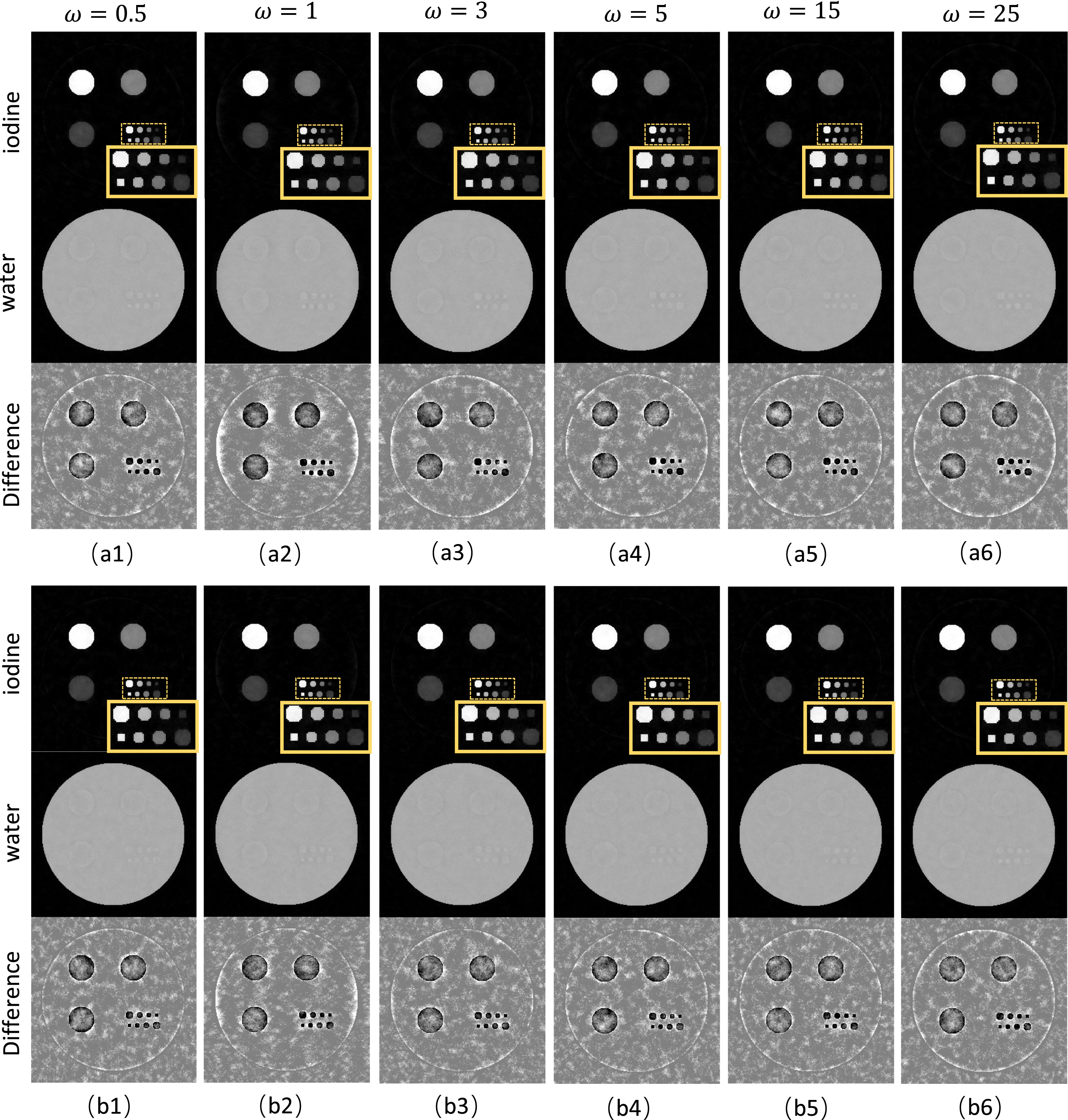
Decomposition results of the numerical phantom generated under the stationary beam filtration. (a1)-(a6) are obtained from uniform tube potential modulation, and (b1)-(b6) are obtained from weighted tube potential modulation. The display windows are [0, 20] mg/ml, [0, 1.5] g/ml and [-1, 1] mg/ml for the iodine bases, water bases and iodine difference images, respectively.
In addition, the material decomposition results obtained with the dynamic filtration are shown in Fig. 5. Similar results are observed for this special slow kVp switching scheme. For example, the decomposed water bases and iodine bases exhibit similar quality with respect to different modulation frequency ω and modulation type. Compared to the results of stationary filtration, results indicate that the dynamic filtration slightly improves the decomposition accuracy, see the difference images in Fig. 4 and Fig. 5. As a result, the image quality does not show significant difference with respect to varied modulation frequency ω and beam filtration scheme.
Decomposition results of the numerical phantom generated under the dynamic beam filtration. (a1)-(a6) are obtained from uniform tube potential modulation, and (b1)-(b6) are obtained from weighted tube potential modulation. The display windows are [0, 20] mg/ml, [0, 1.5] g/ml and [-1, 1] mg/ml for the iodine bases, water bases and iodine difference images, respectively.
Quantitative measurements of the iodine densities are shown in the bar plots, see Fig. 6. It is observed that the decomposed iodine concentrations are close to the ground truth, indicating the viability of generating accurate dual-energy decomposition in slow kVp switching DECT imaging applications.
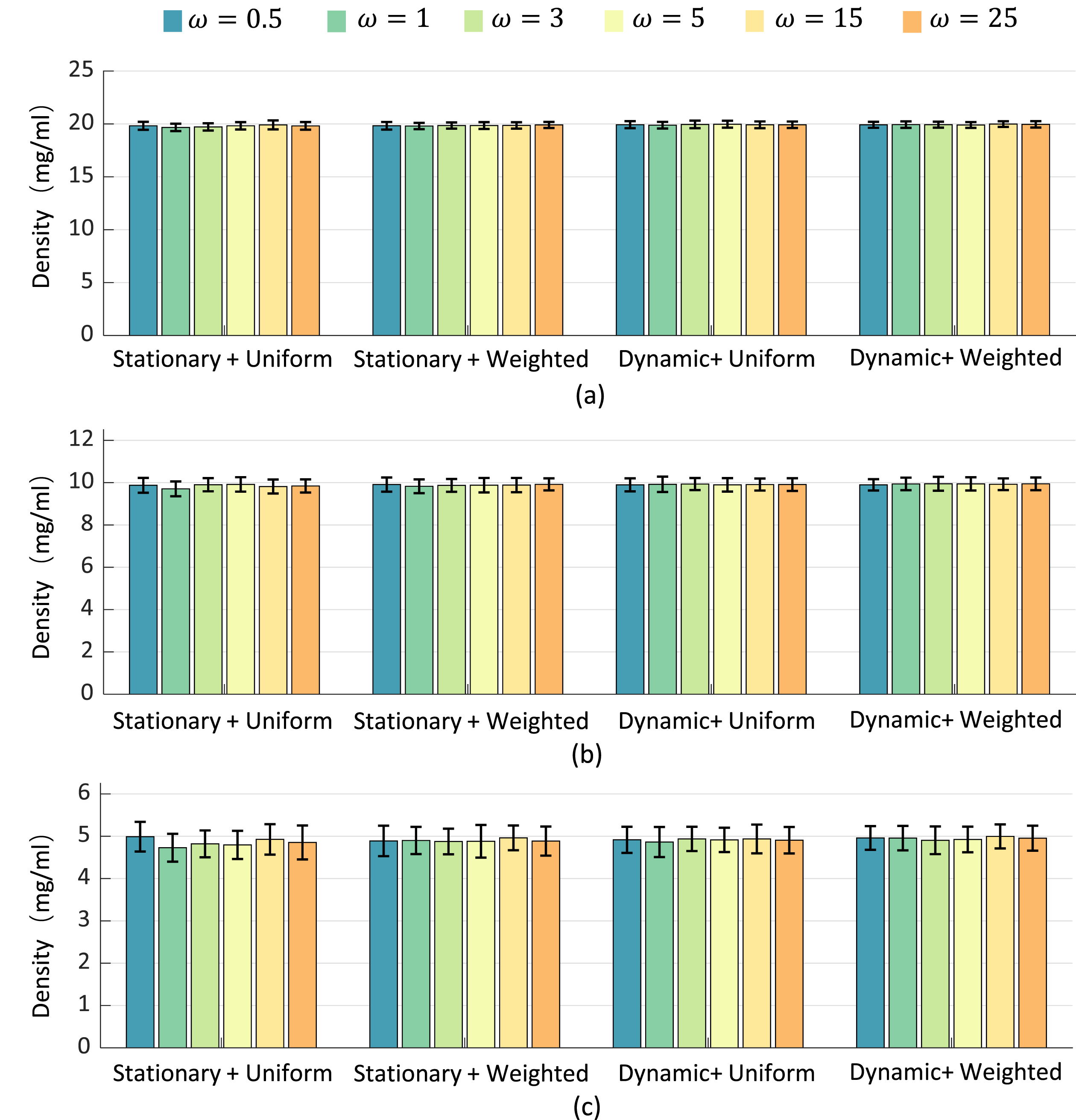
Quantitative comparison results of the iodine density: (a) 20 mg/ml, (b) 10 mg/ml, (c) 5 mg/ml, for varied slow kVp switching settings.
The experimental DECT imaging results of the iodine phantom with uniform tube potential modulation under different ω are shown in Fig. 7. The decomposed basis images are presented in the top two rows of Fig. 7(a1) to (a6). The virtual monochromatic CT images (VMI) are synthesized at 70 keV. As seen, the iodine material can be well separated from water, and the synthesized VMI images have high quality. The quantification results measured on the four iodine inserts are plotted in Fig. 7(a7). For a wide range of frequency ω from 0.5 to 25, the estimated iodine concentrations agree well with the ground truth. Additionally, the experimental DECT imaging results of the iodine phantom with weighted tube potential modulation under different ω are shown in Fig. 8. The decomposed basis images are presented in the top two rows of Fig. 8(a1) to (a6). The virtual monochromatic CT images (VMI) are synthesized at 70 keV. Again, the iodine material can be well separated from water, and the quantification plots in Fig. 8(a7) demonstrate high accuracy of the basis decomposition. Due to the slightly mismatched imaging geometry among the six repeated CT scans, minor decomposition residuals are remained on the iodine basis images.
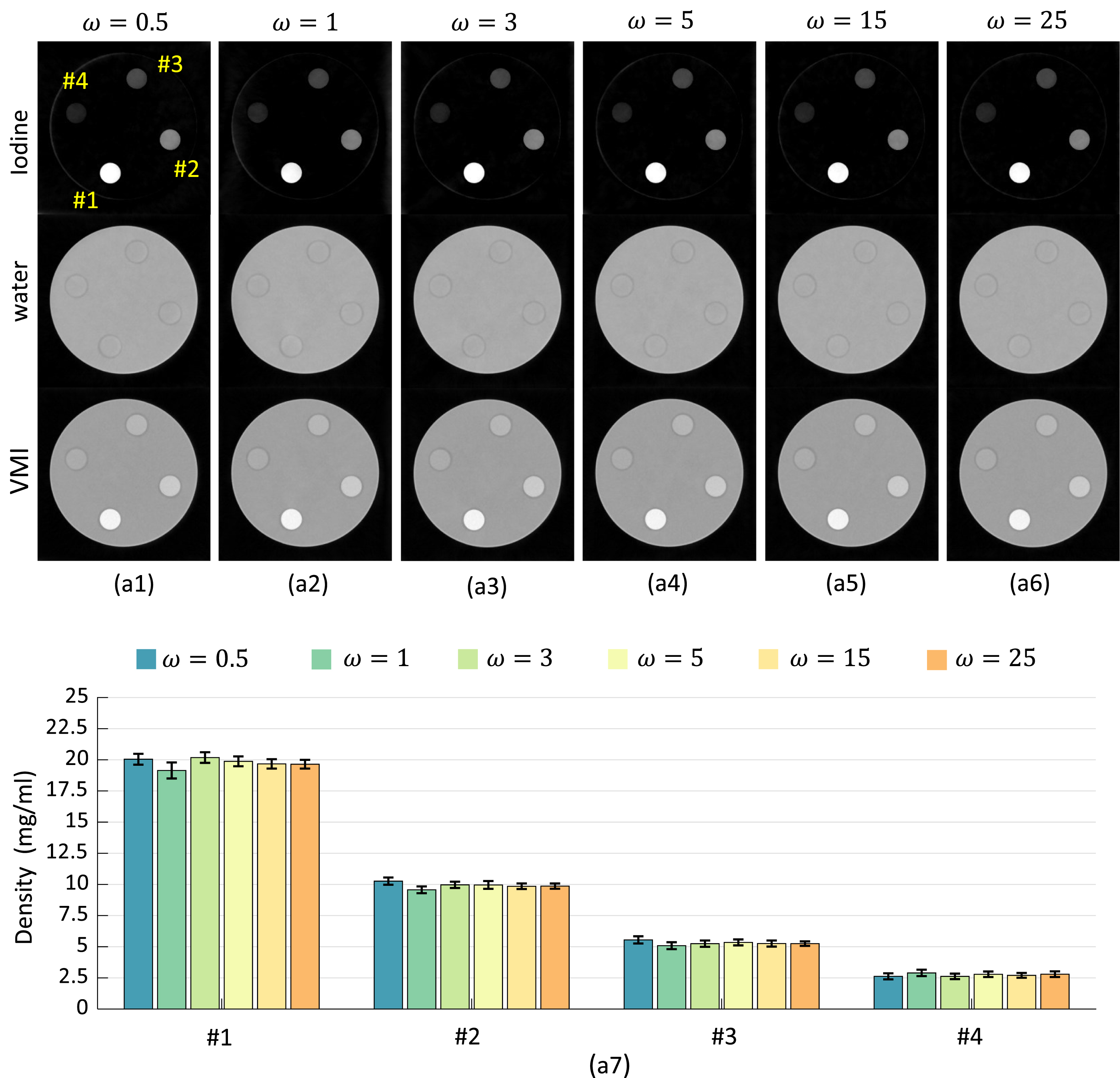
Experimental results of the iodine phantom generated with uniform tube potential modulation are depicted in (a1)-(a6). The display windows for the iodine bases, water bases and VMI images are [0, 20] mg/ml, [0, 1.5] g/ml and [0, 0.3] cm-1, respectively. The measured iodine densities are plotted in (a7).
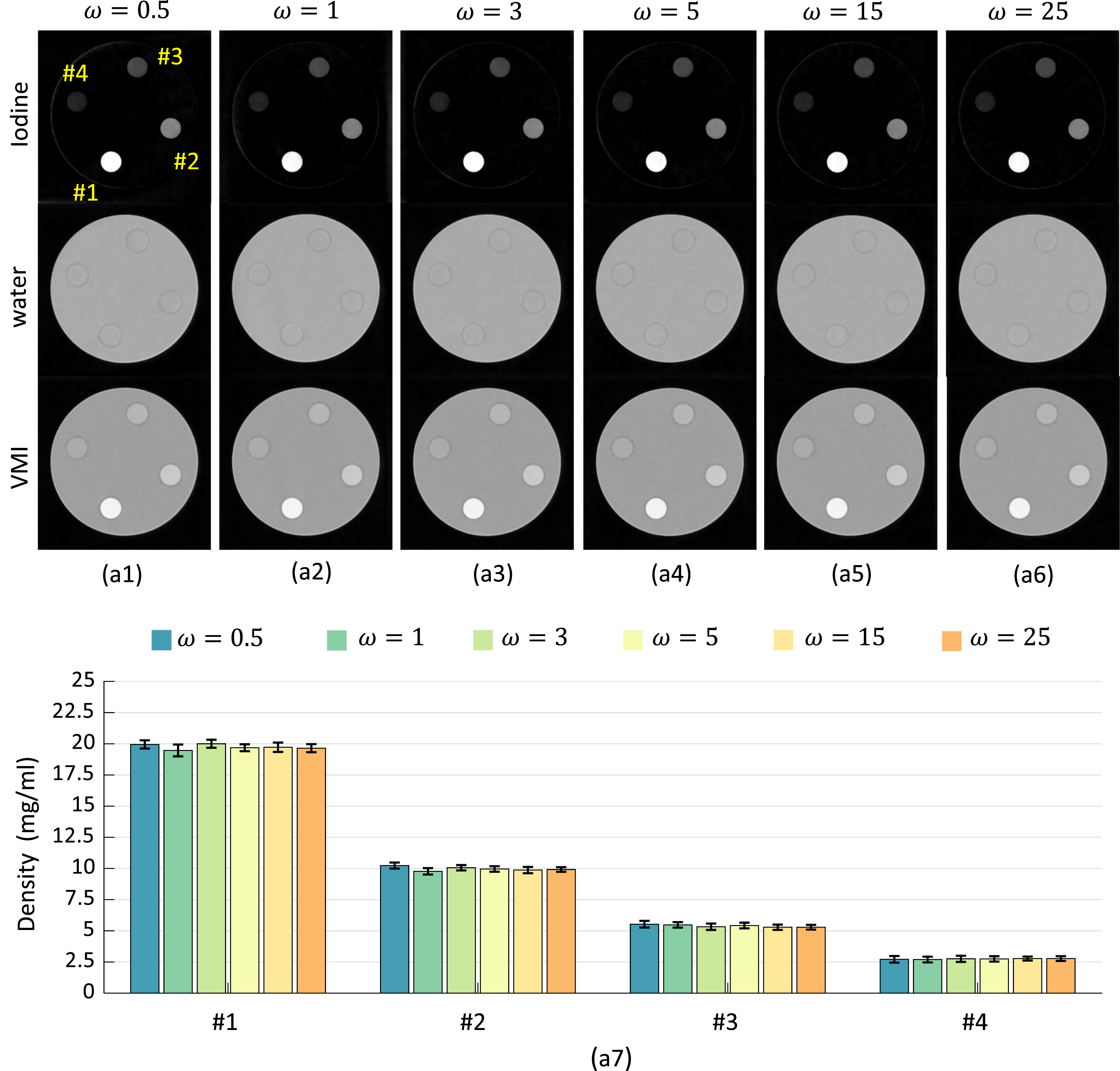
Experimental results of the iodine phantom generated with weighted tube potential modulation are depicted in (a1)-(a6). The display windows for the iodine bases, water bases and VMI images are [0, 20] mg/ml, [0, 1.5] g/ml and [0, 0.3] cm-1, respectively. The measured iodine densities are plotted in (a7).
Finally, the decomposition results of the pork specimen are shown in Fig. 9. The basis images in the top two rows of Fig. 9 are obtained with uniform tube potential modulation, and the basis images in the bottom two rows of Fig. 9 are obtained with weighted tube potential modulation. For a specific voltage modulation frequency ω, it is observed that the both tube potential modulation schemes can generate similar decomposition results. For instance, the fine bony structures can be well separated from the soft tissues.
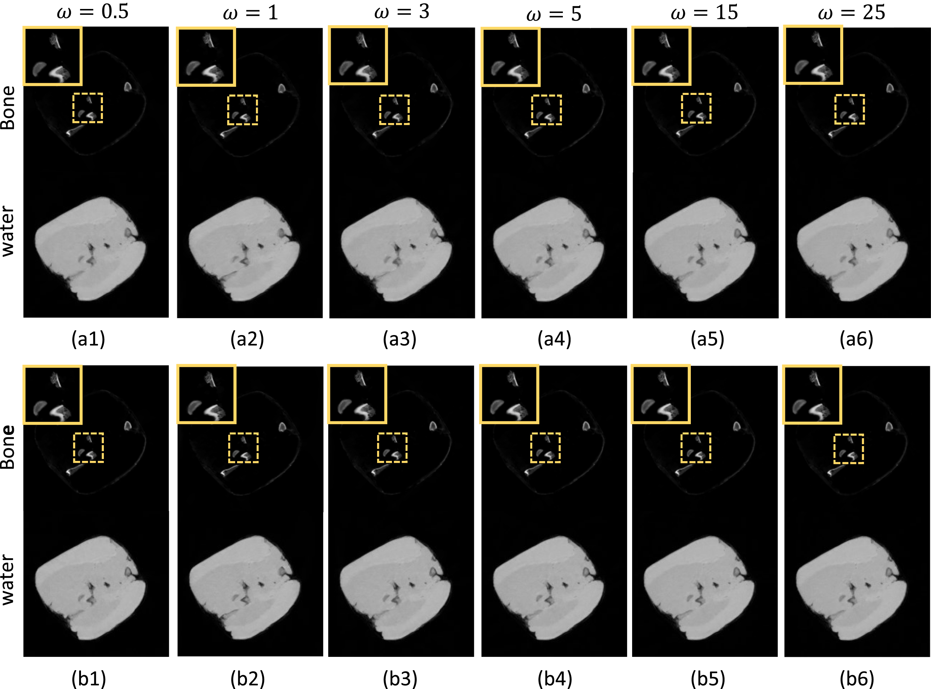
Decomposition results of the pork specimen generated with stationary beam filtration. Images in (a1)-(a6) are obtained with uniform kVp modulation, and images in (b1)-(b6) are obtained with weighted kVp modulation. The display window for all images is [0, 1.5] g/ml.
In this work, the DECT imaging performance under different slow kVp switching settings (protocols) are investigated. A couple of key system parameters such as the beam filtration, the potential modulation scheme, and the tube potential switching frequency are studied rigorously. Both numeral and experimental results demonstrate that the final decomposition performance is slightly impacted by the slow kVp switching protocols having different settings. In other words, the slow kVp switching technique is quite a robust DECT imaging approach.
Compared to the stationary beam filtration, intuitively, varying the beam filtration is able to obtain X-ray beam spectra with larger energy separation. However, results in this study demonstrate that similar material basis maps can be generated from the two beam filtration schemes. In addition, it seems like that assigning longer duration for the lowest and highest tube voltages over a complete modulation period provides better beam energy separation, whereas, results in this study show that the final outcome is barely impacted by the X-ray beam modulation schemes. Compared to the sparse-view low-dose CT image reconstruction tasks, the sparse-spectral DECT image reconstruction under slow kVp switching scenario seems to be less challenging. This is because the completeness of the structural information is well reserved in the slow kVp switching DECT imaging task, and the beam spectra can be well defined in prior in the utilized iterative DECT image reconstruction algorithm. Another interesting finding of this study is the less dependency of the beam modulation frequency ω. Different from the ultra-fast kVp switching technique, which requires super high-end generator and X-ray tube, such observation indicates that high quality DECT imaging could be achieved with very low-cost generator and X-ray tube. This would significantly reduce the system manufacture complexity and cost of the DECT imaging scanners.
The current study has several limitations: First, experiments with a real slow kVp switching X-ray source was not performed due to the lack of such a hardware in our laboratory. Second, the total image reconstruction time is long with this iterative DECT image reconstruction algorithm. To accelerate, the latest deep learning techniques [33, 34] might be investigated in the future. To do so, convolutional neural network (CNN) with certain architecture needs to be designed for the slow kVp switching DECT imaging applications. Third, the radiation dose reduction performance in this slow kVp switching DECT imaging technique was not compared. It would be a very interesting research topic to study in the future. Fourth, this study is mainly focused on the two-material decomposition tasks, i.e., water-iodine decomposition and water-bone decomposition, rather than multi-material (≥3) decomposition. For the latter decomposition tasks, more investigations are needed in the future to evaluate the imaging performance of slow kVp switching techniques.
Conclusion
In conclusion, the DECT imaging performance of slow kVp switching was evaluated comprehensively under a variety of tube potential modulation settings. Both numerical and experimental results demonstrate that the material-specific quantitative DECT imaging performance is less sensitive to the spectral separation and modulation frequency. In the future, low-cost and robust DECT systems could be developed with slow kVp switching technique.
Footnotes
Acknowledgments
This research was funded by National Natural Science Foundation of China (62201560, 52005085), Guangdong Basic and Applied Basic Research Foundation (2021TQ06Y108), Beijing Natural Science Foundation (7234374), Shenzhen Science and Technology Program (JSGGKQTD20210831174329010).
Appendix
The manual selections of regularization parameters γ and λ
b
are shown in ![]() . As observed, the variations of parameters γ and λ
b
lead to different image resolution and noise levels. Based on the manually adjusted results, eventually, γ
water
= 10-1, γ
iodine
= 10-3, and λ
water
= 75, and λ
iodine
= 2 ×10⁵ are selected to balance the image resolution and noise level during the numerical experiments. For the physical experiments, similar procedures are applied to determine the parameters γ and λ
b
.
. As observed, the variations of parameters γ and λ
b
lead to different image resolution and noise levels. Based on the manually adjusted results, eventually, γ
water
= 10-1, γ
iodine
= 10-3, and λ
water
= 75, and λ
iodine
= 2 ×10⁵ are selected to balance the image resolution and noise level during the numerical experiments. For the physical experiments, similar procedures are applied to determine the parameters γ and λ
b
.