
Abstract
BACKGROUND:
The gene that encodes BRCA1-associated protein 1 (BAP1) has been reported to be dysregulated in several human cancers such as uveal melanoma, malignant pleural mesothelioma, hepatocellular carcinoma, thymic epithelial tumors, and clear-cell renal cell carcinoma (ccRCC). The gene is located on the human chromosome 3p21.3, encoding a deubiquitinase and acts as a classic two-hit tumor suppressor gene. BAP1 predominantly resides in the nucleus, where it interacts with several chromatin-associated factors, as well as regulates calcium signaling in the cytoplasm. As newer therapies continue to evolve for the management of RCC, it is important to understand the role of BAP1 mutation as a prognostic and predictive biomarker.
OBJECTIVE:
We aimed to systematically evaluate the role of BAP1 mutations in patients with RCC in terms of its impact on prognosis and its role as a predictive biomarker.
METHODS:
Following PRISMA guidelines, we performed a systematic literature search using PubMed and Embase through March 2021. Titles and abstracts were screened to identify articles for full-text and then a descriptive review was performed.
RESULTS:
A total of 490 articles were initially identified. Ultimately 71 articles that met our inclusion criteria published between 2012–2021 were included in the analysis. Data were extracted and organized to reflect the role of BAP1 alterations as a marker of prognosis as well as a marker of response to treatments, such as mTOR inhibitors, VEGF tyrosine kinase inhibitors, and immune checkpoint inhibitors.
CONCLUSIONS:
Alterations in BAP1 appear to be uniformly associated with poor prognosis in patients with RCC. Knowledge gaps remain with regard to the predictive relevance of BAP1 alterations, especially in the context of immunotherapy. Prospective studies are required to more precisely ascertain the predictive value of BAP1 alterations in RCC.
INTRODUCTION
BRCA1-associated protein 1 - BAP1 gene is a tumor suppressor located on the human chromosome 3p21.3 and encodes ubiquitin carboxy-terminal hydrolase. It is considered a classic two-hit tumor suppressor gene. [1] Within the nucleus, BAP1 acts as a chromatin scaffold for chromatin-remodeling complexes and hence regulates cell proliferation by deubiquitylating host cell factor 1 (HCF1). [2, 3] Cytoplasmic BAP1 is localized to the endoplasmic reticulum, where it stabilizes type 3 inositol-1,4,5-trisphosphate receptor (IP3R3), [3] which via calcium mediated cytochrome-c release from the mitochondria leads cell apoptosis. [1, 4]. Moreover, BAP1 has been shown to play a role in the metabolic activity of cells. For example, a study that evaluated plasma from sixteen BAP1 + /–individuals from 2 families carrying various germline BAP1 mutations and compared with thirty BAP1 wild-type (wt) controls from the same families [5]. They observed increased glycolysis and increased reduced aerobic mitochondrial respiration in BAP1 + /–, as compared to BAP1wt members, thus concluding that Warburg effect was seen in cells from individuals carrying heterozygous germline BAP1 mutations, much like cancer cells and these mutations may be the reason for a higher incidence of cancer among them [5].
Germline BAP1 mutations were observed in patients with familial mesotheliomas [6] and familial melanocytic tumors [7, 8]. A meta-analysis of all the published studies with BAP1-mutated families showed an increased association of BAP1 mutations with malignant mesothelioma, uveal melanoma and cutaneous melanomas, the etiology of a novel BAP1 cancer syndrome [8]. Analysis of clear-cell renal cell carcinoma (ccRCC), revealed the prevalence of BAP1mutations at 14% [9, 10]. Some studies showed correlation between loss of BAP1 activity and higher grade tumors [10, 11], and a molecular subtype of ccRCC with mutations in VHL and BAP1 was proposed. Since then, several studies have analyzed the role of BAP1 in clear-cell RCC prognosis as well as in defining responsiveness to various treatment modalities. However, here is paucity of data defining the role of BAP1 alterations in the context of response to immunotherapy in patients with ccRCC.
This systematic review was designed to evaluate the existing literature with regard to the clinical utility of BAP1 mutations in patients with metastatic ccRCC.
Diagnosis of BAP1 status
The use of immunohistochemistry (IHC) to identify BAP1 protein has previously been described in a cohort of 176 ccRCC tumor samples [10]. Of these, 148 had wild-type for BAP1 and 150 tumors showed the presence of nuclear BAP1 protein by IHC. Twenty-two samples carried the BAP1 mutation while 25 samples were negative for the BAP1 protein by IHC. This study showed that the positive and negative predictive values of the IHC for detection of BAP1 protein in ccRCC were > 98%. There is now a Clinical Laboratory Improvement Amendments (CLIA)–certified IHC test available for BAP1 protein available for use in clinical practice. More recently, non-invasive techniques such as radiomic features from CT scans are being evaluated to predict genomic status, including BAP1 mutations, in kidney tumors [12–14].
Variability in prevalence of BAP1 mutations
Unlike ccRCC, the role of BAP1 mutations is not as prominent in tumors of non-clear cell histology. In a cohort of patients with 186 ccRCC and 79 non-ccRCC, loss of BAP1 expression was seen in 9% (17/186) of the ccRCC tumors but only in 1% (1/79) of the non-ccRCC tumors (p = 0.016) [15]. Analysis of the TCGA dataset revealed prevalence of BAP1 mutations at 5.6% in papillary RCC (compared to 11% in ccRCC) [16]. Also BAP1 mutation correlated with decreased OS in the entire cohort (p = 0.0002) and within the ccRCC group (p = 0.0035); however, BAP1 mutation did not correlate with survival in papillary or chromophobe RCC. However, BAP1 mutations seem to have higher prevalence in patients with sarcomatoid RCC. In a cohort of 99 patients with sarcomatoid RCC and 906 patients with ccRCC, BAP1 mutations were found in 16% vs 9% respectively [17]. Recently, analysis of tumors from a total of 208 patients with sarcomatoid and rhabdoid RCC revealed significant enrichment for BAP1 somatic alterations when compared to classic ccRCC samples [18].
Sex/gender differences in BAP1 expression
There are differences in BAP1 expression between sexes and races. a higher prevalence of BAP1 mutations as reported in females vs. males and in black vs. white patients, however larger studies to validate these differences are required because results are variable between studies [19, 20]. A comprehensive analysis presented by Rickets and Linehan included a total of 628 sequenced samples (414 males and 214 females) with contributions from a US TCGA ccRCC cohort (424 total samples: 277 males and 147 females), a Japanese cohort (106 total samples: 78 males and 28 females) and a Chinese cohort (98 total sequenced samples: 59 males and 39 females) [21]. Of the total 10 genes analyzed, only BAP1 mutation rate was seen to be higher in tumors derived from female patients in a statistically significant manner (p = 0.0042). On the other hand, in a study analyzing 166 patients reported by Minardi et al, no significant correlation was observed between BAP1 expression and sex (p = 0.155) and age (p = 0.250) [22].
METHODS FOR SYSTEMATIC REVIEW
Evidence acquisition
Search strategy
A systematic literature search was conducted according to the PRISMA (Preferred Reporting Items for Systematic Reviews and Meta-Analyses) guidelines to identify studies reporting on BAP1 as a prognostic and/or predictive biomarker in RCC between 2010 and March 2021 [23]. The PubMed database was searched along with a free-text hand search using one or several combinations of the following items: BAP1, BRCA1-Associated Protein 1, ubiquitin carboxy-terminal hydrolase, ubiquitin thiolesterase AND clear cell renal cell carcinoma, ccRCC, conventional renal cell carcinoma, clear cell renal carcinoma, carcinoma, renal cell. The selection process was conducted in two stages; the first stage was used for initial screening of the title and abstract to identify eligible publications. The second stage was done via full-text reading including a manual search of publications in journals not listed in PubMed to further avoid missing any eligible study. For this systematic review, we excluded (I) non-English articles, (II) non-original articles (i.e., review articles with or without systematic review or meta-analysis), (III) editorials or case reports (IV) repeated publications on the same cohort to avoid publication bias.
Data extraction
A CONSORT diagram for the selection process of included studies is provided in Fig. 1. The literature was searched for records focused on ccRCC and BAP1. Search strategies were created and run by a librarian using a combination of keywords and controlled vocabulary in the databases: PubMed and Embase.com. No filters or limits were applied to this search. ASCO, ASCO-GU, and ESMO conference proceedings were also searched using the same keywords. All search strategies were completed and run on April 25, 2021. Records were added to and deduplicated through EndNote and then uploaded and rechecked for duplicates in Rayyan. The final total was 384 unique records. After removal of abstracts without full text descriptions and removal of duplicates, we had a total of 71 that were then analyzed.
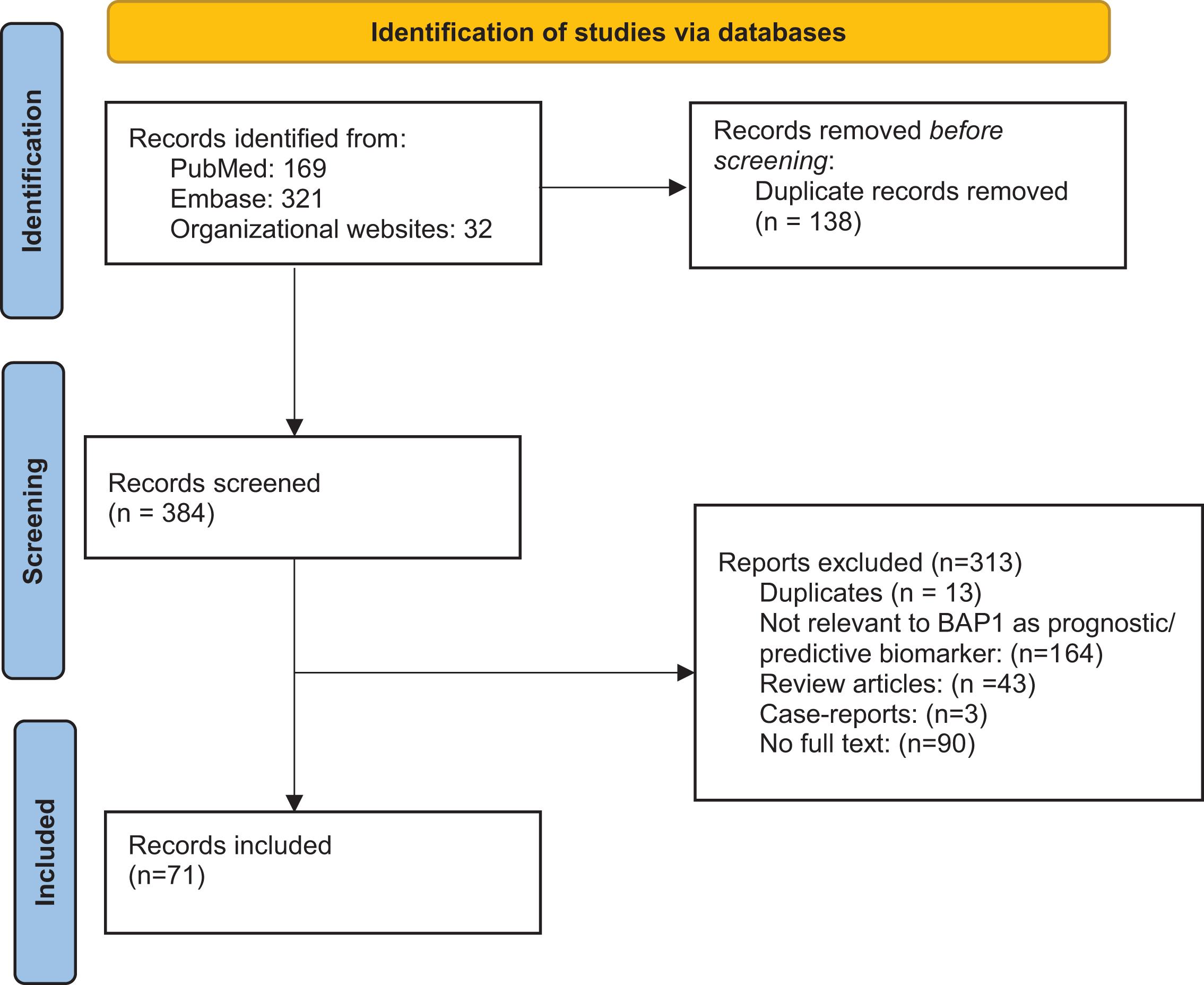
PRISMA Flowchart.
RESULTS
Database search yielded a total of 522 citations, of which the title and abstract were screened for relevance. From these citations, 71 were subjected to full-text review, resulting in articles that met criteria for inclusion (Fig. 1).
Analysis was done in 2 categories. The first category includes studies where the role of BAP1 mutations is described as a prognostic biomarker by defining their association with tumor size, grade, stage at presentation, pathologic features of the tumor, and survival in localized as well as metastatic ccRCC (Table 1a 1b). The second category (Table 2) includes studies where the role of BAP1 mutations is described as a predictive biomarker of responses to treatment regimens (TKI, mTOR inhibitors, nivolumab) in the metastatic setting.
Studies defining the role of BAP1 as a prognostic biomarker in localized ccRCC
Role of BAP1 mutations in metastatic RCC
Responsiveness to treatments in metastatic tumors
Frequency and impact of BAP1 mutations on tumor characteristics and prognosis
We found an overall prevalence of BAP1 mutations in patients with non-metastatic early stage ccRCC between 6–24%. A review of all studies cited here uniformly depicted tumors with mutated BAP1 as carrying poor prognosis. Peña-Llopis et al were the first group to describe in a discovery cohort the correlation between BAP1 mutation and occurrence of high grade tumors [10]. Soon thereafter, Kapur et al combined 2 cohorts of almost 470 patients (including 327 tumors from the TCGA database) to describe in detail the correlation between BAP1 mutations and aggressive features on tumors including higher grade, sarcomatoid and rhabdoid features and coagulative tumor necrosis [24]. This was followed by several other studies that correlated BAP1 mutations with high-risk tumor characteristics as well as with adverse cancer related outcomes such as overall survival (OS), cancer specific survival, presence of metastatic disease at the time of diagnosis (Table 1). Studies identified a higher prevalence of BAP1 mutations, up to 31%, in patients that presented an IVC thrombus at the time of diagnosis, supporting the association with poor prognosis [25–27].
Studies have also looked specifically at patients with small renal masses (< 4 cm). A study that analyzed 70 samples from T1 tumors (of which 20% had BAP1 mutated tumors) found a significant association between BAP1 and high grade tumors [28]. Another cohort of 203 small renal tumors, found a correlation between BAP1 mutations and poor survival in unadjusted analysis (P = 0.050), however the difference became insignificant after adjustment for multiple factors (adjusted P = 0.100) [29].
As further studies continued to evolve, the absence of BAP1 protein has also been correlated with early metastasis in patients that were followed after initial nephrectomies [19, 31]. Studies describing an evaluation of primary and metastatic lesions in patients with ccRCC for BAP1 mutations are shown in Table 1b. Overall, > 80% concordance was found between the primary and metastatic sites for BAP1 mutations in most studies [32–34]. One study by daCosta et al showed ∼45% discordance between BAP1 in primary vs metastatic tumor sites [35]. Interestingly, metastatic lesions to the pleura were enriched for BAP1 mutations [36] while these mutations were infrequent in patients with pancreatic metastasis, supporting an indolent course for the latter [37].
BAP1 as a predictive biomarker
Management of kidney cancer has undergone a paradigm shift with the approval of many new therapies over the last two decades. However, we have not yet been able to identify molecular targets to predict response to specific therapies. The role of BAP1 mutation as a predictor of responsiveness to targeted agents has been described from analysis of the RECORD-3 and COMPARZ phase-III clinical trials. Previous retrospective studies found an association between BAP1 mutations and mTOR pathway activation [24, 10]. No such data are currently available from prospective immunotherapy trials and thus there remains an existing knowledge gap.
i) Response to mTOR inhibitors: A study conducted by Lim et al. that included several cancers treated with the mTOR inhibitor, everolimus, included 15 patients with metastatic RCC; mutated BAP1 was noted only in 2 patients without a response to everolimus. The results from RECORD-3, a phase -III study comparing first-line everolimus followed by sunitinib (VEGF- TKI) at progression with the opposite sequence in patients who experienced progression [51]. For everolimus-treated patients, those with BAP1 mutated cancers were seen to have a higher risk of progression than those with wild-type BAP1 (median PFS first line (IL) of 4.9 vs 10.5 months; hazard ratio (HR): 1.84; 95% CI: 1.1, 3.2). Similarly, in the sunitinib arm tumors with mutated BAP1 had a higher risk of progression than tumors with wild-type BAP1 (median PFS 1L 8.1 vs 11.0 months); the HR here was not significant (HR: 1.69; 95% CI: 0.9, 3.2). This indicated that cancers with BAP1 mutants had a poor prognosis regardless of treatment regimen (mTOR inhibitor vs. TKI) used in the front-line setting. Contradictory to these two studies, the Spanish Oncology Genitourinary Group (SOGUG) presented data on 77 patients with kidney cancer (of whom 87% had clear cell RCC). In these everolimus- (79%) or temsirolimus-(21%) treated patients, lack of IHC expression for BAP1 was associated with better mTOR inhibitor response, even on multivariable analysis [52].
ii) Response to VEGF-TKIs: As stated above, in the RECORD-3 clinical trial, even though not significant, sunitinib treated patients had a higher risk of progression in the presence of a BAP1 mutations as compared to wild type BAP1 (median PFS 1L 8.1 vs 11.0 months; HR: 1.69; 95% CI: 0.9, 3.2). A retrospective study from an institutional cohort of patients at MSKCC (n = 105) that included 24% patients with BAP1 mutations showed a shorter time to treatment failure for patients with mutated BAP1 in response to VEGF-TKIs (median 6.4 months vs 11.0 months; p = 0.01) as well as a shorter overall survival (median 28.7 months vs. not reached; p = 0.02) [53].
iii) Response to immunotherapy-based therapy: Even though data from larger clinical trials using immune checkpoint inhibitors is lacking, smaller studies have tried to dissect this relationship. The NIVOREN GETUG-AFU 26 study included 324 patients who had received the programmed death-1 (PD-1) inhibitor nivolumab [54]. The investigators reported no association of BAP-1 loss with PFS or OS (p = 0.6 and 0.9 respectively). Braun et al described 592 patients on clinical trials- CheckMate 010/009 (phase-II) and CheckMate 025 (a phase III trial that demonstrated an OS benefit with nivolumab over the mTOR inhibitor everolimus in previously treated patients with ccRCC) [55]. The study included 261 patients treated with PD-1 inhibitor and 193 patients treated with mTOR inhibition along with predominantly localized ccRCC tumors from the TCGA dataset. Overall, they reported prevalence of BAP1 mutation at around 19% in advanced ccRCC and no association was reported between BAP1 mutation and response to nivolumab or mTOR inhibition.
DISCUSSION
In this systematic review, we summarize the studies investigating prognostic and predictive role of BAP1 mutations in RCC. In most studies included in this review, BAP1 alterations portended a worse prognosis as compared to the patients without these alterations. In most of the studies, BAP1 mutations correlate with tumor characteristics such as higher-grade, presence of necrosis, larger size, and sarcomatoid/ rhabdoid change. Importantly, tumors with mutated BAP1 seem to have worse prognosis regardless of treatment regimen, although controversies exist between trials. In large phase-III clinical trials, COMPARZ and RECORD-3; BAP1 mutations led to poor outcomes when treated with VEGF-inhibitors. However, a similar analysis from the phase-III S-TRAC trial (using adjuvant sunitinib for patients with stage III ccRCC) did not show an impact of BAP1 alterations on DFS. Only small studies as shown in Table 2, have tried to correlate these mutations with outcomes when treated with immune checkpoint inhibitor and have not found an association. Previous studies have, however, shown BAP1 mutations as potentially related to markers of responsiveness to immune checkpoint inhibitors. For example, BAP1mutation prevalence has been seen to be higher in sarcomatoid and rhabdoid tumors and these tumors are known to be more responsive to immunotherapy drugs [18]. Another study by Pal et al on samples from 648 patients has shown the average tumor mutation burden to be higher in ccRCC samples with co-occurring BAP1 and PBRM1 mutations [61]. Further studies have shown an association between an inflammatory tumor microenvironment at BAP1 loss as well [62]. Wang et al. identified an “inflamed” subtype of RCC, which was enriched for BAP1 mutations while the “non-inflamed” subtype was enriched for angiogenesis-related genes. Similar findings were reported from a real-world patient population of 316 patients, where a higher prevalence of BAP1 mutations was found in the “inflamed” or T-effector subgroup (18.6% vs. 3.0%, p = 0.0035) [63]. The role of BAP1 alterations non- clear cell RCC is much less studies due to the lower prevalence of these tumors as compared to the clear-cell type.
This systematic review has several limitations. While performing the search, it was evident that there were not enough studies that reported numerical data on survival and response to treatment in the context of BAP1 mutations. Due to paucity of available data, a merged analysis of outcomes was not possible. Moreover, while we meticulously tried to focus on metastatic RCC and their responsiveness to immune checkpoint inhibitors, some studies presented included non-metastatic patients as well. Thus, the role of BAP1 as a predictive marker based on the available data could not be optimally defined.
Further studies to evaluate the role of BAP1 alterations as part of a broader need to find the optimal biomarker in RCC are required. As a variety of effective therapies become available and more trials are ongoing for patients with kidney cancer, it is imperative to discover better predictive and prognostic biomarkers. This has eluded RCC so far. It seems that a single biomarker such as a single gene mutation or a single gene expression signature will likely not be helpful in predicting risk of recurrence or response to treatments. Instead, it is imperative to study various biomarkers as part of a “composite predictive and prognostic biomarker” in ccRCC [64]. We recommend integrating a stratifying approach in forthcoming clinical trials for localized as well as advanced RCC, where a multidimensional integrated biomarker is incorporated; such that it involves tumor genomic features, including mutations such as BAP-1, transcriptomic profiles and other biomarkers of interest such as those related to response to various therapies.
Footnotes
ACKNOWLEDGMENTS
The authors would like to acknowledge support as listed below:
FUNDING
The authors report no funding received for this work.
AUTHOR CONTRIBUTIONS
SG, PNL: conception, design, interpretation of results, manuscript review and revision. MP helped with extraction of studies. SG conducted the statistical analysis and drafted the manuscript.
CONFLICT OF INTEREST
SG reports no relevant conflicts of interest
MP reports no relevant conflicts of interest
PNL reports no relevant conflicts of interest
PNL is an Editor-in-Chief of this journal, but was not involved in the peer-review process of this paper, nor had access to any information regarding its peer-review.