
Abstract
BACKGROUND:
Trunk muscle activity during isometric exercise is altered by external-focus instruction.
OBJECTIVE:
To check alterations in trunk muscle activity during side plank exercise both with and without instructions to refrain from crushing an item (external-focus instruction method).
METHODS:
Twenty-one healthy men aged 20–49 participated in this study. Ten trunk muscle activities were measured using surface electromyography during side plank exercises both with and without external-focus instruction. The unpaired t-test or Mann–Whitney U test was used to compare differences between exercise tasks and between sides.
RESULTS:
Side plank exercise with external-focus instruction increased activity of the upper trapezius, lower trapezius, latissimus dorsi, medial head of the triceps, and internal oblique on the supported side when compared with that without external-focus instruction (
CONCLUSIONS:
Adding the external-focus instruction method to the conventional side plank exercise may be effective in increasing the trunk muscles’ activity.
Introduction
Rehabilitation professionals have used plank (bridge) exercises to activate the trunk muscles in healthy individuals and those with low back pain [1, 2, 3]. Furthermore, trunk stability training is practical for sports performance, enhancing vertical takeoff velocity with leg strength training or combining trunk stability and leg strength training [4].
Side planks effect oblique and lumbar paraspinal recruitment and have been analyzed with and without a Swiss ball [5]. Trunk neuromuscular activity is significantly higher with additional motor task perturbations when side planks are performed on unstable surfaces compared to when performed on stable surfaces [6, 7]. Most studies have examined changes in muscle activation upon limb or position variations [3, 5].
Murofushi et al. introduced a novel isometric exercise, an external-focus instruction method performed while exerting control to avoid crushing a paper balloon maintaining a static position and holding the soft paper balloon during a chest squeeze exercise [8]. This method significantly activates the lower trapezius muscle without involving any complicated movements It causes agonist and antagonist muscle co-contraction between the lower trapezius (LT) and upper trapezius (UT), latissimus dorsi (Lat) and clavicular part of pectoralis major (PM) muscles. It is thought that adding external-focus instruction to the side plank exercise, which increases the activity of the trunk muscles [9], can induce a co-contraction and further increase the activity of the trunk muscles [8, 9]. However, no reports have examined how the activity of upper limb and trunk muscles changes when external-focus instruction is added to the side plank exercise using a paper balloon Also, no reports examine activation on the unsupported side during the side plank when external-focus instruction is added.
This study explored changes in trunk muscle activity during the side plank exercise by adding external-focus instruction for pressing a paper balloon with the palm toward the ground. This study aimed to verify whether this method can be adopted for trunk muscle activation training through side plank exercises. We hypothesized that 1) trunk muscle activity during side plank on the supported side would be significantly higher in the external-focus instruction method using paper balloons than in the conventional exercise condition, and 2) trunk muscle activity during side plank on the unsupported side would be significantly higher in the external-focus instruction method using paper balloons than in the conventional exercise condition.
Methods
Participants
Twenty-one healthy male adults aged 20–49 participated in this study (Table 1). All participants were physically active, participated in three practice periods per week as part of a regular exercise program, and had experience in regular side plank exercise (without using a paper balloon). Before starting the experiment, participants who had a clear history of orthopedic disease within the past three months or were restricted or inhibited from participating in sports for medical reasons at the medical interview were excluded. In addition, anyone who felt pain on the examination day was excluded. The participants were instructed to stop if they felt pain during any phase of the test. None of the participants stopped treatment due to injury or pain during the examination.
Participants’ characteristics
Participants’ characteristics
BMI, body mass index;
This study used a within-participant or repeated-measures design. Muscle activity was the dependent variable, and the form of exercise was the independent variable. The study was approved by the research ethics committee of the participating institution (approval number: M2019-295) on March 4, 2020 and followed the principles of the declaration of Helsinki (52nd World Medical Association General Assembly, Edinburgh, Scotland, October 2000) for medical research involving human subjects. All participants provided written informed consent for participating in the study before beginning the trials.
Procedures
Side plank exercises were performed, and wireless surface electromyography (EMG) was used to analyze changes in muscle activation and its variability within the same participants and period. Muscle activation when performing the isometric method using a soft paper balloon [10] with control exerted to avoid crushing the item with the hand towards the ground was compared with that achieved by regular side plank exercise.
Exercise setup and prescribed instructions for PBM and CSPM
Exercise setup and prescribed instructions for PBM and CSPM
PBM, paper balloon method; CSPM, conventional side plank method.
a: Paper balloon method (PBM) hand setup, b: Conventional side plank method (CSPM) hand setup, I: Right PBM, II: Left PBM, III: Right CSPM, IV: Left CSPM.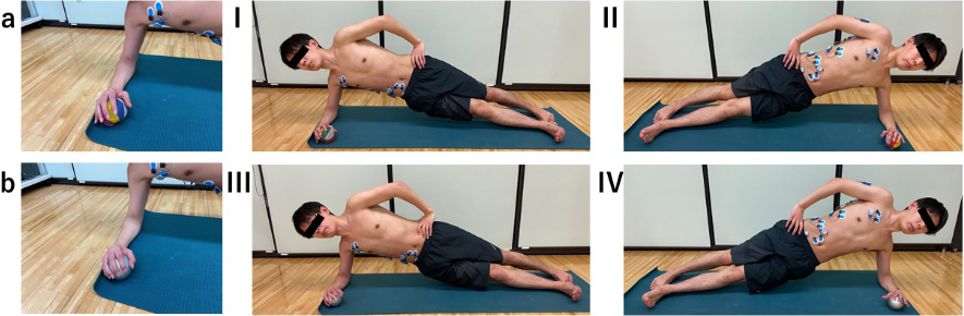
We requested the participants to keep their body position static and hold the item while exerting control to avoid crushing the soft paper balloon (Fig. 1a) and identified this activity as the paper balloon method (PBM). For the regular isometric exercise, we chose a steel ball to be placed in the hand (Fig. 1b) and identified this activity as the conventional side plank method (CSPM).
For PBM, we chose a soft paper balloon called kamifusen (UTF8min紙風èˆ) in Japanese, a classic Japanese toy balloon with a small hole made from rice paper (configuration of the paper balloon:
The participants completed four trials, with two trials each measuring muscle activities on the supported and unsupported sides of the trunk during PBM and CSPM, respectively. Each trial was conducted randomly. The continuation of the trials was regulated based on a prior study and the participants’ potential fatigue due to maximum muscle effort during the exercise task. The participants were given 90-s intervals between each performance to rest [11]. During the task, the examiner visually assessed the posture to see if it was held correctly.
The participants were instructed to remain in the same position during the side plank exercises. The participants were instructed to lie down and bear weight on one side with the shoulder abducted, elbow flexed at 90
Verbal instructions were issued to each participant before the trials to ensure proper pressing of the equipment for PBM. For PBM, participants were instructed to push without crushing the paper balloon for 10-s with maximum exertion using the supported hands to push with maximum force, being careful not to crush the paper balloon (Table 2). For PBM, the joint angle was maintained by preserving the shape of the paper balloon and the position of the hands while using maximal effort (Fig. 1-a, I, II). Participants instantly recognized when the paper balloon was crushed due to the sound of the paper collapsing, indicating when the hands were not maintaining the position. The instructions were created such that participants would focus on an external item relevant to the performed task (external-focus instruction) during the exercise.
Placement of the EMG electrodes
Placement of the EMG electrodes
EMG, electromyography; ASIS, anterior superior iliac spine; PSIS, posterior superior iliac spine.
The electrode application site for electromyography. I, posterior view of the upper back; II, posterior view of the lower back; III, lateral view; IV, frontal view of the chest; V, frontal view of the abdomen; A, upper trapezius; B, medial head of the triceps brachii; C, lower trapezius; D, multifidus; E, latissimus dorsi; F, serratus anterior; G, clavicular part of pectorals major; H, external oblique; I, rectus abdominis; J, internal oblique.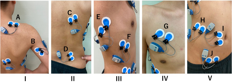
For CSPM, the participants were instructed to place their hands on a steel ball instead of a paper balloon, maintaining the same posture as in PBM (Fig. 1-b, III, IV).
We did not instruct participants to focus on a specific body part or muscle area (internal-focus instruction). None of the participants had experience with the PBM exercise, but each watched a video for guidance before coming to the laboratory. Furthermore, on the examination day, participants had the opportunity to experiment for 5–10-min in the laboratory to acquaint themselves with the exercises.
Muscle activity was measured during the exercise task with surface EMG (Ultium EMG, EM-U810M8, Noraxon USA Inc., Scottsdale, AZ, USA). It was recorded at 2000 Hz with band-pass filtering (10–500 Hz) on a laptop computer (EM-P5, Noraxon) with a receiver (EM-U880, Noraxon). Before affixing the electrodes, the skin was shaved, abraded, and cleaned with alcohol. The electrode application site for EMG was determined according to prior studies [12, 13] and the guidelines of Surface ElectroMyoGraphy for the Non-Invasive Assessment of Muscles (SENIAM;
The activity of each muscle in PBM and CSPM with and without support
The activity of each muscle in PBM and CSPM with and without support
PBM, paper balloon method; CSPM, conventional side plank method.
Differences in muscle activity between exercise tasks. * vs CSPM (Support side), 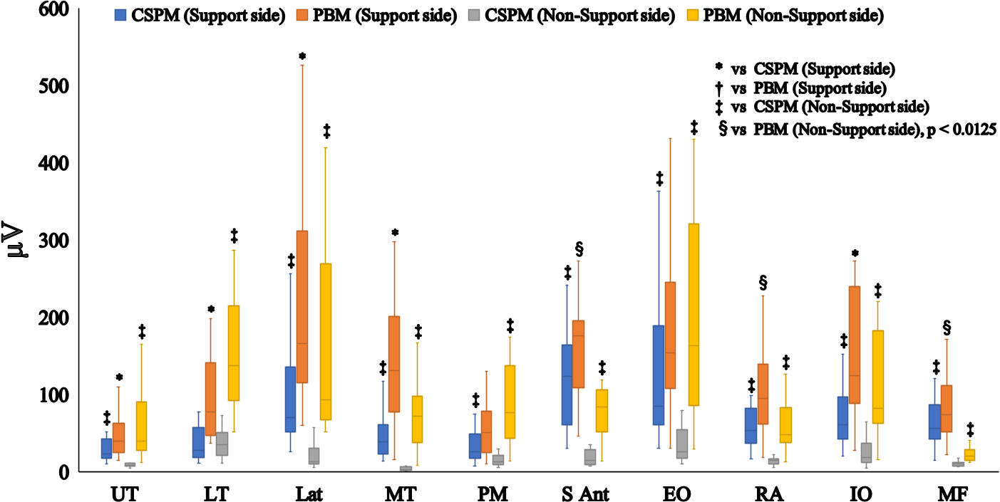
Statistical analyses were performed using IBM SPSS (version 27.0; IBM Corp., Armonk, NY, USA). The Shapiro–Wilk test was performed to determine normality. Depending on the normality of the distribution, the unpaired
Results
The muscle activity in each exercise and the statistical analysis results are shown in Fig. 3 and Tables 4 and 5 respectively. On the supported side, side plank exercise with external-focus instruction significantly increased activity of the upper trapezius (
Results of the statistical analysis
Results of the statistical analysis
Footnotes
Acknowledgments
We thank Editage (
Conflict of interest
The authors have no conflicts of interest to report.