
Abstract
BACKGROUND:
Exercises that stretch the gastrocnemius (GCM) lead to greater ankle dorsiflexion (DF). GCM stretching combined with talar self-stabilisation has been reported to increase ankle DF range of motion (ROM). However, excessive subtalar and midtarsal pronation during GCM stretching may compensate for reduced ankle DF. Thus, this study examined the combined immediate effect of stretching GCM and stabilising talus and subtalar supination on limited ankle DF.
OBJECTIVE:
This study aimed to investigate the immediate effect of GCM stretching alone, GCM stretching with talar self-stabilisation and GCM stretching with talar self-stabilisation and subtalar supination on ankle kinematics in subjects with limited ankle DF.
METHODS:
Fifteen subjects with limited ankle DF were recruited. The subjects performed the three abovementioned methods.
RESULTS:
GCM stretching with talar self-stabilisation and subtalar supination significantly increased open-chain and closed-ankle DF ROM compared with GCM stretching alone. Moreover, GCM stretching combined with talar self-stabilisation and subtalar supination significantly increased open-chain ankle DF ROM with knee straight compared with pre-intervention (
CONCLUSIONS:
The preferred method to increase ankle DF ROM is GCM stretching combined with talar self-stabilisation and subtalar supination.
Introduction
Limited ankle dorsiflexion (DF) is associated with overuse and multiple chronic injuries of the lower extremities, including iliotibial band friction synd- rome [1], patellofemoral pain syndrome [2], Achilles tendinopathy [3, 4], plantar fasciitis [5] and calf muscle tightness [6]. These conditions are usually attributed to irregular tibial advancement over the foot during the mid-stance phase and an early heel-off strategy that brings the body over the foot during the stance phase of the gait cycle [7, 8, 9]. During closed-chain movements such as gait, the talus slides forward and medial during subtalar eversion and pronation. The tibia will move relatively more than the talus and anteriorly glide over the talus as the tibia moves over the foot and the ankle dorsiflexes and pronates. In addition, conjunct external rotation is required to achieve the end range of DF. Several factors may cause limited ankle DF, such as tight calf muscles, loss of mobility in the soft tissue or capsular ligament or a reduction in the flexibility of the talocrural, subtalar or midtarsal joints [10]. In particular, inadequate accessory motion or reduced posterior glide of the talocrural joint is likely influenced by an arthrokinematic restriction of the subtalar joint.
Many previous studies have investigated methods for improving the open- and closed-chain movements in ankle DF range of motion (ROM), including stretching of the gastrocnemius (GCM), talocrural joint mobilisation and GCM stretching combined with talocrural joint mobilisation [11, 12, 13, 14]. Of these methods, stretching of the GCM leads to greater open-chain ankle DF ROM owing to changes in pain onset and increased tolerance of the stretch [14]. The combination of GCM stretching and talocrural joint mobilisation increased the open-chain ankle DF ROM (posterior talar glide) significantly more than GCM stretching alone in subjects with limited ankle DF [12]. Previous studies have demonstrated that anterior-to-posterior talar mobilisation increased open-chain ankle DF active ROM because of the mechanical correction of the bony positioning fault in patients with chronic ankle instability [13]. In addition, studies have used talocrural joint mobilisation to fix the talus at the limit of the posterior talar glide, applying pressure to stabilise the ankle and facilitating relative tibial advancement over the fixed foot during closed-chain activities [12, 15]. However, in this study, we modified talocrural joint mobilisation using Kinesio taping, instead of sustaining the mobilisation by hand, to allow the talus to self-stabilise. In addition, excessive subtalar and midtarsal pronation during weight-bearing activities may compensate for reduced ankle DF [7, 16, 17, 18, 19]. To prevent excessive compensatory pronation in individuals with limited ankle DF, subtalar joint supination may be included in interventions to increase both open- and closed-chain ankle DF ROM. However, no study has examined the effect of subtalar joint supination or the combined use of the three methods (GCM stretching alone, talar stabilisation and subtalar joint supination) on limited ankle DF.
Furthermore, various methods have been used to assess ankle DF as an outcome measure of the effectiveness of therapeutic interventions such as open- (non-weight-bearing) and closed-chain (weight- bearing) conditions [10, 20, 21]. Open-chain measurements typically attempt to better isolate talocrural joint motion by preventing subtalar joint motion [22]. Compared with these, closed-chain measurements have several advantages such that it is relatively easy to perform and involves a more functional position in that greater force from the body weight itself is applied to the ankle joint, thereby stressing the joint through its full excursion [23]. Therefore, this study included open-chain ankle DF ROM with the knee straight and bent and closed-chain ankle DF ROM with the knee straight.
In this study, we aimed to prevent unwanted excessive subtalar joint motion such as eversion or pronation during GCM stretching by allowing the talus to self-stabilise through subtalar supination. Thus, we conducted a navicular drop (ND) test, which is a simple clinical method for assessing foot eversion and pronation [24].
This study aimed to investigate the immediate effect of GCM stretching alone, GCM stretching with talar self-stabilisation and GCM stretching combined with talar self-stabilisation and subtalar supination on ankle kinematics (open-chain ankle DF ROM with the knee straight, open-chain ankle DF ROM with the knee bent, closed-chain ankle DF ROM with the knee straight and ND) in subjects with limited ankle DF. We hypothesised that the three ankle DF ROMs and ND would differ between the three methods in subjects with limited ankle DF.
Subjects’ demographic information (mean
SD)
Subjects’ demographic information (mean
Abbreviations: CI, confidence interval; DF, dorsiflexion; ROM, range of motion.
Subjects
In total, 15 subjects with limited ankle DF participated in this study, including five from the pilot study (Table 1). Subjects with
Procedure
Prior to the interventions, ankle DF ROM was measured three times to determine the demographic information of subjects in previous studies and those in the present study. First, limited ankle DF was determined by measuring the open-chain ankle DF maximum ROM with the knee straight in the prone position using a 14-inch stainless steel goniometer. Second, open-chain ankle DF maximum ROM with the knee bent in the sitting position and closed-chain ankle DF maximum ROM with the knee straight in the standing position was measured using digital inclinometer (Gain Express Holdings Ltd., Shenzhen, China). All measurements were performed twice; the goniometer and inclinometer were removed between the first and second measurements. The mean of two maximum values was used for data analysis.
For the three stretching exercises, the subjects were instructed by the principal investigator (PI) on the correct posture and performance of the stretching exercises. For warm-up, the subjects were allowed to practice the exercises for approximately 10 min. They were allowed to perform stretching in less than three repetitions. A 5-min rest period was allowed after the familiarisation period and before data collection. All subjects randomly performed all the three methods (GCM stretching alone, GCM stretching with talar self-stabilisation and GCM stretching with talar self-stabilisation and subtalar supination) to avoid bias caused by learning or fatigue. At the start of the intervention, the subjects pulled out numbers written on a paper from a small box to determine the order in which they were to perform the tasks (GCM stretching alone in 4, GCM stretching with talar self-stabilisation in 5 and GCM stretching with talar self-stabilisation and subtalar supination in 6). Each stretch was held for 30 s and performed five times. The subjects were allowed a 10-s rest period between stretches [28]. Based on previous studies, the subjects had a washout period of 15 min between the methods to avoid bias due to the effects of stretching (5 repetitions
Measurement of dependent variables
Open-chain ankle DF ROM with the knee straight
Open-chain ankle DF ROM with the knee straight was measured using the goniometer. The subjects were put in the prone position, with feet extending beyond the end of the treatment table. The PI maintained a neutral subtalar joint position and applied force to the plantar surface of the forefoot and midfoot until further movement was firmly resisted. A second investigator confirmed the neutral position of the subtalar joint. The PI marked the three points on the lateral malleolus, fibular head and fifth metatarsal bone using a pen. The three marks were kept the same until the end of the study [30]. The fulcrum of the goniometer was placed over the lateral malleolus, and the stationary and moving arms were aligned with the fibular head and parallel to the fifth metatarsal bone, respectively. A previous study had reported good intrarater (0.96–0.97) and interrater reliability (0.91–0.92), with standard errors of measurement (SEM) of 0.70
Open-chain ankle DF ROM with the knee bent
The talar swing test was performed to assess talocrural joint posterior glide and conjunct external rotation of the talus during passive ankle DF. However, we did not use this method to directly measure talar swing, posterior talar glide and the conjunct external rotation of the talus. Thus, for interpretation accuracy, we used “open-chain ankle DF ROM with the knee bent” instead of “talar swing test” to describe the test performed in this study. The subjects were instructed to sit, with their thighs completely supported on the edge of the treatment table. The test foot was aligned with the subtalar joint in a neutral position parallel to the floor. The second investigator visually monitored the starting and end positions of the measurement. The PI’s thumbs were positioned on the dome of the anterior talus to push the talus posteriorly while the foot was kept parallel to the floor with the subtalar joint in a neutral position until further movement was firmly resisted [10, 21, 31]. The second investigator placed an inclinometer just distal to the tibial tuberosity and recorded the angle of knee flexion when the tibia had stopped moving. After one measurement of open-chain ankle DF ROM with the knee bent, the subjects stood in a comfortable position and then returned to the sitting position for the next trial. Good intrarater reliability (0.99) was reported for the talar swing test in a previous study [31].
Closed-chain ankle DF ROM with the knee straight
The subjects were instructed to stand and step forward with the non-test leg while keeping the test foot in line with the long axis of the leg, knee in extension and heel on the ground. The subject leaned forward and flexed the hip and knee of the non-test leg, as needed, to allow for maximal ankle DF of the test. The PI marked three points on the fibular head, lateral malleolus and half point using a pen. The inclinometer was positioned on the half point in line with the fibular head and lateral malleolus. The maximal position was the point at which the heel began to lift from the floor. The second investigator visually monitored this motion to lift the heel from the floor. The DF ROM was recorded, and the subject returned to the starting position for the next trial. Intrarater reliability values ranged from 0.88 to 0.89, with a SEM of 2.2
ND test
ND was assessed while the subjects were in the sitting position. The PI marked each subject’s navicular tuberosity in the subtalar neutral position using a pen. Then, the investigator measured the difference between the vertical position of the navicular tuberosity (distance between the navicular tubercle and floor) when the subject was in the sitting position and that when the subject was in the standing position, in millimetres using a ruler (i.e. a modified Brody approach) [33, 34, 35]. A previous study reported using the average measurement to define neutral foot (ND, 5–9 mm), pronated foot (ND,
Gastrocnemius stretching alone.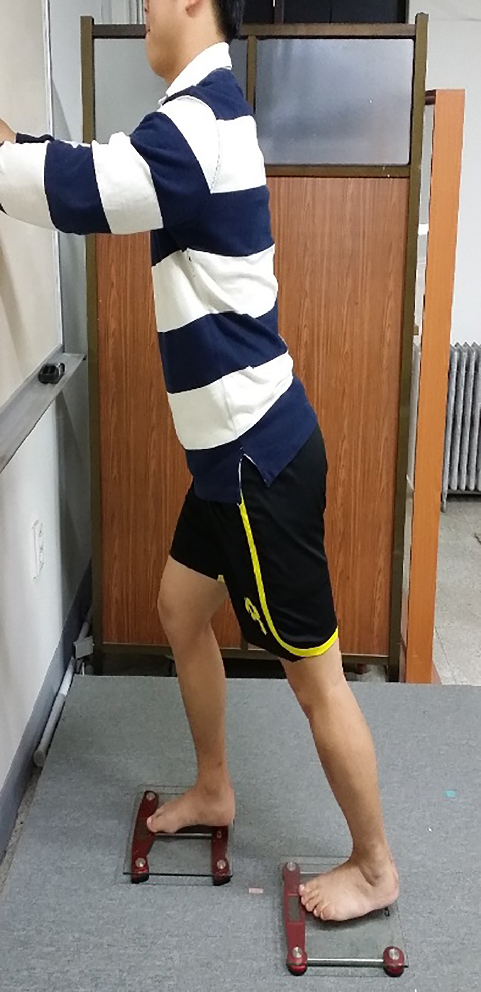
GCM stretching alone
The subjects were instructed to stand on two scales to ensure that they leaned forward by the same amount each time. The subject placed the test foot over the line that bisected the heel and the second toe on the scale to maintain the neutral foot position. The weight on the test leg (side with limited ankle DF ROM) with the knee extended was maintained at 60%
Gastrocnemius stretching with self-stabilising talus.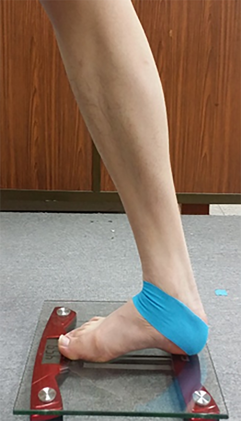
The PI applied a 20-cm long Kinesio tape to stabilise the talus during DF. The single layer of the tape was fully stretched and directly applied to the skin, with the subjects in the sitting position. The tape was applied starting from the anterior aspect of the talus and extended to the lateral malleolus, calcaneus and back to the anterior aspect of the talus [38]. The tape went once around the ankle. The second investigator tactilely confirmed the tape attachment and visually monitored the tape removal during the stretch. After the tape was applied, the subjects performed the GCM stretching in the same way as before, but including talar self-stabilisation this time (Fig. 2).
GCM stretching with talar self-stabilisation and subtalar supination
The PI applied the Kinesio tape for talar self-stabilisation. Then, another 20-cm-long Kinesio tape was fully stretched and directly applied on the skin. The tape was applied starting from the fibula and extended to the lateral malleolus, calcaneus and middle third of the medial tibia [39]. The tape went once around the ankle. The second investigator tactilely confirmed the tape attachment and visually monitored the tape removal and heel contact during the stretch. After the tape was applied, the subjects performed the GCM stretching in the same way as with talar self-stabilisation, but including subtalar supination this time (Fig. 3).
Gastrocnemius stretching with talar self-stabilisation and subtalar supination.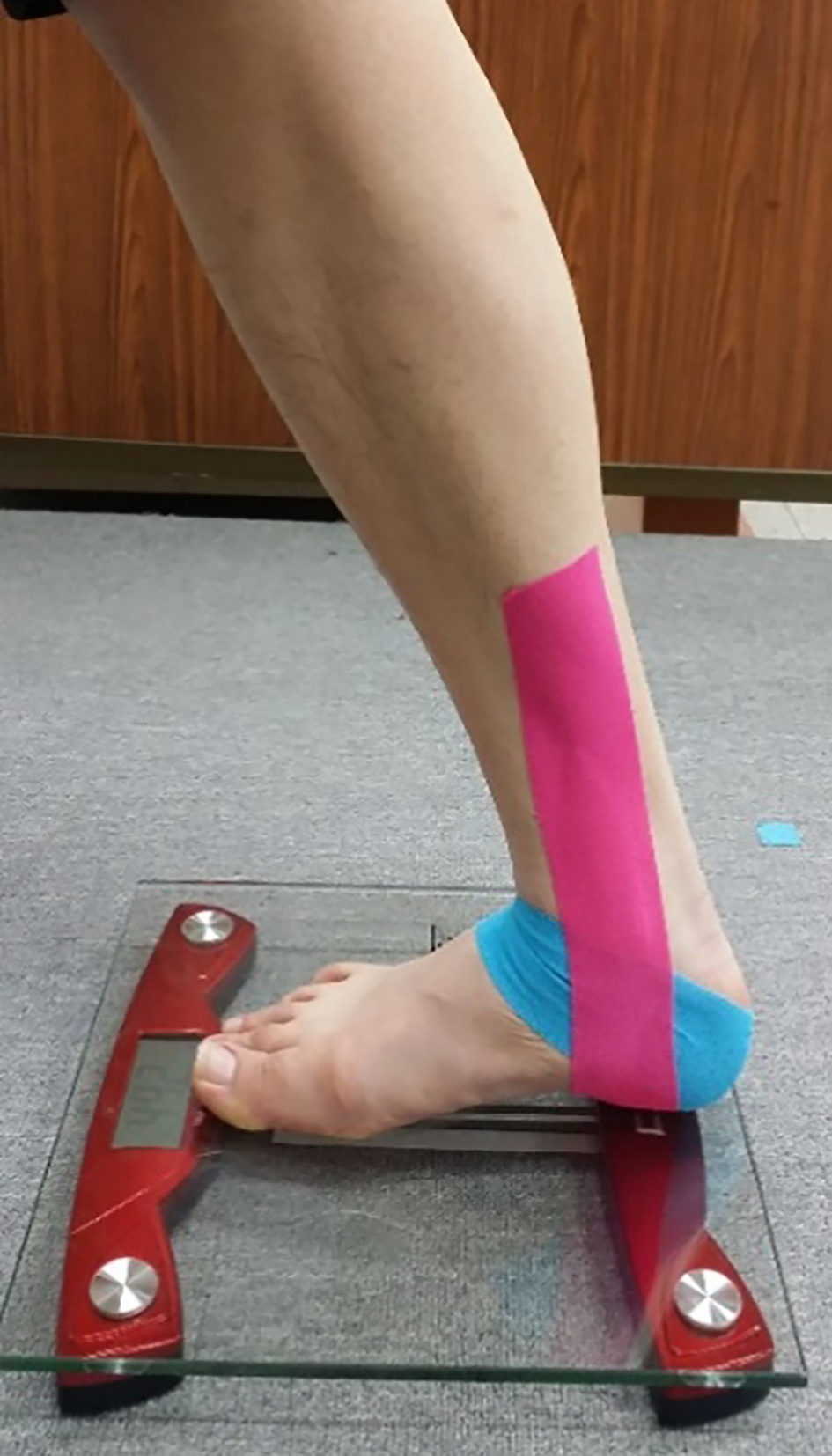
Descriptive statistics by methods (mean
Abbreviations: GCM, gastrocnemius; DF, dorsiflexion; ROM, range of motion.
The G*Power ver. 3.1.6 software package (Franz Faul, Kiel University, Kiel, Germany) was used to calculate the sample size and conduct post power analyses. Using data obtained from a pilot study of five subjects, we calculated the necessary sample size to achieve a power of 0.80 and an effect size (ES) of 0.85 and found that four subjects are needed (calculated from the partial
Results
The Kolmogorov-Smirnov tests showed no significant differences in the total ankle DF ROM and ND data among the three methods (open-chain ankle DF ROM with knee straight:
The two-way ANOVA revealed significant time-by-method interactions for open-chain ankle DF ROM with the knee straight (
There were no significant time-by-method interactions for open-chain ankle DF ROM with the knee bent (
There were significant time-by-method interactions for closed-chain ankle DF ROM with the knee straight (
There were no significant time-by-method interactions for ND (
Discussion
This study investigated the immediate effect of GCM stretching alone, GCM stretching with talar self-stabilisation and GCM stretching with talar self-stabilisation and subtalar supination on ankle DF and ND in subjects with limited ankle DF. Compared with GCM stretching alone, GCM stretching together with talar self-stabilisation and subtalar supination significantly increased open-chain and closed-chain ankle DF ROM. Moreover, compared with pre-intervention, GCM stretching with talar self-stabilisation and subtalar supination significantly increased the open-chain ankle DF ROM with knee straight.
Our results revealed significantly greater ankle DF ROM with the knee straight in GCM stretching combined with talar self-stabilisation and subtalar supination than in GCM stretching alone, increasing by 4.07
We found that the open-chain ankle DF ROM with the knee bent was significantly greater in GCM stretching with talar self-stabilisation and subtalar supination, increasing by 1.8
No significant differences in ND were observed among the three methods. This finding does not support the original research hypothesis. The hypothesis was based on the theory that reduced ankle DF may be compensated for by subtalar and midtarsal pronation during weight-bearing activities [7, 16, 17, 18, 19]. Accordingly, GCM stretching may lead to a pronated foot in subjects with limited ankle DF. Although the present study did not reveal a decrease in ND, different results may have been observed if ND had been measured while GCM stretching was being performed. We measured ND immediately after the stretches. Thus, over the long term, GCM stretching alone might contribute to a change in foot position. In addition, the study subjects were in the neutral foot group (mean ND before intervention, 0.52) according to the definition of previous researcher [35] (neutral foot: ND, 5–9 mm). The results would change if the subjects had pronated feet. Finally, we used the Kinesio tape to prevent subtalar pronation during GCM stretching. The most popular taping methods are the McConnell and Kinesio taping techniques [48]. In general, McConnell taping has been used to correct joint alignment because a rigid tape exerted no tension on the skin of the patient where it is applied. However, this study used the Kinesio tape because its favourable adhesive properties facilitate easy use and prevent allergic reactions and stretch tolerance can be sufficiently altered via sensory mechanisms; hence, the clinical use of Kinesio taping is widespread [49]. The results with Kinesio taping could imitate those obtained with McConnell taping. However, a previous study has reported that Kinesio taping reduces patellofemoral pain syndrome-associated pain but does not change patellar alignment. Previous researchers believed that Kinesio taping can be applied at 50%–85% tension on the skin to restrict partial or full joint motion, but this taping tension was insufficient to correct patellar alignment [50]. Thus, this study applied 120%–130% maximal tension (full-stretch tape) on the ankle joint. In addition, previous results have indicated that McConnell taping corrects patellar alignment and tracking [51] but does not improve the motor function and proprioception of patients with patellofemoral pain syndrome [52, 53]. In addition, conjunct external rotation of the talus occurs near the end range of DF [10].
The goal of this study was to prevent excessive pronation and anterior glide of the talus on the calcaneus by using the Kinesio tape. Complete blocking of these motions by rigid taping such as with the McConnell tape would also appear to limit the conjunct talar external rotation that occurs with normal pronation during DF. Thus, this study used Kinesio taping to maintain joint alignment. However, this result could not prove that the Kinesio tape did work or not. Thus, future studies should examine the long-term effect of various GCM stretching techniques for controlling subtalar movement using both Kinesio and rigid taping in subjects with limited ankle DF and pronated feet.
This study has several limitations. First, owing to its cross-sectional design, this study could not determine the long-term effects of GCM stretching with talar self-stabilisation and GCM stretching with talar self-stabilisation and subtalar supination on ankle kinematics. Second, we measured only open-chain ankle DF ROM with the knee straight, open-chain ankle DF ROM with the knee bent, closed-chain ankle DF ROM with the knee straight, and ND. We did not measure other gait parameters such as heel-off time, although a previous study has reported that heel-off time was significantly earlier in subjects with limited ankle DF than in those without it [54]. Future studies should be conducted to investigate the long-term effects of GCM stretching with talar self-stabilisation and GCM stretching with talar self-stabilisation and subtalar supination on heel-off time during the gait cycle. Third, we were not blinded to the measurements; thus, investigator bias might have affected our results. The bias might have been more likely during the measurement of open-chain ankle DF ROM with the knee straight than during closed-chain measurement because we did not use a force controller to minimise errors, resulting from the application of unequal force. Future studies should use a force controller such as a handheld dynamometer.
Conclusions
This study investigated the immediate effect of GCM stretching alone, GCM stretching with talar self-stabilisation and GCM stretching with self-stabilisation and subtalar supination on open-chain ankle DF ROM with the knee straight, open-chain ankle DF ROM with the knee bent, closed-chain ankle DF ROM with knee straight and ND on subjects with limited ankle DF. The results showed that GCM stretching with talar self-stabilisation and subtalar supination produced a significantly greater open- and closed-chain ankle DF ROM than GCM stretching alone. Stabilisation of the talus by taping both at the talocrural and subtalar joints may restrict excessive foot pronation in subjects with limited ankle DF, leading to increased ankle DF. Thus, GCM stretching combined with talar self-stabilisation and subtalar supination is superior to GCM stretching alone in subjects with limited ankle DF, when performed immediately.
Footnotes
Conflict of interest
None to report.