
Abstract
BACKGROUND:
Tribbles pseudokinase 3 (TRIB3) is a member of the tribbles-related family, which is involved a lot of cellular processes and multiple cancers, such as breast cancer, colorectal cancer, renal cell carcinomas, and lung cancer. However, the expression pattern and biological function of TRIB3 in hepatocellular carcinoma (HCC) has not yet been completely elucidated.
METHODS:
The expression of TRIB3 and clinicopathological characteristics were evaluated by HCC tissue microarray and qPCR analysis. Lentivirus packaging and transfection were employed to establish cell lines with TRIB3 overexpression or knockdown. The biological functions of TRIB3 in the growth of HCC were determined using MTT and crystal violet assays. Tumor growth was monitored in a xenograft model in vivo.
RESULTS:
The expression of TRIB3 was upregulated in HCC tissue samples compared to paired normal tissues in 45 patients examined by qPCR assay. TRIB3 expression was significantly correlated with HCC tumor size and prognosis in postoperative patients by analysis of the TRIB3 expression data and HCC clinical features. Forced expression of TRIB3 significantly promoted HCC growth in vitro. In contrast, downregulation of TRIB3 inhibited cell growth in vitro. Moreover, knockdown of TRIB3 suppressed tumorigenesis of HCC cells in vivo.
CONCLUSION:
TRIB3 promotes growth abilities of HCC cells both in vitro and in vivo and predicts poor prognosis of HCC patients, which serves as a prognostic marker and might provide a potential therapeutic candidate for patients with HCC.
Introduction
Hepatocellular carcinoma (HCC) is one of the most common malignant tumors, ranking the fifth most common cancer and the second largest cause of cancer-related death worldwide [1, 2]. China has a higher incidence and mortality of HCC than other countries, and the frequent metastases, recurrence, and early blood vessel invasion of HCC are responsible for poor prognosis of patients with HCC [3, 4]. Moreover, the prognosis of HCC is very poor, with the 5-year survival rate
Tribbles pseudokinase 3 (TRIB3), a member of the tribbles-related family, contains the substrate-binding domains but lacks the conserved amino acid catalytic motifs essential for kinase activity [7, 8]. A series of studies have demonstrated that TRIB3 is involved in a lot of cellular processes, such as cell proliferation and differentiation, epithelial-to-mesenchymal transition, cellular stress response, and glucose and lipid metabolism [9, 10, 11]. Currently, emerging evidence suggests that TRIB3 serves as a crucial oncoprotein. The biological function of TRIB3 has been observed in multiple cancer cell lines and primary tumors, including breast cancer, colorectal cancer, renal cell carcinomas, and lung cancer [12, 13, 14, 15]. However, the function role of TRIB3 and its expression pattern in HCC are still poorly understood.
In the current study, we found that TRIB3 expression was upregulated in human HCC tissues and was significantly associated with prognosis and clinicopathologic parameters by tissues array analysis. We further demonstrated that TRIB3 promoted growth abilities of HCC cells both in vitro and in vivo. Our study confirmed TRIB3 to be a prognostic marker and a potential therapeutic candidate for patients with HCC.
Materials and methods
HCC tissue samples and tissue microarray analysis
After obtaining written informed consent, HCC tissues were obtained from the patients who underwent liver resection radiation at the Shandong Provincial Hospital Affiliated to Shandong University, Jinan, Shandong, China. Forty-five pairs of HCC tissues and the corresponding paired normal tissues were applied to RNA extraction for quantitative real-time PCR (qPCR) analysis. To evaluate the expression pattern of TRIB3, we examined the tissues array which was constructed from 176 paraffin-embedded primary HCC tissues and adjacent non-tumor liver tissues and analyzed the relationship between TRIB3 expression pattern and clinical features. The protocol for tissue collection was approved by the Ethics Committee of Shandong Provincial Hospital Affiliated to Shandong University. The study was performed in accordance with the Declaration of Helsinki and the guidelines of the committee.
Immunohistochemistry (IHC)
Paraffin-embedded tissue sections were deparaffinized, rehydrated, subjected to antigen retrieval, and the endogenous peroxidases were blocked. After washing the section thrice with 0.01 mol/L PBS, the sections were blocked in 0.01 mol/L PBS containing 0.3% Triton X-100 and 5% BSA. Next, sections were incubated with anti-TRIB3 (1:100) antibody (13300-1-AP, ProteinTech) at 4
RNA preparation and qPCR
The total RNA of 45 pairs of tumor tissues and adjacent noncancerous tissues and HCC cells was extracted using TRIzol reagent (Invitrogen). Total RNA (2
Cell culture
HCC cell lines HepG2, Hep3B, YY-8103, PLC/PRF/ 5, LM3, Huh7, and SNU-398 were purchased from the Cell Bank of Type Culture Collection of the Chinese Academy of Sciences. All cell lines were maintained at 37
Plasmids and cell transfection
The coding sequence for the full length of TRIB3 was cloned into the p23-ZsGreen plasmid (Invitrogen) to generate the TRIB3 expression vector. Lentiviral short hairpin RNA (shRNA) targeting TRIB3 and Snail were designed using software provided by Qiagen (Va- lencia, CA, USA). HepG2 and Hep3B cells were infected with p23-ZsGreen-TRIB3 lentiviral. SNU-398 and Huh7 cells were infected with pLKO.1-shRNA. Overexpressed and silenced cells were sorted using flow cytometry or selected using puromycin (4 ug/mL) for 4 days, respectively.
The sequences of shTRIB3 were as follows: sh1 5’-CCGGGATCTCAAGCTGTGTCGCTTTCTCGAGAA AGCGACACAGCTTGAGATCTTTTTG-3’ (forward) and 5’-AATTCAAAAAGATCTCAAGCTGTGTCGC TTTCTCGAGAAAGCGACACAGCTTGAGATC-3’ (reverse); sh2 5’-CCGGGCACTTAAGAAAGGACCA AATCTCGAGATTTGGTCCTTTCTTAAGTGCTTTT TG-3’ (forward) and 5’-AATTCAAAAAGCACTTAAG AAAGGACCAAATCTCGAGATTTGGTCCTTTCTT AAGTGC-3’ (reverse).
Western blot analysis
Cells samples were extracted using RIPA Lysis Buffer and PMSF (Thermo Scientific) for at least 30 min on ice. Protein concentration was determined using the Bradford reagent (Sigma) in accordance with the manufacturer’s instructions. The lysates were centrifuged at 12,000 rpm for 15 min at 4
MTT assay
The cell growth in vitro was measured using MTT assay. In the MTT assay, 1000 cells per well were seeded into 96-well plates. Twenty microliters of a 5 mg/mL MTT solution was added into the medium, and the plate was cultured at 37
Crystal violet assay
The cell growth in vitro was also measured by crystal violet assay. One thousand cells per well were seeded into 6-well plates, and the cells were cultured in a medium with 10% FBS. The medium was changed ev- ery 3 days, and cells were stained with 1 mL 0.5% crystal violet solution in 20% methanol after 2 weeks. Next, the fixed cells were washed with PBS and photographed. Glacial acetic acid (1 mL) was added to dissolve the cells, and the absorbance value was detected at 570 nm (OD 570) using an automatic microplate reader.
Tumorigenesis in vivo
Male nude mice were raised under standard conditions. Cell suspensions (1
Statistical analysis
All the results are shown as mean
TRIB3 expression is increased in HCC tissues and correlates with clinical prognosis. (A), (B) The mRNA level of TRIB3 in 45 pairs of HCC tumor samples (T) and matched normal liver tissues (N) was determined by quantitative real-time PCR. The expression of TRIB3 was normalized to GAPDH. (C) Immunohistochemistry staining of TRIB3 in of two paired N and T tissues samples. (D) H-scores of the TRIB3 staining intensity in N (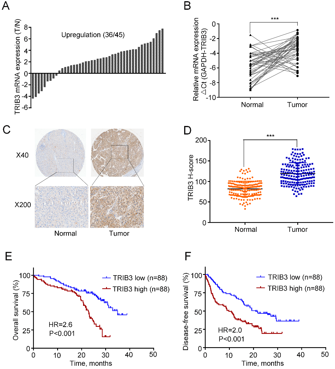
Clinical significance of TRIB3 expression in HCC
First, we quantified the mRNA level of TRIB3 in 45 pairs of HCC tissues and their matched normal counterparts using qPCR, and the results showed that upregulation of TRIB3 was observed in 36 pairs, accounting for 80% of the total specimens examined (Fig. 1A). Meanwhile, TRIB3 expression was significantly higher in tumor samples compared to that in paired normal tissues (
Moreover, to further explore the clinical significance of TRIB3 in patients with HCC, we examined TRIB3 expression in an HCC tissue microarray containing 176 specimens by immunohistochemical staining and staining intensity was scored in a standard manner as described previously [16]. Higher protein expression of TRIB3 in HCC tissues than in normal tissues was further confirmed by immunohistochemical staining, and TRIB3 was distributed in both the nucleus and cytoplasm (Fig. 1C). In addition, we found that the H-scores of the HCC tissues were higher than those of the normal tissues (H-scores: 119.2
Relationship between the TRIB3 expression and the clinicopathologic features of HCC
Relationship between the TRIB3 expression and the clinicopathologic features of HCC
Abbreviation: HBsAg: Hepatitis B surface antigen; AFP:
Univariate and multivariate cox regression analyses of overall survival
Abbreviation: HBsAg: Hepatitis B surface antigen; AFP:
Univariate and multivariate cox regression analyses of disease-free survival
Abbreviation: HBsAg: Hepatitis B surface antigen; AFP:
Furthermore, we explored the relationship between TRIB3 expression and clinicopathologic parameters in 176 patients with HCC. As shown in Table 1, high TRIB3 expression was associated with HBsAg positive (
TRIB3 overexpression promotes growth of HCC cells in vitro. The protein levels of TRIB3 in seven HCC cell lines (A) and stably transfected HCC cells (B) were examined by western blotting. The effect of TRIB3 overexpression on the viability of HepG2 (C) and Hep3B (D) cells was assessed by MTT assay. The effect of TRIB3 overexpression on the colony formation of HepG2 (E) and Hep3B (F) cells was assessed by crystal violet assay. Left panel: representative images of the crystal violet assay. Right panel: quantification of the crystal violet assay. Data are presented as mean 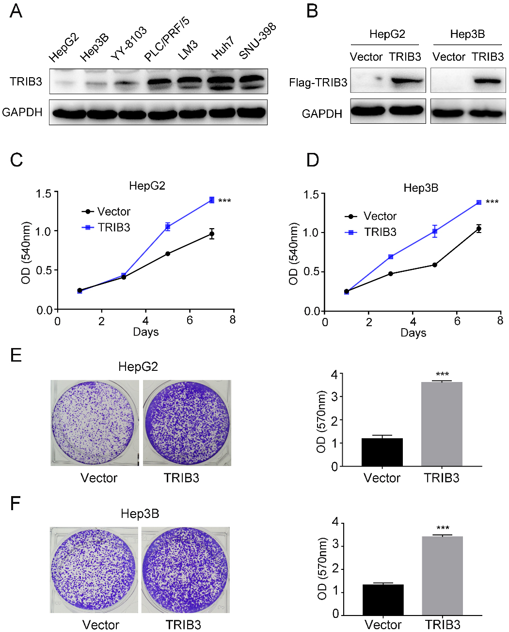
TRIB3 knockdown inhibits growth of HCC cells in vitro. The protein levels of TRIB3 in stably transfected SNU-398 (A) and Huh7 (B) HCC cells were examined by western blotting. The effect of TRIB3 knockdown on the viability of SNU-398 (C) and Huh7 (D) cells was assessed by MTT assay. The effect of TRIB3 knockdown on the colony formation of SNU-398 (E) and Huh7 (F) cells was assessed by crystal violet assay. Left panel: representative images of the crystal violet assay. Right panel: quantification of the crystal violet assay. Data are presented as mean 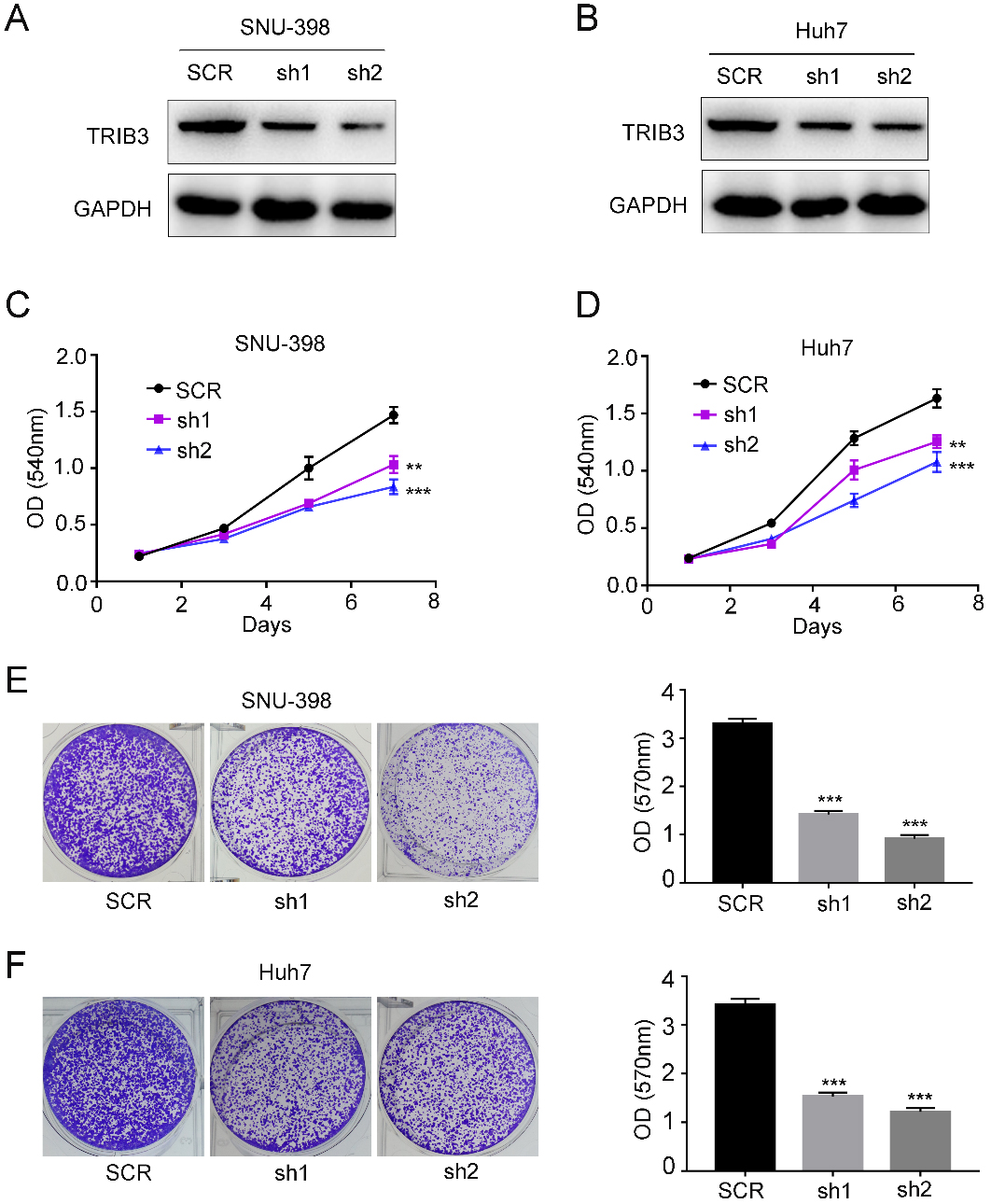
The above clinical observations encouraged us to investigate the biological function of TRIB3 in HCC growth in vitro. To this end, we first examined the TRIB3 expression level in a panel of HCC cell lines (HepG2, Hep3B, YY-8103, PLC/PRF/5, LM3, Huh7, and SNU-398) (Fig. 2A). Next, we exogenously overexpressed Flag-tagged TRIB3 in HepG2 and Hep3B cells, which showed relatively low expression levels of exogenousTRIB3 (Fig. 2B). Considering that high TRIB3 expression levels were associated with large tumor size in the clinical data, we speculated that TRIB3 might influence HCC cell growth. Hence, we measured the effect of exogenous TRIB3 overexpression on cellular growth by using MTT and crystal violet assays. Indeed, the MTT assays showed that the absorbance values of the HepG2 cells after transfection with TRIB3 overexpression vectors were significantly higher than those of the vector controls (
TRIB3 knockdown suppresses tumorigenesis of HCC cells in vivo. (A) Representative images of the tumors generated by TRIB3 con and TRIB3 knockdown Huh7 cells (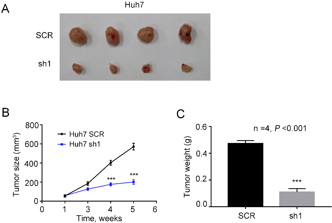
To further evaluate the biological function of TRIB3 in HCC cells, we knocked down the expression of TRIB3 in SNU-398 and Huh7 cells using two independent shRNAs, which showed relatively high expression of TRIB3 (Fig. 3A and B). As expected, the MTT assays showed that the absorbance values of the SNU-398 cells after downregulation of TRIB3 using two independent shRNAs were significantly lower than those of the scramble controls (
Knockdown of TRIB3 suppresses tumorigenesis of HCC cells in vivo
The above results show that TRIB3 promotes HCC cell growth in vitro, which prompts us to verify this result in vivo. Next, control and shTRIB3 Huh7 cells were injected subcutaneously into the flanks of 5-year-old nude mice (
Discussion
Currently, a series of studies have explored the roles of TRIB3 in different cancers [17, 18, 19]. Yu et al. demonstrates that expression of TRIB3 positively associates with breast cancer stemness and progression, which provides insights into breast cancer development and confers a potential therapeutic strategy against TRIB3-overexpressed breast cancer [20]. TRIB3 is also reported to interact with
Based on the clinical sample results, this study revealed TRIB3 was upregulated in HCC tissues compared with that in their paired normal counterparts. Patients with HCC with high TRIB3 expression yielded worse overall survival and disease-free survival than those with low TRIB3 expression. In addition, the expression levels of TRIB3 correlated with the clinical characteristics of the patients, such as HBsAg positive, AFP, tumor size, and satellite nodules. Moreover, high TRIB3 expression, as well as high level of AFP and large tumor diameter, was an independent risk factor for overall survival and disease-free survival, suggesting that it might play a positive role in the growth of HCC. The functional analysis for TRIB3 in the progression of HCC cells revealed that overexpression of TRIB3 could promote the growth of HCC cells in vitro. In contrast, knockdown of TRIB3 inhibited the growth of HCC cells. Moreover, TRIB3 downregulation also inhibited tumorigenesis of HCC cells in nude mice. To data, this is the first study to reveal the functional roles and expression patterns of TRIB3 in HCC.
In summary, we have shown that TRIB3 is upregulated and exerts an oncogenic role in HCC. TRIB3 is a strong indicator of aggressive malignant behavior and poor clinical outcome in HCC, which might provide a potential therapeutic target for HCC. Additional research aimed at elucidating the mechanisms by which TRIB3 regulates the growth patterns of HCC cells.
Footnotes
Conflict of interest
The authors declare that they have no competing interests.