
Abstract
BACKGROUND:
Phyllodes tumor (PT) is a rare tumor showing various malignant potential. The histological grade of PT is related to clinical outcome, but its relationship between gaining of malignant potential and underlying mechanism including cancer stem cell factor was not understood yet.
OBJECTIVE:
The main purpose of this study was to determine the expression pattern of cancer stem cell marker in PT and to understand its clinical and pathological implications.
METHODS:
CD44, CD166, ALDH1, and Ki-67 immunohistochemistry were performed on a tissue microarray from 185 cases of PT specimens (138 benign, 32 borderline, 15 malignant). The immunohistochemistry result and clinicopathological parameter of each cases were compared to analyze the implications of cancer stem cell markers on PT.
RESULTS:
Borderline/malignant PT showed higher CD44 expression of the stromal component than benign PT (
CONCLUSIONS:
The cancer stem cell markers, CD44 and CD166, are expressed in both the epithelial and stromal components of phyllodes tumor. Besides, ALDH1 is only expressed in stromal component. In the stromal component, expression of cancer stem cell markers increases with higher PT histologic grade. In the epithelial component, the absence of cancer stem cell marker expression is related to poor clinical prognosis.
Introduction
Phyllodes tumor (PT) is a rare tumor which accounts for 0.3–1.5% of all breast neoplasms and may exhibit malignant characteristics such as recurrence or metastasis [1]. Grossly, it appears as circumscribed, firm, bulging mass. Histologically, it typically exhibits prominent intracanalicular growth with leaf-like stromal projections. The epithelial component of PTs includes bland-looking luminal epithelial cell and myoepithelial cells which may be accompanied by hypercellular stromal component. PTs are histologically classified as benign, borderline, or malignant by the World Health Organization (WHO) classification [2]. The tumor shows aggressive clinical features as the histologic grade increases. There is no established regimen for metastatic phyllodes tumor, but recent studies revealed combined chemotherapy can be useful for metastatic phyllodes tumor, rather palliative radiotherapy and resection have limited roles. Thus, close follow up is required in patients with high probability of distant metastasis [3].
Cancer stem cells are defined as cells with ability of self-proliferation and differentiation in tumors, thus enabling cancer to develop, recur, and metastases. The concept of the cancer stem cell was first proposed to explain the tumorigenesis of acute myeloid leukemia (AML), in animal models [4]. The leukemic stem cells in this study expressed specific cell surface markers, such as CD34
Previous immunohistochemical analysis of human malignant PT and mouse xenografted human PT revealed that malignant PT possesses mesenchymal stem cell-like features such as CD44, CD166, CD90, CD177, and disialoganglioside (GD2) positivity. In addition, ALDH
Materials and methods
Patient selection
This study was approved by the Institutional Review Board of Yonsei University Severance Hospital. PT tissue samples were obtained from patients diagnosed at the Department of Pathology of Severance Hospital from 2000 to 2010. All tissues were fixed in 10% buffered formalin and embedded in paraffin. All archival hematoxylin and eosin (H&E)-stained slides for each case were reviewed by two pathologists (JS Koo and SI Kim). The histologic grading of PT was based on the WHO Classification of Tumours of the Breast 4
Tissue microarray
The representative area was selected by H&E slide review, and the corresponding area was marked on its paraffin block. The selected area was punched out with a biopsy needle, and the 5-mm tissue core was placed in a 5
Immunohistochemistry
The antibodies used for immunohistochemistry are listed in Table 1. Each 3-
Source, clone, and dilution of antibodies used in this study
Source, clone, and dilution of antibodies used in this study
Immunohistochemical findings were assessed by light microscopy (BX53 Upright microscope, Olympus). The findings were quantified based on the proportion and intensity of stained cells. Proportions of stained cells were graded as 0: negative, 1: less than 30% positive cells, and 2: more than 30% positive cells. Intensities were graded as 0: negative; 1: weak, 2: moderate, and 3: strong. If the product of the two grades was 0–1, we diagnosed the sample as negative, and if 2–6, we diagnosed the sample as positive [7]. Ki-67 immunohistochemical staining was performed by using anti-Ki-67 (30-9) Rabbit Monoclonal Primary Antibody and immunostained slides were scanned by ROCHE VENTANA iScan HT slide scanner. Stromal component of each case of phyllodes tumor was designated and Ki-67 Labeling index was calculated by Virtuoso v. 5.6.1 software. If Ki-67 labeling index of one case overs median value of all cases it was considered as high Ki-67 labeling index. In case of Ki-67 labeling index lesser than median value, it was considered as low Ki-67 labeling index. The range of number of counted cells was from 1000 to 7000 due to variable range of stromal cellularity according to histologic grade of PT.
Clincopathologic characteristics of patients with phyllodes tumor
Heat map of the expression of cancer stem cell-related proteins according to phyllodes tumor (PT) histologic grade. (red, positive, green, negative, E, epithelial component, S, stromal component).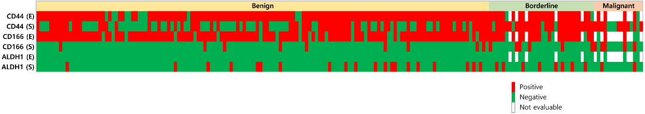
Expression of cancer stem cell-related proteins according to phyllodes tumor (PT) histologic grade. (a)–(c) H&E stain of PT cases (X200). Stromal cellularity and atypia are increased as the histologic grade increases. (d)–(f) CD44 immunohistochemical stain (X200). CD44 expression in the stromal component is higher in borderline/malignant PT than in benign PT. (g)–(i) CD166 immunohistochemical stain ((g)–(h); X200, (i); X400). CD166 expression in the epithelial component increases as the histologic grade decreases. (j)–(l) ALDH1 immunohistochemical stain ((j)–(k); X200, (l); X400). ALDH1 was only expressed in stromal component and its stromal expression is increased as the histologic grade increases. (m)–(o) Ki-67 immunohistochemical stain (X200). Ki-67 Labeling Index was high in high grade phyllodes tumor. It is associated with stromal CD166 expression.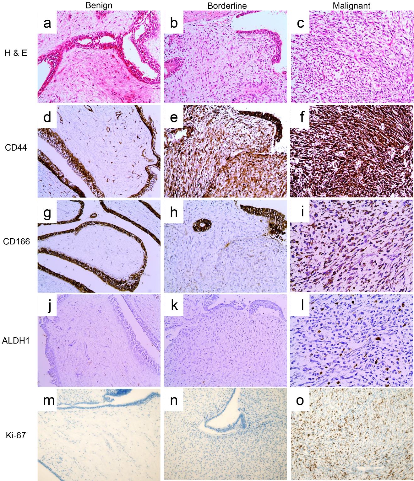
Data were analyzed using SPSS for Windows, Version 12.0 (SPSS Inc., Chicago, IL, USA). For determination of statistical significance, Student’s
Results
Basic characteristics of PT
The basic characteristics of the 185 patients enrolled in this study are shown in Table 2; 138 cases were diagnosed as benign, 32 were borderline, and 15 were malignant. Patient age and tumor size increased with higher histologic grades (
Expression of cancer stem cell-related proteins in phyllodes tumor according to histologic grade
Expression of cancer stem cell-related proteins in phyllodes tumor according to histologic grade
The expression levels of CD44 in the stromal component (
Expression of cancer stem cell-related proteins according to Ki-67 labeling index in stromal component
The relationship between expression pattern of cancer stem cell-related protein and Ki-67 labeling index was investigated. There was statistical significant relationship between level of stromal CD166 expression and Ki-67 labeling index value. There was high portion of CD166 positive cases in high stromal Ki-67 labeling index group. (
Expression of cancer stem cell-related proteins in phyllodes tumor according to Ki-67 labeling index in stromal component
Expression of cancer stem cell-related proteins in phyllodes tumor according to Ki-67 labeling index in stromal component
The correlation between cancer stem cell marker expression in breast PT and pathologic parameters was found. The number of positive cancer stem cell markers in the epithelial component was correlated with stromal overgrowth. As the number of positive cancer stem cell markers in the epithelial component increased, stromal overgrowth was rarely detected (
Impact of the expression of cancer stem cell-related proteins on patient prognosis
There was no clinical factor showing statistical significant relationship to cancer stem cell marker expression in PT (Table 5). However, in subgroup analysis, the CD44 status (
Univariate analysis of the impact of CAF-related proteins in the stromal component of phyllodes tumor on patient prognosis using the log-rank test
Univariate analysis of the impact of CAF-related proteins in the stromal component of phyllodes tumor on patient prognosis using the log-rank test
E, epithelial component, S, stromal component.
Correlation between the number of positive markers for cancer stem cells in phyllodes tumor (PT) and pathological parameters. As the number of positive cancer stem cell markers in the epithelial component increases, stromal overgrowth is rarely detected. As the number of positive cancer stem cell markers in the stromal component increases, higher histologic grades, increased stromal cellularity, increased stromal atypia, and increased stromal mitosis are observed.
Impact of the expression of cancer stem cell-related proteins on patient prognosis. CD44 status and number of positive markers for cancer stem cells in the epithelial component are correlated with disease-free survival (DFS) in benign phyllodes tumor (PT). CD44 negativity and decreased number of positive markers for cancer stem cells in the epithelial component are correlated with shorter DFS in benign PT.
The purpose of this study was to investigate cancer stem cell marker expression in breast PT and its clinical implications. First, we found that the expression pattern of cancer stem cell markers was different in PT of different histologic grades. Previously, there was study about malignant PT possesses stem cell like feature, such as CD44, CD166, CD90, CD177 and GD2 positivity [6]. In our study, CD44 expression was elevated in the stromal component in higher histologic grade PT, an observation consistent with previous study. Second, our study revealed differences in cancer stem cell marker expression patterns between the epithelial and stromal components of PT. In high histologic grade PT, CD44 expression and ALDH1 expression in the stromal component was elevated, whereas CD166 expression in the epithelial component was decreased. Also, the correlation of CD166 expression and histologic grade is confirmed by Ki-67 labeling index. Previously, the association of increased ALDH1 expression in stromal component with high histologic grade has been studied [8]. Additionally, ALDH
The genes related to malignant transformation of PT include TP53, PIK3CA, RB1, NF1, PTEN, BRAF, and EGFR [13, 14, 15, 16, 17]. Interestingly, CD44-positive cancer stem cells are known to have activated EGFR signaling [18, 19, 20], TP53/PTEN mutations [21], and a hyperactive phosphatidylinositol 3-kinase pathway [22]. Therefore, it can be hypothesized that the CD44-positive stromal component in PT achieves malignant transformation by autonomous stromal growth. We propose that the CD166-positive epithelial component contributes to tumor progression in low histologic grade PT, and after gaining autonomous growth potential, the CD44-positive stromal component becomes involved in malignant transformation. Further studies are required to test this hypothesis.
In this study, the stromal overgrowth in PT increases in prominence as the number of positive markers for cancer stem cells in the epithelial component decreases. Moreover, we demonstrated that in benign PT, lower levels of CD44 and a decreased number of positive markers for cancer stem cells in the epithelial component results in shorter DFS. These findings suggest that the cancer stem cell feature of the epithelial component is lost during malignant transformation of PT. In addition, an increase in the number of positive markers for cancer stem cells in the stromal component correlates with a higher histologic grade, increased stromal cellularity, increased stromal atypia, and increased stromal mitosis. These results indicate that the cancer stem cell phenotype in the stromal component is related to tumor aggressiveness.
This study has clinical implications for the treatment of PT. Previous studies have found that CD44 [23, 24, 25], CD166 [26], and ALDH1 [27] targeted therapies were effective in suppressing cell proliferation in various cancer cell lines. We are hopeful that stem cell marker targeted therapy can also be effective in PT, although further studies are required to confirm this clinical application.
In conclusion, we have shown that the cancer stem cell markers, CD44, CD166, and ALDH1, are expressed in both the epithelial and stromal components of PT. As the histologic grade of PT increases, the predominance of cancer stem cell marker expression shifts from the epithelial component to the stromal component. Lastly, the expression of cancer cell markers in the epithelial component is correlated with the clinical prognosis in PT. The limitation of this study is on limited explanation of detailed activated molecular pathway of cancer stem cells in PT. The further studies may be required to show each molecular pathway is activated in cancer stem cells of each histologic grade and stromal/epithelial component of PT.
Footnotes
Conflict of interest
The authors have no relevant affiliations or financial involvement with any organization or entity with a financial interest in or financial conflict with the subject matter or materials discussed in the manuscript. This includes employment, consultancies, honoraria, stock ownership or options, expert testimony, grants or patents received or pending, or royalties.