
Abstract
BACKGROUND:
Increasing evidence reveals that aberrant microRNAs (miRNAs) expression play a crucial role in the tumorigenesis of cancers, including hepatocellular carcinoma (HCC), whereas the role of miR-654-3p in HCC remains unclear. This study aimed to investigate the role of miR-654-3p in HCC.
METHODS:
Real-time quantitative PCR was performed to detect miR-654-3p expression in HCC tissues and cell lines. The association of miR-654-3p expression with clinical characteristics of HCC patients were analyzed. And the prognostic value of miR-654-3p was examined using Kaplan-Meier curve and Cox regression analysis. CCK-8 and Transwell assays were used to observe the effects of miR-654-3p on proliferation, migration, and invasion of HCC cells.
RESULTS:
The miR-654-3p expression was downregulated in both HCC tissues and cell lines, which was significantly associated with lymph node metastasis and TNM stage. Downregulation of miR-654-3p predicted poor prognosis of HCC patients. Overexpression of miR-654-3p inhibited HCC cell proliferation, migration, and invasion, while knockdown of miR-654-3p promoted these cellular behaviors in vitro.
CONCLUSION:
Our study suggested that miR-654-3p expression was downregulated in HCC and might serve as a potential prognostic marker and therapeutic target for the survival of HCC patients. miR-654-3p might exert a suppressor role in HCC through inhibiting tumor cell proliferation, migration, and invasion.
Introduction
Hepatocellular carcinoma (HCC) is one of the most common and aggressive malignancy and is the third leading cause of cancer-related mortality worldwide [1]. In China, the incidence of HCC is relatively high and accounts for approximately half of the total number of all cases [2, 3]. Till now, despite therapeutic strategies have been widely used and improved, the prognosis of major patients with HCC is still very poor due to early metastasis and high frequency of recurrence [4]. Thus, it is urgently needed to explore more novel cancer-associated molecular biomarkers and therapeutic target for the HCC treatment.
MicroRNAs (miRNAs or miRs) is a cluster of small, single-strand, and noncoding RNAs that can post-transcriptionally regulate the corresponding target gene expression by binding to 3’-UTR regions [5, 6]. Extensive studies have indicated to emerge as essential regulators in various cellular processes, such as differentiation, proliferation, migration, invasion, and apoptosis [7, 8, 9]. MiRNAs have been implicated to function as either oncogenes or tumor suppressor genes during cancer development in various malignancies, including HCC [9, 10, 11]. Previous studies have found that miR-654-3p, a member of the 14q32 region, was aberrantly expressed in several tumors, such as natural killer/T-cell lymphoma [12] and ovarian cancer [13]. In a study by Murilo Vieira Geraldo and co-workers indicated that miR-654-3p played tumor suppressor properties in thyroid cell lines and could regulate some genes expression, such as PTEN, RB1, MMP-9, and FGFR4 [14]. A recent study revealed that several miRNAs were abnormally expressed in HCC and hepatocellular adenomas compared with adjacent normal liver, including miR-654-3p [15]. However, the potential role of miR-654-3p in HCC remains unclear.
In the present study, we firstly explored the expression of miR-654-3p in HCC clinical tissue samples and cell lines by quantitative real-time PCR (qRT-PCR). Then, the association of miR-654-3p expression with clinical characteristics and patients’ prognostic information was evaluated. In addition, the functional role of miR-654-3p in HCC cells was investigated.
Materials and methods
Patients and tissue samples
Paired 117 HCC tissue samples and adjacent normal liver tissue samples were collected from patients who were pathologically diagnosed with HCC and underwent surgery at Affiliated Hospital of Weifang Medical University between June 2010 and December 2013. All fresh tissue samples were immediately put into liquid nitrogen until use. All patients did not receive any therapies prior to surgery. The clinical characteristics of the patients were collected and listed in Table 1. Written informed consent was provided by each patient participated in the study prior to surgery. The study was approved by the Ethics Committee of Affiliated Hospital of Weifang Medical University.
The relationship between miR-654-3p expression and clinicopathological parameters of HCC patients
The relationship between miR-654-3p expression and clinicopathological parameters of HCC patients
*
The human HCC cell lines SK-HEP-1, PLC/PRF/5, Huh7, SNU-182, and one normal liver cell line HL7702 were purchased from Cell Bank of Chinese Academy of Sciences (Shanghai, China). All cells were cultured in DMEM (Thermo Fisher Scientific, IL, USA) containing 10% FBS (Thermo Fisher Scientific, IL, USA) at 37
The mimic negative control (NC), miR-654-3pmimic, inhibitor NC, and miR-654-3p inhibitor were purchased from GenePharma (Shanghai, China). Lipofectamine 3000 reagent (Invitrogen; Thermo Fisher Scientific, IL, USA) was used for cell transfection following to the manufacturer’s instructions.
miR-654-3p expression is downregulated in HCC that analyzed using qRT-PCR. A. The relative expression of miR-654-3p was significantly downregulated in HCC tissues compared with adjacent normal tissues. Differences were analyzed using a 2-tailed Student’s t-test. B. The expression of miR-654-3p was significantly lower in HCC cell lines compared with normal liver cells. Differences were analyzed using one-way ANOVA followed by Tukey’s post-hoc test. *** 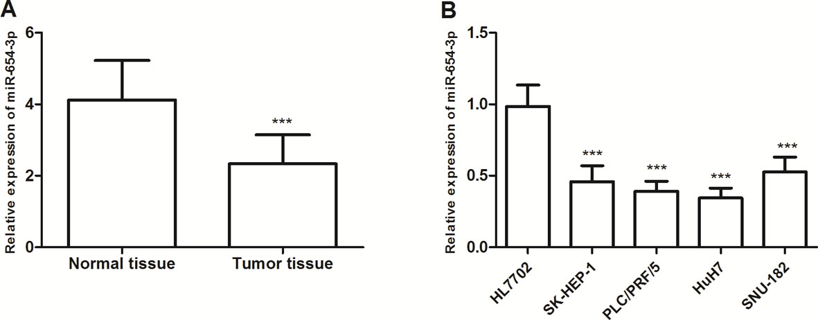
Total RNA was extracted using the TRIzol reagent (Invitrogen; Thermo Fisher Scientific, IL, USA) from HCC tissues or cells according to the manufacturer’s protocol. Then, cDNA was reverse-transcribed using total RNA with the Taqman MicroRNA Reverse Transcription Kit (Applied Biosystems; Thermo Fisher Scientific, IL, USA). The qRT-PCR was performed using a GoTaq qPCR Master Mix (Promega, Madison, WI, USA) on the ABI 7900 System (Bio-Rad) for quantification of miR-654-3p levels. The relative expression levels of miR-654-3p were calculated using 2
CCK-8 assay
CCK-8 assay (Dojindo Molecular Technologies, Inc.) was used to evaluate cell proliferative abilities of HCC cell lines after transfected with miR-654-3p mimic or inhibitor. After transfection, the transfected cells were seeded into 96-well plates at a density of 2
Transwell migration and invasion assays
Transwell chambers (Millipore, Burlington, MA, USA) were used to assess cell migratory and invasive abilities of HCC cells. For Transwell migration assays, transfected HCC cells (5
Statistical analysis
Each experiment was performed at least three times and data were presented as mean
Results
The miR-654-3p expression is downregulated in HCC tissues and cell lines
Firstly, we detected miR-654-3p expression levels in 117 pairs of HCC tissues and the adjacent normal tissues by using qRT-PCR. The results showed the relative expression of miR-654-3p was higher in HCC tissues than in normal tissues (
The relationship between miR-654-3p expression and clinical characteristics of HCC patients
To explore whether miR-654-3p expression was associated with HCC progression, the association between its expression and clinical characteristics of HCC patients was analyzed. Then we divided all the patients with HCC into a low miR-654-3p expression group and a high miR-654-3p expression group based on the mean expression level of miR-654-3p. As shown in Table 1, low levels of miR-654-3p were associated with positive lymph node metastasis (
Decreased expression of miR-654-3p associated with poor prognosis of patients with HCC
Subsequently, we investigated the prognostic significance of miR-654-3p in HCC patients using Kaplan-Meier curve analysis and log-rank test. As shown in Fig. 2, patients with high levels of miR-654-3p had longer overall survival rates compared with those patients with lower miR-654-3p expression (log-rank
Kaplan-Meier curves for overall survival time. Patients in the low miR-654-3p expression group had a shorter overall survival time compared with those in the high expression group (log-rank 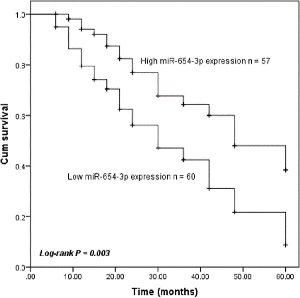
Multivariate Cox analysis for the association between clinical characteristics and overall survival of HCC patients
*
To determine the effect of miR-654-3p on HCC cell behavior, we constructed both miR-654-3p overexpression and knockdown HCC cells using miR-654-3p mimics and inhibitors, respectively, in PLC/PRF/5 and HuH7 cells, which had a relative lower miR-654-3p expression level. The qRT-PCR results confirmed transfection efficiency in PLC/PRF/5 and HuH7 cells (
Overexpression of miR-654-3p inhibited cell proliferation, while knockdown of miR-654-3p promoted cell proliferation, compared with untreated cells. Differences were analyzed using one-way ANOVA followed by Tukey’s post-hoc test. A. Transfection efficiency of miR-654-3p mimic and inhibitor was confirmed using qRT-PCR. B. CCK-8 assays were performed 0, 24, 48, and 72 h after transfection to determine the proliferation of PLC/PRF/5 and HuH7 cells. *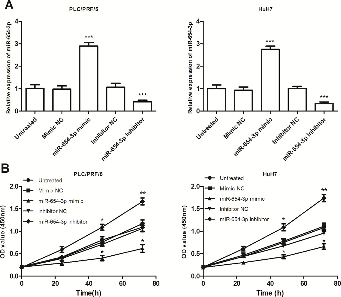
Increased expression of miR-654-3p inhibited cell migration and invasion, while inhibition of miR-654-3p enhanced cell migration and invasion of PLC/PRF/5 and HuH7 cells, compared with untreated cells. Transwell migration (A) and invasion (B) assays were used to explore the effects of miR-654-3p in HCC. Differences were analyzed using one-way ANOVA followed by Tukey’s post-hoc test. **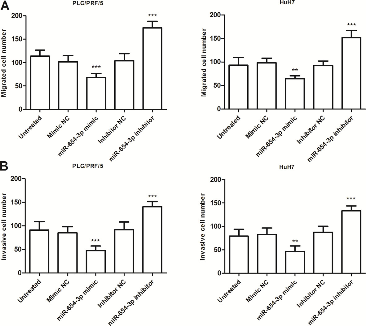
The present study indicated that miR-654-3p was significantly downregulated in HCC tissues and cell lines compared with matching adjacent normal tissue and normal liver cell lines, respectively. The low expression of miR-654-3p was found associated with positive lymph node metastasis and advanced TNM stage. It also demonstrated that low miR-654-3p expression was an independent predictor of poor prognosis in patients with HCC. Functionally, upregulation of miR-654-3p inhibited the proliferation, migration, and invasion, while knockdown of miR-654-3p reversed the suppressor effects of miR-654-3p overexpression. These findings suggest that miR-654-3p may exert its suppressor roles in HCC.
HCC has become one of the most rapid-growing causes of cancer-related death worldwide because of difficult diagnosis and poor prognosis. Accumulating evidence has highlighted the crucial roles of miRNAs in the regulation of tumor progression, and indicating miRNAs may serve as diagnostic and/or prognostic biomarkers and effective therapeutic targets for cancer treatment [16, 17, 18]. For instance, miR-1285–3p was downregulated in osteosarcoma and inhibited the proliferation and invasion of osteosarcoma cells, suggesting miR-1285–3p might be a potential therapeutic target and prognostic biomarker in osteosarcoma [19]. miR-3691–5p was found upregulated in HCC tissues and cell lines and correlated with poor prognosis, as well as functioned as an oncogene in HCC [20]. Hence, identification of HCC specific miRNAs is still critical for defining novel prognostic biomarker and therapeutic targets for the treatment of HCC.
In this study, by using qRT-PCR, the results showed that the expression of miR-654-3p was downregulated in both HCC tissues and cell lines. Consistent with our findings, Zheng and co-workers using miRNA profiling indicated miR-654-3p was one of the downregulated miRNAs in HCC [15]. Notably, low miR-654-3p expression was found to be associated with positive lymph node metastasis and advanced TNM stages. These results suggested that miR-654-3p may play a suppressor role in the progression of HCC. In addition, downregulation of miR-654-3p significantly predicted poor prognosis. Then, multivariate Cox regression analysis using clinical characteristics and 5-year follow-up information of HCC patients showed that low miR-654-3p expression was an independent prognostic predictor for overall survival of HCC patients. Thus, the results indicated that miR-654-3p may be a novel effective prognostic biomarker for HCC, which served as the prognostic role similarly to several miRNAs in HCC, such as miR-454-3p [21], miR-339-5p [22], and miR-664 [23].
MiRNAs can function as tumor oncogenes or suppressors in human cancers, such as miR-221-5p in renal cell cancer [24], miR-203a-3p in colorectal carcinoma [25], and miR-1204 in HCC [26]. Previous studies have demonstrated that miR-654-3p was downregulated in several cancers including ovarian cancer [13], colorectal cancer [27], and non-small cell lung cancer [28]. For instance, miR-654-3p decreased cell proliferation, migration, and induced reprogramming of metastasis-related genes (such as MMP 9 and IL-1
Taken together, we found that miR-654-3p was downregulated and acted as a tumor suppressor in HCC. Moreover, lower miR-654-3p expression was correlated with shorter overall survival of HCC patients. Overexpression of miR-654-3p inhibited proliferation, migration, and invasion, while inhibition of miR-654-3p promoted proliferation, migration, and invasion of HCC cells. Thus, miR-654-3p may serve as a tumor suppressor and a novel potential prognostic biomarker and therapeutic target for HCC.
Footnotes
Acknowledgments
This work was supported by Shandong Medicine and Health Science Technology Development Project [No. 2017WS849; No. 2017WS777].
Conflict of interest
The authors declare that they have no competing interests.