
Abstract
OBJECTIVES:
The purpose of this study was to isolate the secondary metabolites of endophytic fungi from Ginkgo biloba (SMEFGB) and investigate their anti-cervical cancer activity.
METHODS:
SMEFGB were cultured. The secondary metabolites of endophytic fungi was extracted, purified and identified. The effects of secondary metabolites on proliferation, apoptosis and migration of human cervical cancer HeLa cells were determined. In addition, the effects of SMEFGB on growth of Hela implanted tumor in mice were investigated.
RESULTS:
In 9 stains of endophytic fungi successfully isolated from the leaves of Ginkgo biloba, the stain J-1, J-2 and J-3 could produce podophyllotoxin. These 3 stains were identified by molecular biology. The secondary metabolites of stain J-1, J-2 and J-3 markedly inhibited the proliferation of HeLa cells, promoted their apoptosis and blocked their migration. In addition, the secondary metabolites of stain J-1, J-2 and J-3 significantly attenuated the growth of HeLa implanted tumor in mice.
CONCLUSIONS:
Our results indicated that SMEFGB had obvious anti-cervical cancer activity in vitro and in vivo.
Keywords
Introduction
Cervical cancer is a common malignant tumor in women, and its incidence ranks second among women’s multiple tumors, which is second only to breast cancer [1]. It is reported that there are 500,000 new cases each year worldwide. It usually occurs in women aged 40–60 years and is a disease caused by human papillomavirus infection [2]. The main symptoms of the disease are vaginal bleeding and leucorrhea abnormalities [3]. The treatment methods of cervical cancer in clinical mainly include surgery, radiotherapy, chemotherapy, and targeted therapy. Among them, radiotherapy plays a very significant role in the treatment of cervical cancer. At present, radiotherapy and chemotherapy have become the main treatment strategies for cervical cancer.
Although radiotherapy and chemotherapy can improve the clinical symptoms and survival rates of patients to a certain extent, these programs will damage the immune function of patients and cause many complications. Therefore, it is urgent to seek more safe and effective treatment for patients with cervical cancer. In recent years, some scholars have found that traditional Chinese medicine can not only promote the apoptosis of cervical cancer cells, but also regulate the immune function of patients, reduce the complications of chemotherapy, etc. Ginkgo biloba is a special medicinal plant in China. It is found that Ginkgo biloba contains more than 100 kinds of chemical constituents, in which flavonoids, terpenoids and ginkgolic acids have been proved to have obvious antitumor activity [4, 5]. However, the growth of ginkgo is slow, which has restricted the development of its medicinal values. In recent years, the endophytic fungi from Ginkgo biloba (SMEFGB) has become a hot research topic. It is expected to become a new way for seeking new sources of medicines or biological active substances from Ginkgo biloba [6, 7]. However, there are only few studies on SMEFGB, especially on their antitumor activities [8].
Therefore, investing the active substances of SMEFGB has important theoretical significance and potential application value for understanding the distribution of Ginkgo biloba endophytic fungi resources and developing the related medicines. In this study, the secondary metabolites of SMEFGB were isolated and identified. The in vitro and in vivo activities of secondary metabolites on human cervical cancer HeLa cells were investigated to provide a practical basis for further development of Ginkgo biloba resources and natural anti-tumor substances.
Materials and methods
Culture of SMEFGB
Endophytic fungi were isolated from the roots, stems, leaves and bark tissues of Ginkgo biloba (College of Life Science, Dezhou University, Dezhou, China). The strains were inoculated to plate containing potato dextrose agar medium (Sigma-Aldrich Corp., MO, USA), followed by culturing at 28
Extraction and purification of SMEFGB
After fermentation of endophytic fungi, the mycelium and fermentation broth were collected. The fermentation broth was concentrated, and extracted with ethyl acetate (Sigma-Aldrich Corp., MO, USA) for two times. The mycelium was dried, crushed, and extracted with ethyl acetate for two times. The ethyl acetate extract of mycelium and fermentation broth were combined. After removing the solvent, the crude extract was obtained. The crude extract was dissolved in methanol (Sigma-Aldrich Corp., MO, USA), and then was filtered and mixed with column chromatography silica gel. After evaporating the solvent, the mixture was placed in the silica gel column, followed by gradient elution using petroleum ether/ethyl acetate (7:3–3:7; Sigma-Aldrich Corp., MO, USA). The various parts of the eluent were collected. After removing the solvent, the final SMEFGB product was obtained. High performance liquid chromatography (HPLC) was used to analyze the compositions of product.
Determination of SMEFGB effect on proliferation of human cervical cancer HeLa cells
Human cervical cancer HeLa cells (Cell Bank of the Chinese Academy of Sciences, Beijing, China) in logarithmic growth phase (2
Determination of SMEFGB effect on apoptosis of HeLa cells
HPLC of extract from mycelium and fermentation broth of Ginkgo biloba endophytic fungi. A: Control substance; B: J-1; C: J-2; D: J-3; 1: podophyllotoxin.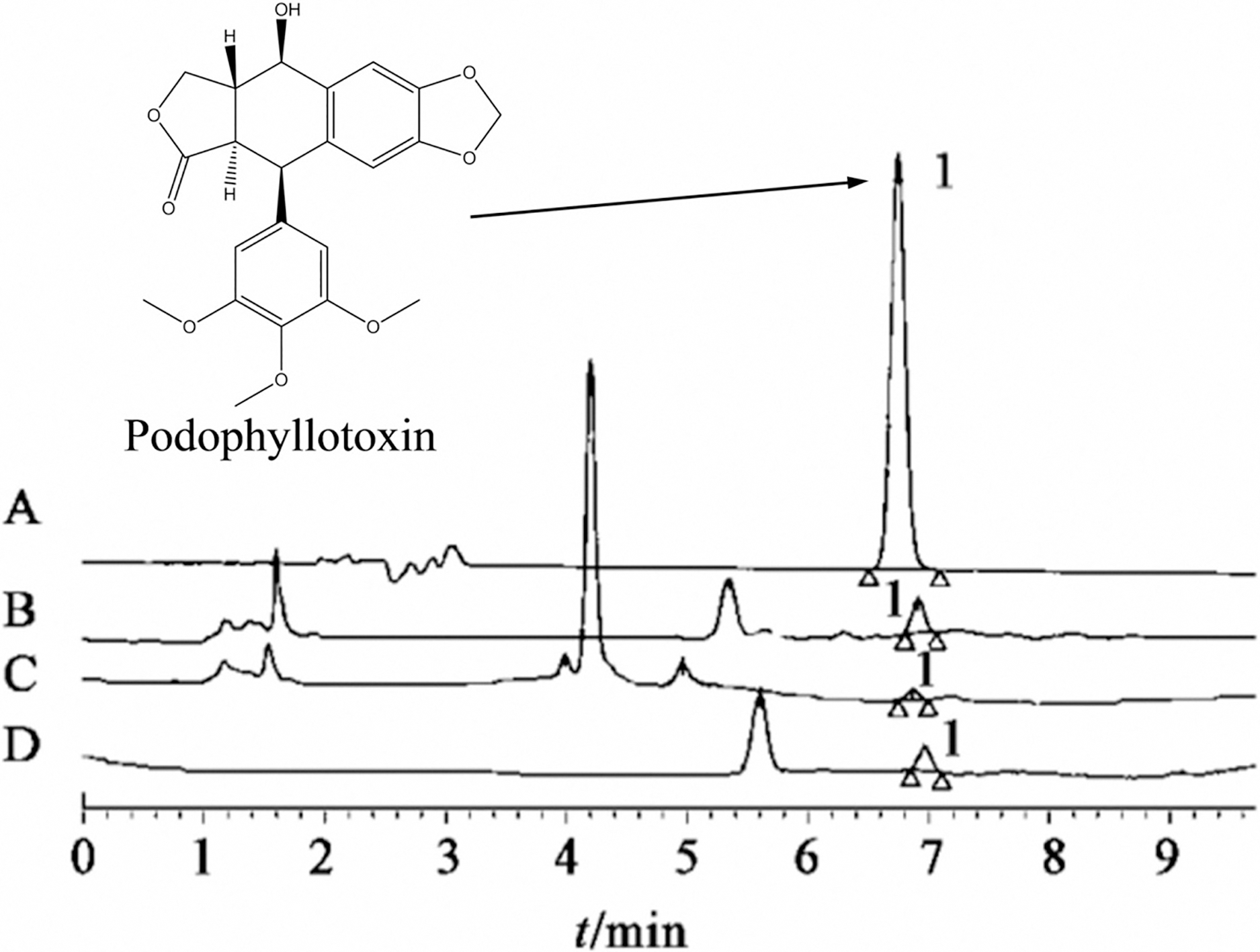
HeLa cells in logarithmic growth phase (2
Macroscopic and microscopic (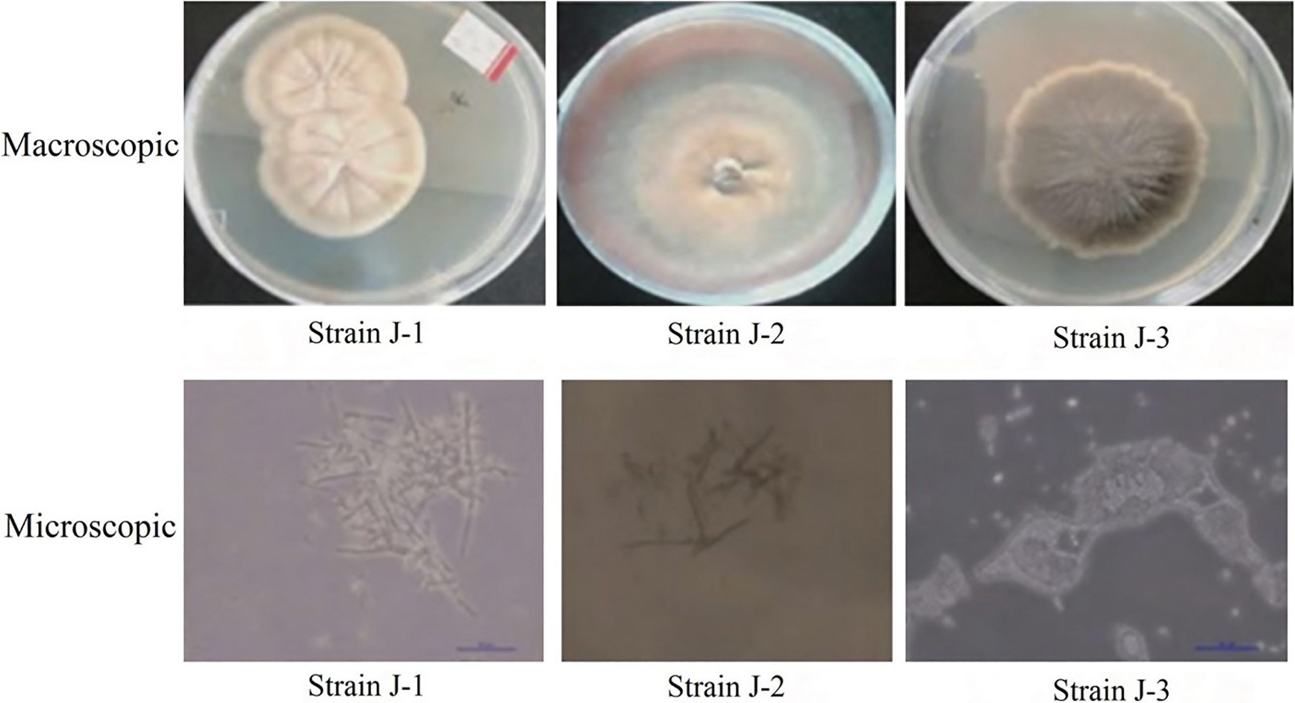
The migration of HeLa cells was determined by in vitro scratch assay. HeLa cells in logarithmic growth phase (2
Establishment of mice model with HeLa implanted tumor
HeLa cells in logarithmic growth phase were taken, and the single-cell suspension (5
Determination of SMEFGB effect on growth of HeLa implanted tumor in mice
Fifty mice were randomly divided into control, CY and strain J-1, strain J-2 and strain J-3 groups with 10 mice in each group. The mice in later 4 groups were intraperitoneally injected with 3 mg/kg DDP, and 50 mg/kg SMEFGB from strain J-1, strain J-2 and strain J-3, respectively. The mice in control group were intraperitoneally injected with equal volume of normal saline. The administration was performed once per two days and lasted for 20 days. At the end of experiment, all nude mice were executed by cervical dislocation, and the tumor block was stripped, the maximal diameter of the tumor (a) and the short diameter (b) were measured using Vernier caliper. The tumor volume was calculated as follows: tumor volume
Effect of SMEFGB on the proliferation of Hela cells
Effect of SMEFGB on the proliferation of Hela cells
Each experiment was performed for 3 times. The data were presented as mean
Results
Isolation and identification results of SMEFGB
A total of 9 stains of endophytic fungus were successfully isolated from the leaves of Ginkgo biloba. After activation and culture, the mycelium and fermentation broth were collected. HPLC was used to analyze the composition of the endophytic fungi. The results showed that strains J-1, J-2 and J-3 could produce the podophyllotoxin compound (Fig. 1). The macroscopic and microscopic morphological characteristics of endophytic fungi producing podophyllotoxin were shown in Fig. 2.
Molecular identification was performed on the endophytic fungi producing podophyllotoxin. Results showed that the amplified fragment base sequence of strain J-1 had 100% similarity with the sequences of Chaetomium globosum strain MF564. In addition, the amplified fragment base sequence of strain J-2 had 100% similarity with the sequences of Chaetomium sp.4RF3. Moreover, the amplified fragment base sequence of strain J-3 had 100% similarity with the sequences of Pseudallescheria sp. T55. The phylogenetic tree of 18S rRNA sequence of strains J-1, J-2 and J-3 and corresponding sequences of other species was shown in Fig. 3.
Phylogenetic tree of 18S rRNA sequence of strain J-1, J-2 and J-3.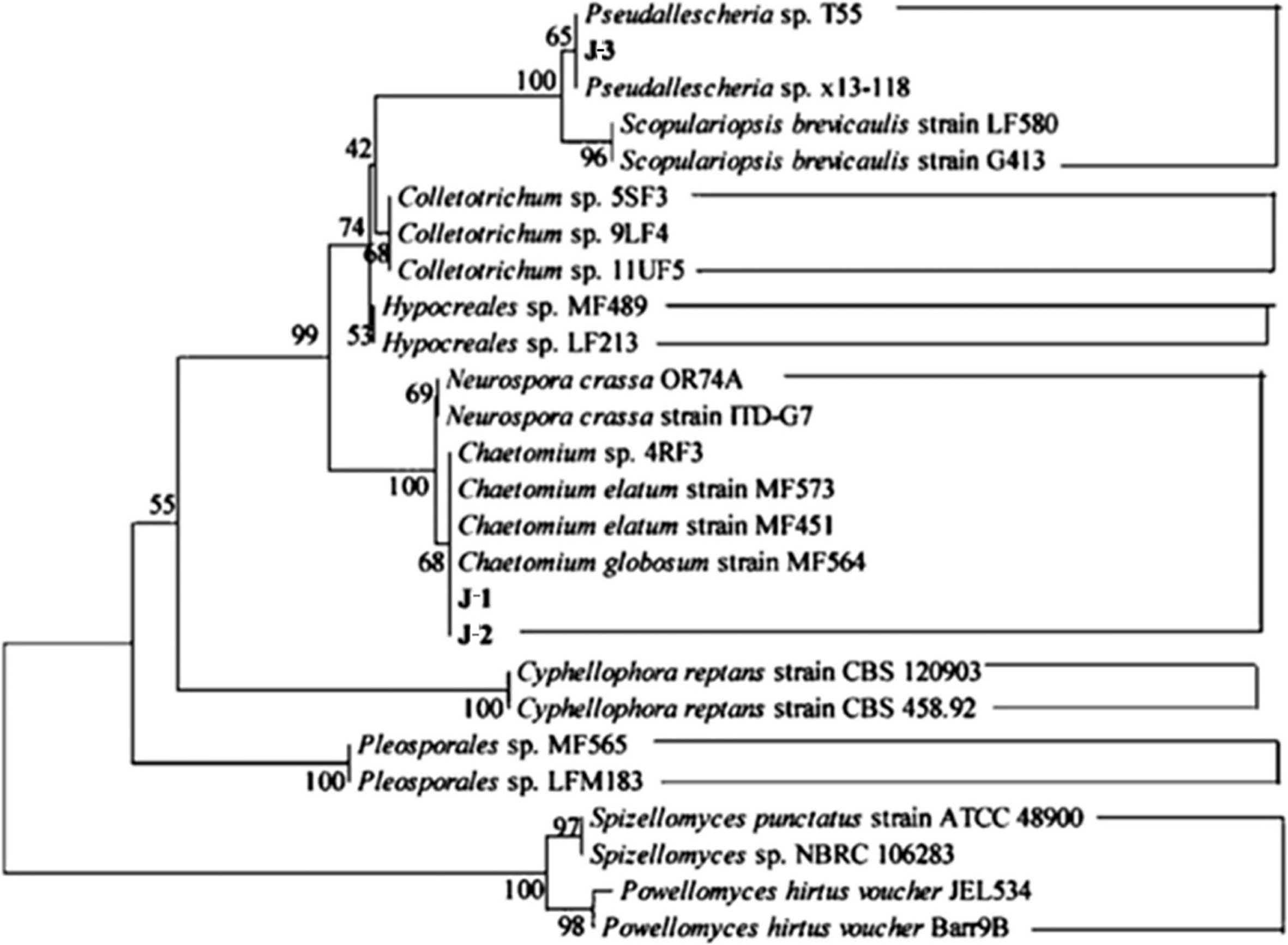
MTT results showed that after treatment for 24 h, there were no significant differences in OD values among the 5 groups (
Promotion effect of SMEFGB on apoptosis of Hela cells
Flow cytometry results showed that after treatment for 48 h, the apoptosis rates of Hela cells in strain J-1, strain J-2, strain J-3 and CY groups were significantly higher than those in control group, respectively (
Effect of SMEFGB on the apoptosis of Hela cells. 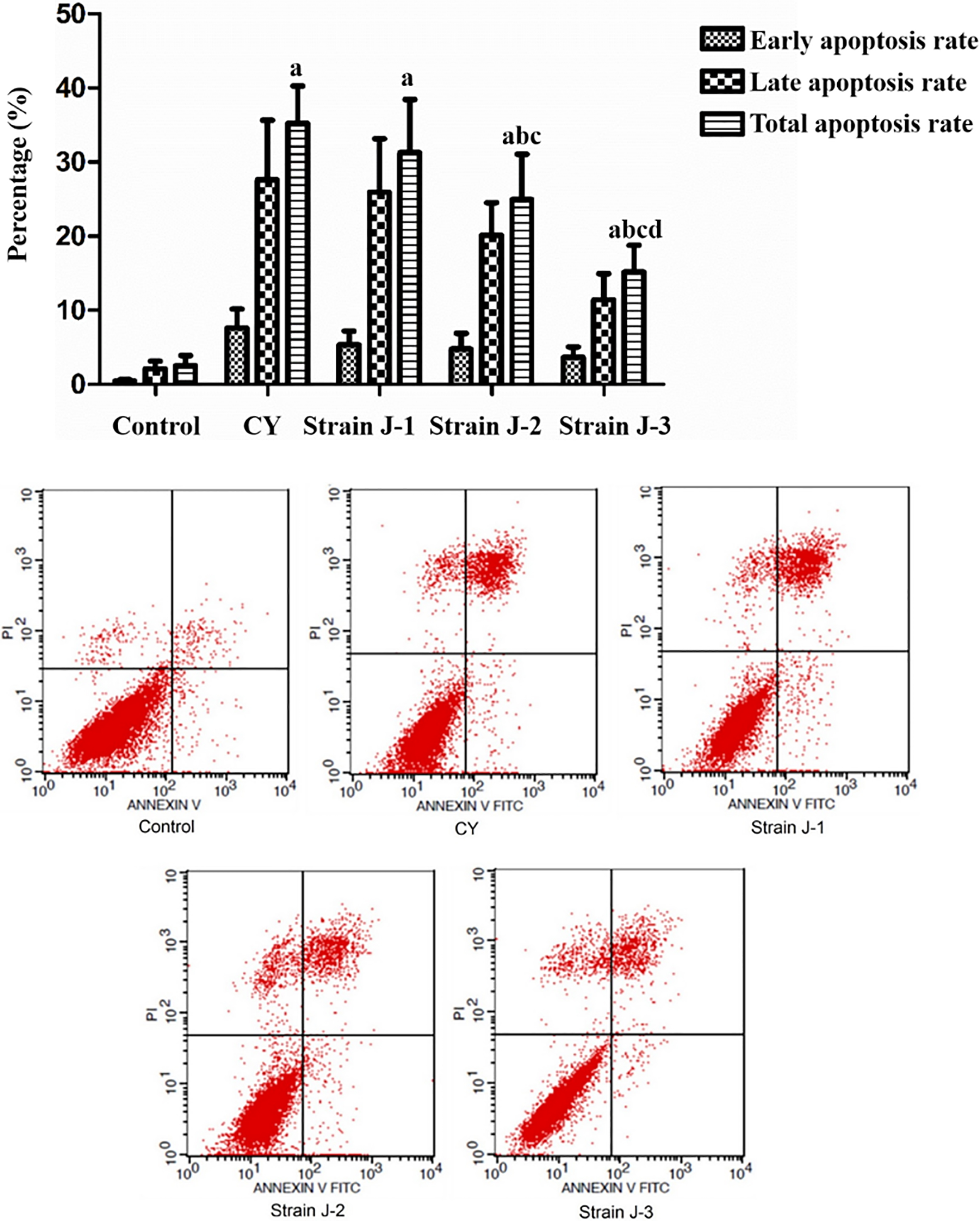
As shown in Fig. 5, SMEFGB could inhibit the migration of Hela cells in vitro. After treatment for 48 h, the scratch migration rates of Hela cells in CY, strain J-1, strain J-2 and strain J-3 groups were 32.20
Effect of SMEFGB on migration of Hela cells. 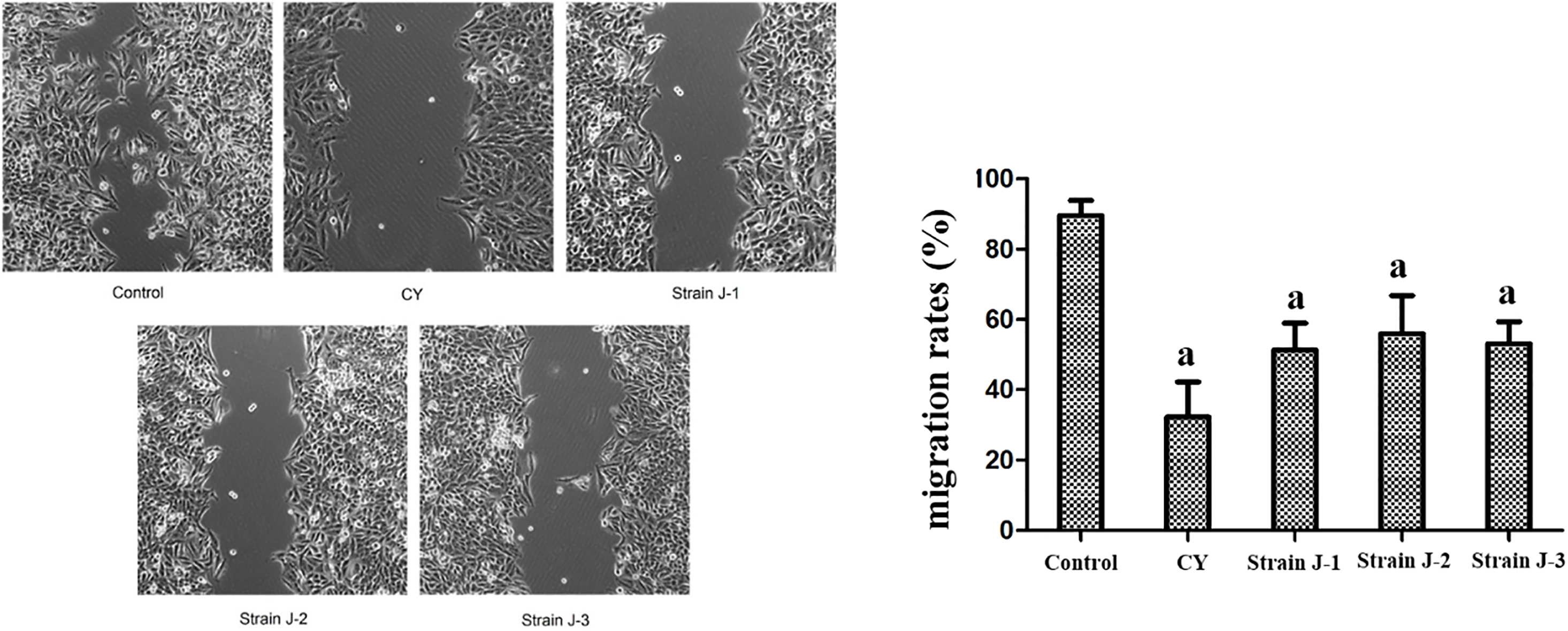
Effect of SMEFGB on growth of Hela implanted tumor in mice. 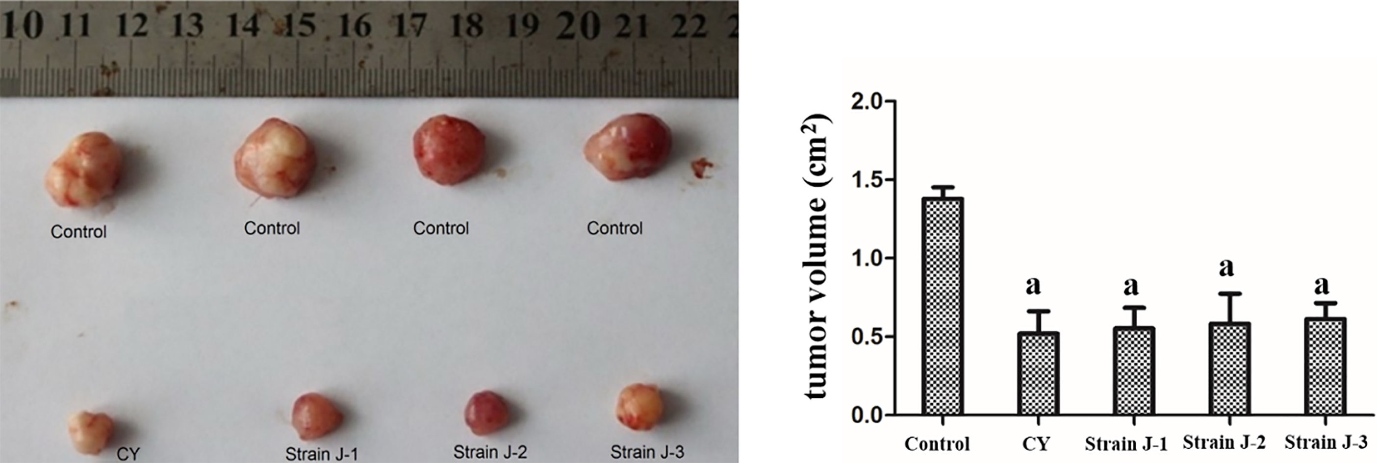
After subcutaneous inoculation of Hela cells, a subcutaneous tumor with a diameter of about 5–10 mm began to appear in mice. The xenograft was removed from the body. The tumor was completely wrapped by the envelop, with clear boundary of the surrounding tissues (Fig. 6). Compared with the control group, the tumor volume in strain J-1, strain J-2, strain J-3 and CY groups was significantly decreased, respectively (
Effect of SMEFGB on growth of Hela implanted tumor in mice
Effect of SMEFGB on growth of Hela implanted tumor in mice
Cervical cancer is currently the only cancer with known cause, which is caused by human papillomavirus (HPV) infection. If the virus continues to infect the cervix for more than two years, it will cause cervical precancerous lesions. If the infected person does not get timely treatment at this time, it will develop into cervical cancer in 5 to 10 years. Therefore, early detection and early treatment are effective strategies for treating cervical cancer. In addition, it has been reported that vaccination and HPV screening can prevent cervical cancer. However, because of poverty and backward ideas, many people are reluctant to spend much money to prevent cervical cancer.
There are more than 50 commonly used antitumor drugs in clinic, but most drugs can only alleviate the disease, not achieve the goal of complete cure. Therefore, the research and development of new anti-cancer drugs have always been the main aspect of drug research fields [9]. In recent years, the epiphytic microbes of animals and plants have been considered as an important source of natural active products. In the medical field, more and more studies have been reported on plant endophytic fungi producing antitumor active substances [10, 11, 12]. Over the past 10 years, more than 100 compounds with cytotoxic activity from endophytic fungi have been reported [13]. The taxol from the taxus is an efficient anticancer drug. As found in 1970s, an endophyte from the taxus plants can produce taxol. This has brought the upsurge of studying the endophyte [14]. However, until now the research level on endophytic fungi is not very deep, and the action mechanism is not very clear. With the improvement and deepening of the technical level and theoretical research, the endophytic fungi will play a great role in the development of drugs and the protection of the natural environment.
Endophytic fungi of Ginkgo biloba exist in different parts of root, stem, leaf, seed, bark tissue of Ginkgo biloba. The diversity of endophytic fungi of Ginkgo biloba has great difference based on origin place, part, plant growth condition and separation time, and the dominant bacteria also have great difference. Endophytic fungi from Ginkgo biloba has extensive antibacterial activity, including anti-plant pathogenic fungi and anti-human pathogens. Guo et al. [15] have isolated 522 strains of endophytic fungi from the Ginkgo biloba Yangli, Shaanxi, China, and determined their effect on 7 kinds of plant pathogenic bacteria. Our results showed that 50.7% strains of endophytic fungi had the antimicrobial activity, including 12 strains with very obvious antibacterial effect, especially for wheat red mildew and Fusarium oxysporum. Gao et al. [16] have found that 8 strains of endophytic fungi from the Ginkgo biloba in Wuhu, Anhui, China have strong DPPH radical scavenging capacity, of which the effects of 4 strains are significantly higher than those of other strains, with scavenging rate of 96.68%, 96.09%, 85.32% and 84.48%, respectively. Miao et al. [17] have isolated 19 strains of endophytic fungi in Ginkgo biloba in Fuyang, Anhui, China and determined their antitumor effects. Our results show that, 3 strains of endophytic fungi have inhibitory effects on the growth of human esophageal cancer EC109 cells. The active components of were mainly distributed in the fermentation broth, and the most active strain was YX. The IC50 of extract from the fermentation broth on human esophageal cancer EC109 cells, human nasopharyngeal carcinoma HONE1 cells and human cervical carcinoma HeLa cells are 18.3, 3.6 and 6.5 g/mL, respectively. In this study, we successfully isolated 9 stains of endophytic fungus from the leaves of Ginkgo biloba. In addition, among them, strains J-1, J-2 and J-3 could produce the podophyllotoxin compound. Similar to others’ findings, our results showed that SMEFGB can significantly prompt HeLa cell apoptosis and inhibit HeLa cell proliferation and migration in vitro experiment.
The metabolites of endophytic bacteria include alkaloids, quinones, flavonoids, phenylpropanoids, terpenoids, steroids, and peptides, etc. Qin et al. [15] have isolated 4 compounds from ethyl acetate extract of Ginkgo biloba endophytic fungi Chaetomium globosum. It has been found that these compounds have strong cytotoxic activity. Li et al. [16] have isolated 19 strains of endophytic fungi and 3 new alkaloids from the methanol extract of Ginkgo biloba endophytic fungi Chaetomium globosum. These alkaloids have strong inhibitor activity on hepatoma HepG-2 cells. Yan et al. [17] have obtained the secondary metabolites of Ginkgo biloba endophytic fungi Aspergilus sp. and find that these metabolites can inhibit the growth of human nasopharyngeal carcinoma cell KB cells, human gastric carcinoma cell SGC-7901 cells, human colon cancer cell SW1116 cells and human lung cancer cells A549 cells. In vivo antitumor studies of endophytic fungi in Ginkgo biloba have not been reported at home and abroad. In this study, we inoculated nude mice with 0.2 ml HeLa single cell suspension and treated them with strains. The in vivo results showed that SMEFGB notably inhibited the growth of tumor transplanted with HeLa cells in nude mice.
In conclusion, SMEFGB were successfully isolated and identified in our study. The in vitro and in vivo experiments showed that SMEFGB had obvious inhibitory effects on human cervical cancer HeLa cells. Our study has provided a practical basis for further development of Ginkgo biloba resources and natural anti-tumor substances. However, the effects of SMEFGB on other tumor cells and the related mechanisms need to be further studied.
Footnotes
Acknowledgments
We acknowledge to the Shandong Provincial Natural Science Foundation for funding this research (Grant ZR2017BH081). The funders had no role in study design, data collection and analysis, decision to publish, or preparation of the manuscript.
Conflict of interest
The authors declare that they have no conflicts of interest.