
Abstract
BACKGROUND:
Transcription factors are commonly deregulated in various cancers. Here, we evaluated role of ELF3 in pathogenesis of bladder carcinoma (BCa).
MATERIALS AND METHODS:
We confirmed ELF3 expression pattern in BCa cell lines using western blot; and in different grades of tumors using Immunohistochemistry. Cell invasion assay was employed to demonstrate potential role of ELF3 in EMT.
RESULTS AND CONCLUSION:
ELF3 showed selective expression in low-grade cell lines and tumor tissues. Overexpression of ELF3 in mesenchymal cell line UMUC3 resulted in reduced invasion and decreased expression of mesenchymal markers. We observed association of low ELF3 expression with increased risk and overall poor survival using publicly available data. ELF3-modulated reversal of EMT might be a useful strategy in the treatment of bladder cancer.
Introduction
Bladder carcinoma is incredibly heterogeneous, comprises multiple disease entities with distinct molecular features and biological characteristics [1]. Low-grade non-muscle-invasive (NMI) tumors have high recurrence frequency of 70% and 20% of them progress to muscle invasive tumors (MI) [2]. Muscle-invasive tumors are generally diagnosed de novo and are involved in metastasis. MI and NMI are two distinct tumors with different clinico-pathological characteristics. NMI carcinomas develop via epithelial hyperplasia whereas MI develop via flat dysplasia and carcinoma in situ (CIS) [1]. The risk of progression of NMI to MI after 5 years ranges from 6% to 45% [3] with less than 15% of 5 year survival rate [4]. Treatments have not advanced beyond cisplatin or cisplatin containing combination chemotherapies for decades [5]. A detailed understanding of bladder carcinoma pathogenesis is an unmet need.
Various studies suggest multifocal occurrence is the characteristic feature of bladder carcinoma. At molecular levels, epithelial to mesenchymal transition (EMT) could play a major role in progression of NMI to MI [6, 7, 8, 9]. This transition in cell phenotype is also associated with aggressiveness, recurrence, and metastases of tumors [10]. However, our understanding about the disease pathogenesis is still not evolved comprehensively. EMT is known to be governed by several transcription factors, such as SNAIL, SLUG, ZEB1, ZEB2, and TWIST among others, that transcriptionally repress epithelial markers [11]. These molecules are often referred as EMT transcription factors (EMT-TFs).
Among families of transcription factors, there are increasing evidence for E-twenty six (ETS) transcription factor family members to regulate EMT in cancer [12, 13, 14, 15]. ETS family members usually expressed by epithelial cells and are largely implicated in matrix metalloproteinase (MMP) regulation [16]. ETS-related transcription factors are also involved in cell proliferation, adhesion, motility/migration, cell survival, invasion, extravasation, micro-metastasis, establishment and maintenance of distant site metastasis and angiogenesis [15, 16, 17]. Member of ETS family, ELF5 (E74 like ETS transcription factor 5) is shown to directly suppress EMT by repressing the transcription of SLUG [12]. ETV1, another member of ETS family also acts to promotes snail expression to induce EMT in gastric cancer [18]. Likewise, ELF3 (E74 like ETS transcription factor 3) also known by ESE-1, EPR-1, ESX, and ERT a member of ETS family is restricted to epithelial tissue expression [19, 20]. Higher expression of ELF3 was reported in epithelial rich tissues such as colon, small intestine and negligible expression in epithelial poor tissues such as brain, heart, spleen, thymus and skeletal muscles [19, 20]. Furthermore, expression of ELF3 is negligible in non-epithelial origin cells like lymphocytes, monocytes, and endothelial cells [20].
Mutation profile studies have identified recurrent anomalies in multiple genes and pathways that are potential key drivers of bladder carcinoma [21, 22, 23]. A study by Yachida et al., identified ELF3 as significantly mutated driver gene in ampullary carcinoma. The mutations were also significantly correlated with loss of ELF3 expression [24]. A large-scale genomics study from The Cancer Genome Atlas (TCGA) identified 58 genes as significantly mutated across 412 muscle invasive bladder carcinoma [25]. ELF3 was reported with frequency of 12% non-synonymous mutation in these tumors. However, the prognostic and functional significance of ELF3 are yet to be explored thoroughly in bladder carcinoma. Here we demonstrate the inverse correlation of ELF3 expression with EMT dynamics along with its association with overall survival. We also studied the effect of ELF3 overexpression on bladder carcinoma invasiveness using cell based invasion assay.
Methods
Cell culture
Five bladder carcinoma cell lines UMUC3, J82, SW780, RT112, T24 were acquired from ATCC and cultured in Dulbecco’s modified eagle medium supplemented with 10% fetal bovine serum, 1% sodium pyruvate and 1% penicillin-streptomycin mixture. Cell lines were grown and maintained in humidified 5% CO
Transient transfection
Routinely maintained UMUC3 cells were transfected by piRES-puro-ELF3 (Addgene cat.no. 25728) using X-tremeGENE HP DNA Transfection reagent, Roche, as per manufacture instructions. For western blotting, cells were harvested 72 hours after transfection. For Invasion assay, cells were seeded 24 hours post transfection into boyden chamber.
Cell invasion assay
The invasive property of UMUC3 was measured using the BD BioCoat™ Matrigel™ Invasion guidelines. Briefly, boyden chamber inserts (Thermo Fisher Scientific, Waltham, MA, USA) were coated with 20
Western blotting
Whole cell extracts from 70–80% confluent cells were prepared using modified RIPA lysis buffer (Merck Millipore, Billerica, MA, USA) containing protease inhibitors (Roche, Indianapolis, IN, USA) and phosphatase inhibitors (Thermo Scientific, Bremen, Germany). Western blot analysis was performed on 10% SDS-PAGE using 30
Immunohistochemistry
Bladder carcinoma formalin fixed paraffin embedded tissue sections were obtained following institutional review board approval and informed consent from Kidwai institute of molecular oncology (KMIO/ MEC/011/24. November. 2016). The sections were deparaffinised and antigen retrieval was carried out using heat-induced epitope retrieval by incubating them for 20 minutes in antigen retrieval buffer (0.01 M Trisodium citrate buffer, pH 6). The quenching of endogenous peroxidases was done by using a blocking solution followed by washes with wash buffer (PBS with 0.05% Tween-20). The sections were incubated with primary antibody overnight at 4
Immunofluorescent staining
Approximately 3
Computational analysis
Normalized intensities of twenty-six ETS family members were compiled from Earl et al., 2015 for sixteen cell lines [26]. These sixteen cell lines were stratified into group of epithelial cell lines (Epi) (
We also analyzed expression of ELF3 using web-based platforms such as UALCAN [29] for Kaplan-Meier overall survival analysis to corroborate our cell-line based findings with patient data-sets from The Cancer Genome Atlas (TCGA).
Computation of EMT score
Meta-cohort of bladder carcinoma dataset Affy- metrix U133A platform was compiled in Tan et al., 2014 [27]. Briefly, bladder carcinoma from GSE31684, GSE7476, and GSE5287 were downloaded from GEO. The data were RMA-normalized and combined using ComBat [30]. Clinical and processed RNA-seq data of TCGA bladder carcinoma cohort was downloaded from GDAC (version 2016_01_28) (Broad2016). The FPKM values of ELF3 were extracted for downstream analysis.
Expression profile of ETS family in bladder carcinoma cell lines. (A) Heatmap representing normalized intensities of ETS family from epithelial (RT4, UMUC9, HT1197, HT1376, BC16.1, CUBIII, SCABER, UMUC6, PSI, 5637, KU7), mixed (HU456 and KK47) and mesenchymal (253JBV, T24, TCCSUP, J82, UMUC13, UMUC3) tumor derived cell lines reported using microarray analysis. (B) Relatively low expression of ELF3 in mesenchymal compared to epithelial tumor derived cell lines.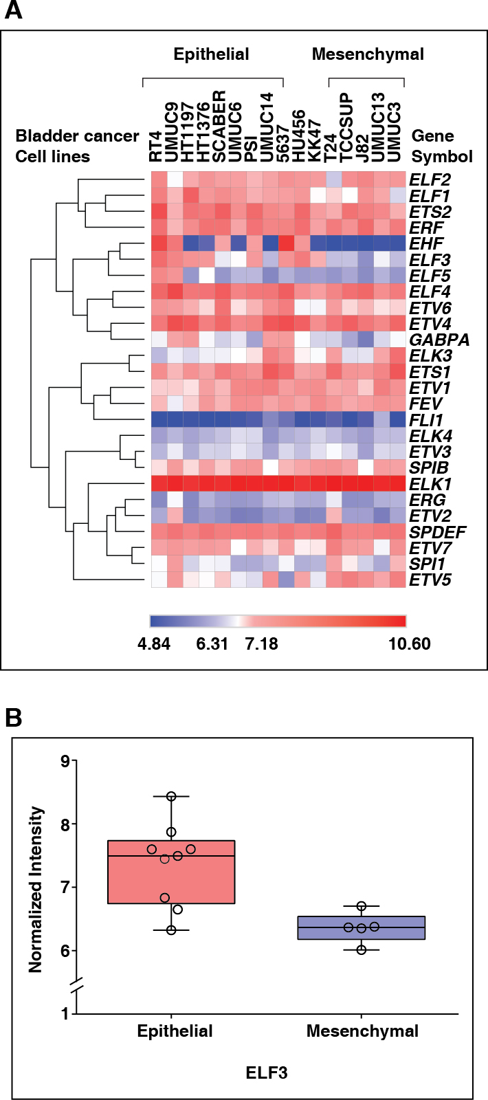
Subcellular localization of ELF3 in bladder carcinoma cell lines. Immunofluorescence staining showed cytoplasmic and nuclear localization of ELF3 in (A) SW780 (B) RT112; and cytoplasmic in (C) T24, (D) J82. (E) Negative control.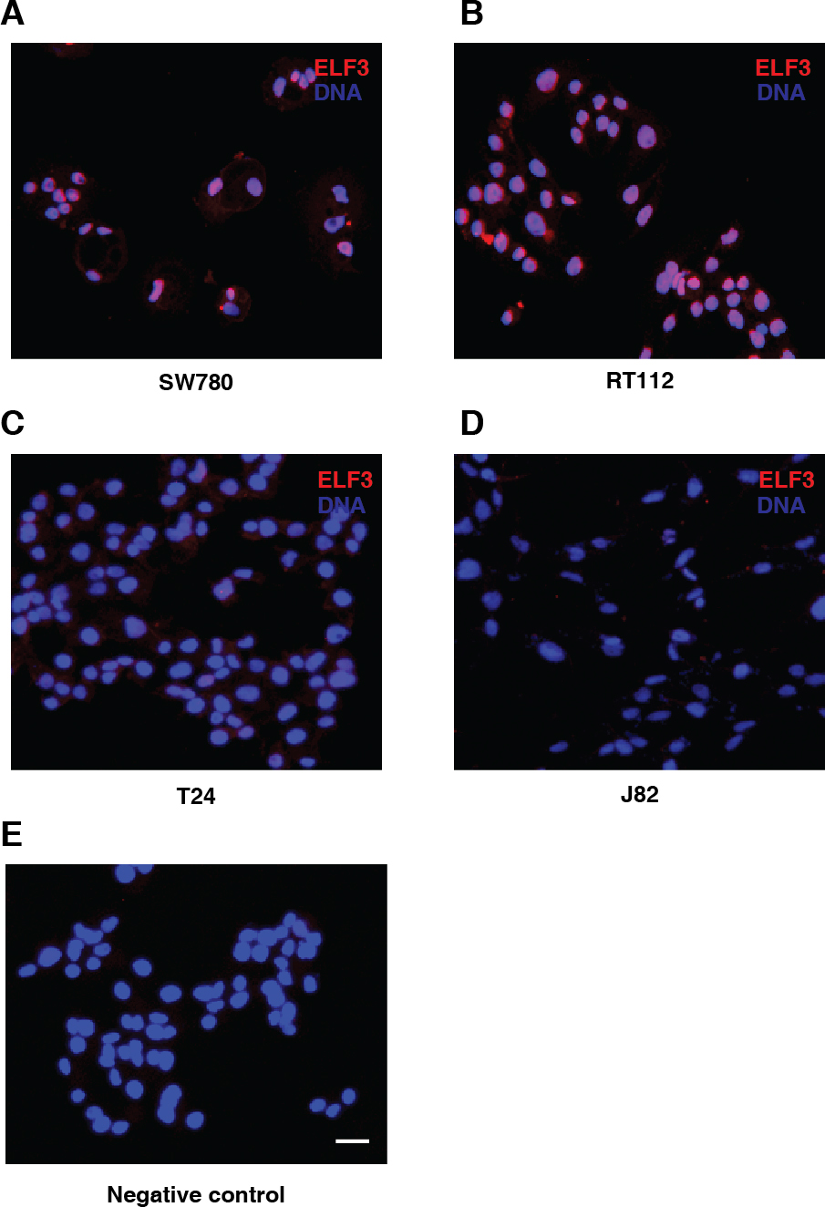
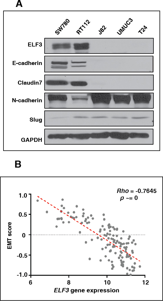
Association of ELF3 expression with EMT signature (A) Western blot analysis depicting higher expression of E-cadherin and Claudin7 in SW780 and RT112; and higher expression of slug in J82, UMUC3 and T24 (B) Spearman’s inverse correlation of EMT signature with ELF3 expression in bladder carcinoma patient data procured from Tan et al.
Overexpression of ELF3 reduces cell invasion in-vitro (A) Transient transfection of ELF3 in UMUC3 lead to decreased invasiveness (B, C) Western blot and image J analysis indicates lower expression of mesenchymal markers and unchanged expression of epithelial markers.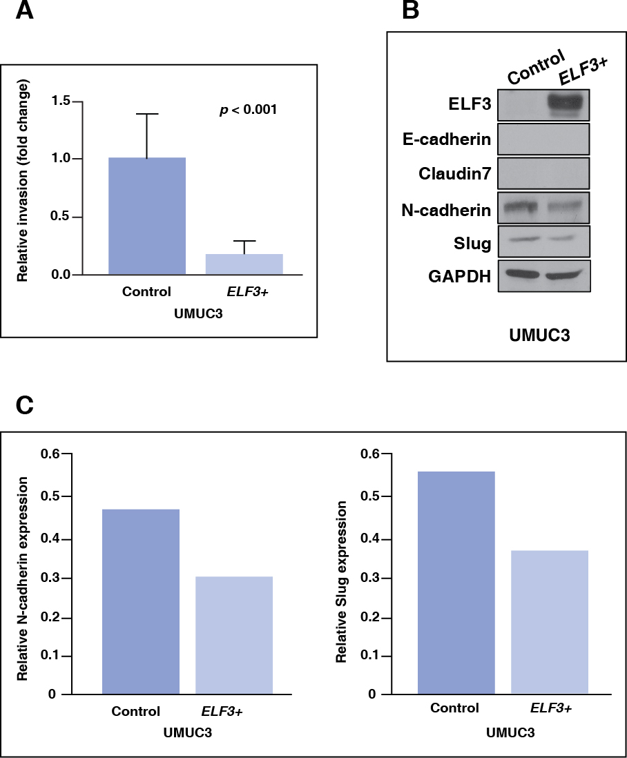
Expression pattern of ELF3 and ETS family members
EMT score computed by Tan et al., 2014 classified RT4, UMUC9, HT1197, HT1376, BC16.1, CUBIII, SCABER, UMUC6, PSI, 5637, KU7, as epithelial cell lines [27]. HU456 and KK47 were stratified as cell lines showing both epithelial and mesenchymal characteristics [27]. Whereas 253JBV, T24, TCCSUP, J82, UMUC13 and UMUC3 showed most mesenchymal characteristics. We further utilized same classification to assess expression pattern of ETS family members from microarray data provided by Earl et al., 2015 across 40 bladder carcinoma cell lines (Supplementary table S1) [26]. We observed that ELF2, ELF1, ETS2, ERF, ELF4, ETV4, ETS1, SPDEF, ELK1, FEV, ETV1 were highly expressed genes across cell lines. Whereas FL1, ELK4, ETV3, ERG and ETV2 showed relatively low expression across cell lines (Fig. 1A). We observed ELF3 expression was relatively low in cell lines characterized as mesenchymal (T24, TCCSUP, J82, UMUC13 and UMUC3) and high in cell lines characterized as epithelial (RT4, UMUC9, HT1197, HT1376, PSI and 5637) (Fig. 1B). Other epithelial cell lines SCABER and UMUC6 presented moderate expression of ELF3 whereas UMUC14 exhibited low expression of ELF3. ELF3 expression difference was assessed using non-parametric unpaired Mann-Whitney test and we observe that it was significantly higher in epithelial cell lines compared to mesenchymal cell lines with
ELF3 is down regulated in cell lines derived from high grade tumors
To confirm the expression pattern of ELF3, western blot was performed across 5 bladder carcinoma cell lines. These cell lines were established from low-grade tumor to high-grade tumors (Supplementary Table S2). As shown in Fig. 2A–E and Fig. 3A, cell lines derived from low grade tumor, SW780 and RT112 expressed ELF3 while other cell lines T24 and J82 derived from high grade tumors and also characterized as most mesenchymal cell lines along with UMUC3 showed negligible expression of ELF3. We also observed varied localization pattern of ELF3 with respect to grade of the cell lines. ELF3 was localized in nucleus and cytoplasm in SW780 and RT112 whereas in T24 and J82, ELF3 was localized in negligible amounts in cytoplasm and absent in nucleus.
ELF3 expression is associated with epithelial to mesenchymal transition in bladder carcinoma cells
To study the functional role of ELF3 in EMT process, western blot analysis was performed in bladder carcinoma cell lines against the expression pattern of various EMT markers. We observed low expression of ELF3 correlated with low expression of epithelial markers E-cadherin and Claudin7 in high grade bladder carcinoma cell lines, J82, T24 and mesenchymal cell line UMUC3. Whereas expression of mesenchymal markers such as N-cadherin and SLUG were higher in high grade compared to low grade bladder carcinoma cell lines (Fig. 3A). Expression of ELF3 might be involved in negative regulation of EMT in bladder carcinoma. To further validate our findings, we compared expression of ELF3 with the generic EMT signature derived from bladder carcinoma patient data sets as described by Tan et al., 2014 [27]. This scoring method was developed to quantitatively estimate the EMT phenotype across clinical samples as well as cell lines using transcriptomics. Based on this in-silico analysis, we identified that low ELF3 expression significantly correlated (Rho
Over expression of ELF3 decreases invasion in mesenchymal bladder carcinoma cell line UMUC3
Expression of ELF3 was downregulated across high-grade bladder carcinoma cell lines. We further sought to identify whether ELF3 overexpression can potentially revert the invasiveness in UMUC3 cell line. We observed significant decreased invasiveness of UMUC3 (Fig. 4A). ELF3 over expression also showed marginal decrease in expression of mesenchymal mar- kers like N-cadherin and SLUG, however there was no change in the expression of epithelial markers (Fig. 4B) as confirmed by western blot.
Immunohistochemistry revealed higher expression of ELF3 in low grade compared to high grade tumors
ELF3 expression in different grades of urothelial tumors. Immunohistochemical staining show comparable expression of ELF3 in (A) adjacent normal and (B) tumor of low grade urothelial carcinoma; whereas decreased expression from (C) adjacent normal to (D) tumor of high grade urothelial carcinoma. (E) IHC image depicts decreased expression of ELF3 in mesenchymal cells compared to epithelial cells (Blue arrows: mesenchymal cells. Red arrows: epithelial cells).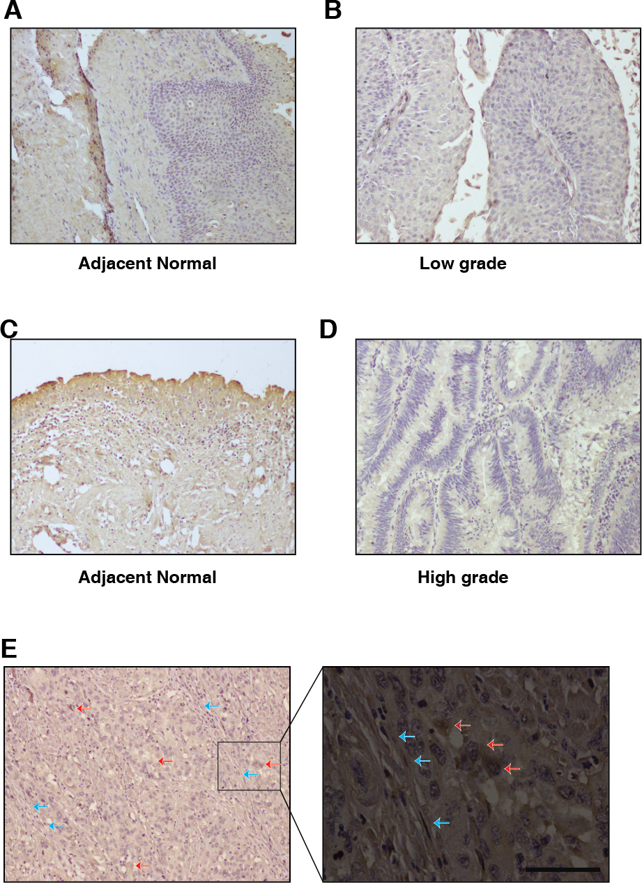
To further investigate the expression pattern of ELF3, we performed immunohistochemical staining on bladder tumors. Patient presented with low grade carcinoma showed unchanged ELF3 expression in tumor compared to adjacent normal (Fig. 5A and B). Whereas patient presented with high grade carcinoma showed decreased level of ELF3 expression in tumor compared to adjacent normal (Fig. 5C and D). We observed considerable difference in the ELF3 expression between low grade and high grade tumors. In addition, we also observed decreased ELF3 expression in mesenchymal cells compared to the epithelial cells in the high grade tumor (Fig. 5E). Our result suggests that ELF3 expression is higher in low grade compared to high grade tumors.
To assess potential risk associated with ELF3 expression in bladder carcinoma we have used five published studies hosted on an online biomarker validation tool SurvExpress [28]. These datasets were classified into low risk and high-risk groups, and
To investigate the association between ELF3 expression and overall survival, we analysed publicly available datasets using Kaplan-Meier method using UALCAN [29]. Survival analysis revealed patients with low ELF3 expression (
Discussion
ELF3 is an epithelial-restricted member of the ETS transcription factor family, and involved in wide range of cellular processes such as differentiation, and inflammatory response [29, 31]. It is one of the critical regulators involved in urothelial cyto-differentiation in normal human urothelial (NHU) cell line [31]. Expression of ELF3 is reported in nucleus as well as in cytoplasm [32]. ELF3 positively regulates expression of TGF
We interrogated ELF3 expression in TCGA datasets using web-based resource UALCAN to corroborate our findings from cell lines in clinical samples. UALCAN is implemented in PERL-CGI to enable web-based gene expression analysis in various stages and molecular subtypes of 31 different cancers [29]. We observed there is lower expression of ELF3 in basal/ squamous (
To further understand the role of ELF3 in EMT, we investigated the expression of ELF3 in bladder carcinoma cell lines. We observed the level of ELF3 was specifically low in mesenchymal cells. Immunohistochemical analysis in low grade and high grade bladder tumors suggests a decrease in the expression of ELF3 from low grade to high grade. In addition, the expression of epithelial markers were low in high grade cell lines and inversely correlated with the expression of mesenchymal markers. It suggests that ELF3 might be involved in regulation of EMT. In this study, we identified similar expression pattern of ELF3 and Claudin7 as reported by Kohno et al., which suggests ELF3 is indeed involved in the formation of epithelial structure in bladder cancer [43]. We have further checked the expression of ZEB1 in bladder cancer cell lines. It has been shown that ELF3 regulates the expression of ZEBs contributing to the EMT phenotype [41]. In our study, we observed that RT112 with high expression of ELF3 showed low expression of ZEB1. While J82, UMUC3 and T24 having low expression of ELF3 showed high expression of ZEB1 (Supplementary Fig. 4).
Various studies have indicated localization of ELF3 affects its function. ELF3 mediates the translocation of snail and
We further sought to understand the process of bladder carcinoma progression. We checked the gain of function of ELF3 in bladder cell lines where negligible expression of ELF3 was observed. We found that upon ELF3 transfection in mesenchymal cell line UMUC3, the invasive ability of the cell line was decreased significantly. Transient transfection also showed a slight decrease in expression of mesenchymal markers N-cadherin and Slug. Subsequently we also checked the effect of ELF3 overexpression and EMT modulation in J82 and T24; however, these cell lines were very difficult to transfect and we could not get more than 10% transfection efficiency.
We observed that low expression of ELF3 correlated significantly with poor survival in bladder carcinoma patients. Our meta-analysis also suggested that patients are at higher risk with low ELF3 expression in 4 out of 5 bladder carcinoma published data-sets as per SurvExpress [28]. Low expression of ELF3 is also reported to have effect on patient’s overall survival and ELF3 over expression could also be used to develop novel therapeutic regimens and could be implicated to improve bladder carcinoma patient survival. Our study delineates the molecular mechanism of ELF3 mediated EMT in aggressive bladder tumors.
Conclusions
Epithelial to mesenchymal transition phenomenon majorly contributes to metastasis and cancer progression, however the molecular mechanisms underlying this transition remains to be fully understood. Our study revealed the role of ELF3 in the cellular plasticity of bladder carcinoma. This study for the first time demonstrates that expression of ELF3 negatively correlates with expression of epithelial to mesenchymal transition markers and ELF3-modulated reversal of EMT can be a valuable strategy in treatment of bladder carcinoma.
Footnotes
Acknowledgments
PK would like to acknowledge funding support from Ramanujan Fellowship awarded by Department of Science and Technology (DST) Government of India (SB/S2/RJN-077/2015). KG is a recipient of Senior Research Fellowship from University Grants Commission (UGC), Government of India. KP is a recipient of Senior Research Fellowship from Council of Scientific Industrial Research, Government of India. In addition, the authors thank the support from Dr. Ramray Bhat, Department of Molecular Reproduction, Development and Genetics, Indian Institute of Sciences, Bangalore for epifluorescence imaging facility.
Supplementary data
The supplementary files are available to download from