
Abstract
BACKGROUND:
Reseptor tyrosine kinases (cMET and EGFR) are important in lung cancer targeted therapy. We believe if we can use them as markers for clinicians to help decide the diagnosis of lung cancer. This parameter will be important in serum samples of patients with lung cancer diagnosis and treatment. The aim of this study is aimed to evaluate the clinical utility of serum protein and circulating mRNA of cMET and HGF in lung cancer patients. We also analyzed the correlation of mRNA expression with clinicopathologic parameters.
METHODS:
We performed enzyme-linked immunosorbent assay (ELISA) to measure and compare serum protein and circulating mRNA of cMET and HGF levels in peripheral blood from 60 lung cancer patients and 40 healthy control group.
RESULTS:
We found that both protein and gene expression levels of serum c-MET, HGF and EGFR were significantly higher in patients with lung cancer than control group. There was no association between HGF, cMET, EGF, EGFR (both protein and gene) expression levels with age, gender, smoking habit, COPD, pathological types or tumor size, stage, metastatic-non metastatic adenocarcinoma-squamous carcinoma, SCLC-NSCLC. As a result of ROC analysis, serum cMET (AUC: 0.892) and HGF protein (AUC: 0.784) were diagnosed in lung cancer patients (Fig. 1). The AUC values of serum EGF and EGFR proteins were calculated to be 0.631 and 0.692, respectively.
CONCLUSION:
To our knowledge this is the first study comparing the levels of protein and mRNA in the serum material of HGF, c-MET, EGF and EGFR parameters in lung cancer patients’ blood samples. Further prospective studies with more participants for better understanding of mechanism and effect for HGF and c-MET inhibitors in lung cancer will help us to identify of these biomarkers role for guiding us to sellect individualized itargeted therapies.
Introduction
Lung cancer is the second most commonly diagnosed cancer worldwide. It is also the leading cause of cancer mortality among men and women. Despite developments in both diagnosis and therapeutic strategies of lung cancer, 5 year survival rate in tumors that are not mutation-driven still doesn’t exceed 20% [1].
Because of its high frequency and high mortality rate, early detection is exteremely important in order to improve the outcome. Lung cancer screening has been a trend debated topic since 1990’s. Many recent studies have focused on the development of a specific, sensitive, convenient and non-invasive techniques, such as circulating biomarkers to detect lung cancer at an earlier stage. Recently, the presence of circulating tumor-specific nucleicacids (cNA) has been validated in plasma, serum, and other body fluids from different type of cancer patients and they have been proposed as a noninvasive source of tumor-derived information.
In addition to DNA, circulating RNA is detectable through its ability escaping from degradation in the blood [2]. This stability is partially explained by the discovery that the circulating RNA is included within small membrane vesicles, i.e. exosomes, which protect the RNA from ribonucleases (RNases) [3]. As deregulated RNA expression is an early event in tumorigenesis and tumor progression, the measurement of circulating RNA might also be useful for early cancer detection and prognosis. Altough there are limited data on the clinical role of cRNA in lung cancer, recently, some studies have been investigated the role of circulating mRNAs of cMET HGF and EGFR EGF in diagnostic and prognostic evaluation of lung cancer.
The aim of this study is aimed to evaluate the clinical utility of serum protein and circulating mRNA of cMET and HGF in lung cancer patients. We also analyzed the correlation of mRNA expression with clinicopathologic parameters.
Material and methods
Sixty patients who were diagnosed with lung cancer and treated at Istanbul University Oncology Institute between January 2015 to January 2016 were included in this retrospective study.
Serum serum protein and circulating mRNA of cMET and HGF levels were studied with ELISA in both the patient group and the control group which consisted of 20 healthy volunteers.
Our study on human materials has been approved by the Istanbul University Ethics Committee. The protocol was consistent with the Declaration of Helsinki (1989). Informed consent was obtained from all study participants.
Blood samples of the patients and control group were obtained by venipuncture and clotted at room temperature. The sera were collected following centrifugation and frozen immediately at
Measurement of serum cMET HGF EGFR EGF levels
A double-antibody sandwich ELISA was used to determine the level of Neural precursor cell expressed developmentally down-regulated protein cMET (Shanghaıyehua biological technology Co, Ltd) in the samples. Serum samples and standards were added to the wells, which were pre-coated with human cMET monoclonal antibody; streptavidin-horseradish peroxidase conjugate was added to form an immune complex and allowed to incubate at 37
Quantification of cMET/HGF EGFR/EGF mRNA in serum
For analyzing cMET and HGF EGFR and EGF specific mRNA in sera of the patients, circulating cell-free RNA was extracted from serum using a monophasic phenol and guanidine thiocyanate solution (Roche, Mannheim, Germany) according to the manufacturer’s protocol. Briefly, 200
The pellet containing RNA is washed with 75% alcohol, dried at RT, and dissolved in 20
cMET and HGF; EGFR and EGF gen expression in serum was quantified semiquantitatively using GAPDH as the internal control and SYBR Green (Roche Mannheim, Germany) as the fluorescence mol- ecule.
Real-time PCR was performed in the LightCycler 480 Instrument (Roche, Mannheim, Germany). The reaction conditions included a hot start of 10 minutes at 95
Patient characteristics and disease status
Patient characteristics and disease status
SPSS for Windows version 18.0 (SPSS Inc. Chicago, IL, USA) was employed for data analysis. Continuous variables were categorized using median values as cutoff point. Assessment of relationships, comparisons between various clinical/laboratory parameters were accomplished using Mann-Whitney U test and Kruskal-Wallis test for two and three groups, respectively. Spearman’s rank order correlation was used for correlation analysis.
Results
Table 1 summarizes the histopathological characteristics, demographic features of 60 lung cancer patients diagnosed with lung cancer who were treated at Istanbul University Oncology Institute From January 2015 to January 2016.
Levels of serum protein levels in the lung cancer patients and healthy controls
Levels of serum protein levels in the lung cancer patients and healthy controls
ROC curves of cMET and HGF.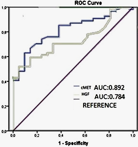
We analyzed circulating cell-free RNA to mesure cMET and HGF EGFR and EGF specific mRNA in the blood serum in 60 patients and 40 healthy controls to determine the role of cRNA as a tumor marker new anticancer targeted therapies for lung cancer. Serum protein HGF levels were significantly higher in patients with lung cancer (872.5 ng/L) as compared to healthy individuals (355.5 ng/L) (
Levels of serum gen expression levels in the lung cancer patients and healthy controls
ROC curves of EGF and EGFR.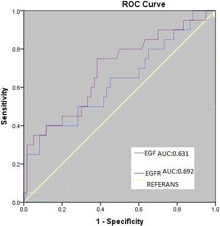
Results of comparisons between serum Cmet, HGF, EGFR, EGF protein and various demographic and disease
cMET-EGFR correlation.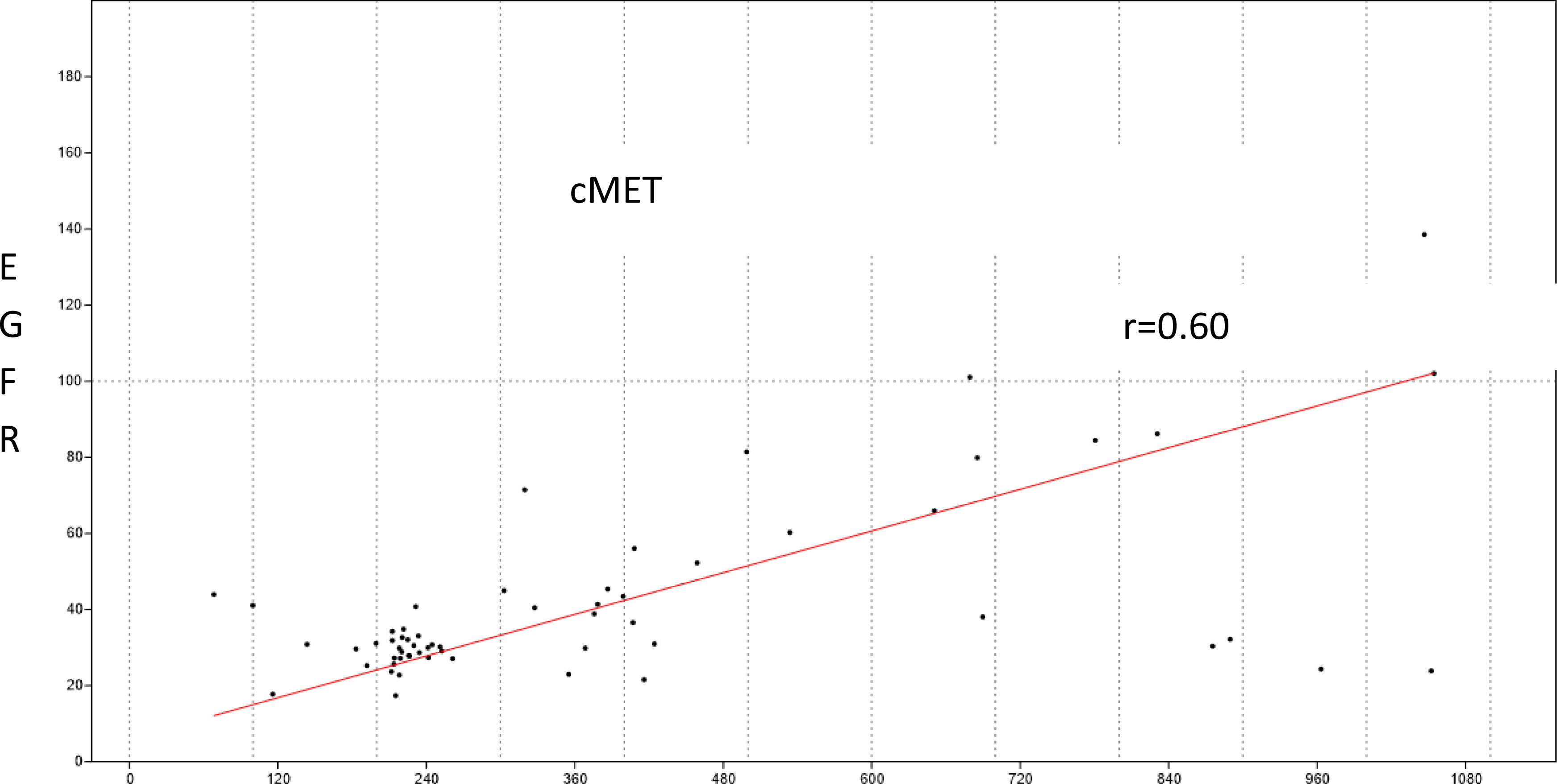
Results of comparisons between serum Cmet, HGF, EGFR, EGF gen and various demographic and disease
Serum protein EGF levels were not istatistically higher in patients with lung cancer than healthy control group (
HGF gene expression levels in lung cancer patients were higher than healthy control group and statistically significant (
The correlation between HGF, cMET, EGF, EGFR at both protein and gene expression levels and clinicopathological characteristics of the patient group is shown in Table 3. There was no association between HGF, cMET, EGF, EGFR (both protein and gene) expression levels with age, gender, smoking habit, COPD, pathological types or tumor size, stage, metastatic-non metastatic adenocarcinoma-squamous carcinoma, SCLC-NSCLC (Tables 4 and 5).
ROC analysis of each test was performed. AUC (Area under Curve) was calculated according to ROC curves. As a result of ROC analysis, serum cMET (AUC: 0.892) and HGF protein (AUC: 0.784) were diagnosed in lung cancer patients (Fig. 1). The AUC values of serum EGF and EGFR proteins were calculated to be 0.631 and 0.692, respectively (Fig. 2). According to AUC, the diagnostic value of cMET and HGF was higher than EGF and EGFR.
Oncoproteins that bind to proteins in the pathway to which protein kinase receptors associated with growth factors are attached block or modify the effects of growth factors. Among the important members of these oncoproteins are c-MET. Oncogenes such as c-MET lead to overproduction of growth factor genes and cause excessive release of the hepatocyte growth factor. Cancer occurs as a result of the altered growth signal [4].
Because of its early invasion and spread to distant organ potential, early detection is extremely important. In order to improve the outcome it is very important to detect these cells early in lung cancer. The fact that c-MET mediated tyrosine kinase signal transduction leads to an excessive increase in oncogenes; it is one of the most remarkable topics today. In recent years, a great deal of information has been obtained about the normal structural properties and biological functions of c-MET. c-MET and HGF have been shown to increase expression in many cancers, including NSCLC [5]. In our study, the expression levels of HGF and c-MET in lung cancer patients were also found to increase which is compatible with the literature.
c-MET pathway activation triggers a program similar to in vivo tumor metastasis that begins with increased cell motility, progressive cell invasion and angiogenesis enhancement by extracellular matrix degradation along with protease production. It is known that HGF changes the migration and invasion capacities of cancer cells by increasing the secretion/expression of cancer cells from matrix and endothelial adhesion and photolytic enzymes [6]. All of these features are thought to be important for invasive phenotype acquisition of the tumor [7].
The developments in molecular biology of lung cancer for the last 10 years and the use of specially targeted agents have been changed the treatment approach. The role of the EGFR receptor in the treatment of lung cancer has been investigated in the majority of EGFR inhibitors, given both the pathogenesis location of the EGF receptor in cancer biology and its role in tumor development [9]. EGFR inhibitor agents block the tyrosine kinase activity of EGFR, which is highly up-regulated in NSCLC, resulting in inhibition of cell activation and proliferation.
Masroor et al. found that EGFR gene expression was 13.5 fold higher in NSCLC than in normal controls and in healthy controls when they performed RT-PCR on serum mRNAs [8]. In this study, which is similar to our study as material and method used, statistical significance was found between NSCLC and healthy control (
Studies about targeted cytokine tyrosine kinase activity in lung cancer are ongoing. c-MET oncogene is a high affinity receptor for HGF. Genetic mutation and gene amplification in c-MET is very common in many cancer types. Over expression of c-MET has also been shown in cases of lung, stomach, liver, and colorectal cancer and is associated with poor prognosis [10]. HGF found in plasma and synthesized from platelets, plays a major role in liver regeneration. It is also detected at a very low level in the normal lung. It induces budding and branching in the embryonic lung. There are some studies showing increased expression of HGF in lung cancer. Consistent with previous data, in our study, we also found significant association with high HGF protein and gene expression levels in NSCLC patients group than control group (respectively 1163.3
Previous studies demonstrate significant correlation between c-MET and EGFR expressions [11]. In our study there is a significant correlation between c-MET and EGFR expressions in serum samples (Fig. 3,
Cheng et al. conducted a study with 45 lung cancer patients. They found that c-MET mRNA levels both in tumor tissue and peripheral blood were significantly high in lung cancer (
Blumenschein and friends did not find statistical significance between the histological sub parameters of serum HGF levels (
This suggests that HGF is a biomarker that can be used clinically in patients with lung cancer, even if histological types are different.
Expression of HGF and its receptor c-MET has been reported to be increased in lung, colon, breast, thyroid, renal carcinoma, melanoma and various sarcomas. Several studies have shown that HGF plays a role in tumor progression. The binding of HGF to c-MET results in the phosphorylation of intracellular tyrosine segments of c-MET. In general, HGF is involved in invasion and metastatic processes in the development and progression of many cancers through its receptor c-MET. While HGF has been reported to play a role in the early stages of cancer, investigations are still underway for reliable and robust evidence in this regard [14]. In the light of these findings, it is reported that C-MET may be one of the oncogenes controlling the metastasis progression of primary cancers. Invasion and metastasis processes of lung cancers are thought to be related to the activation of the motile vacuoles involved in the HGF/c-MET signaling pathway [15]. This hypothesis has guided us to plan our study.
Studies targeting growth factors in lung cancer treatments address the issue of establishing diagnostic or prognostic markers that depend on the amount of these molecules and blocking signal transduction molecules or signaling pathways activated by these signaling molecules in localized tissues [16]. In our study based on this prediction, we determined the relationship between gene expression and protein by determining both protein levels of EGFR/EGF and c-MET/HGF and mRNA levels of these two signaling pathways involved in lung cancer.
Genetic changes in the EGFR and c-MET genes play an important role in the development of lung cancer. Changes in these genes cause cancer to become more aggressive. We think that increasing the number of studies to determine how changes in EGFR/EGF and c-MET/HGF genes play a role in the development of cancer and determining the treatment strategies to be created will provide important contributions to the clinicians in the slowing or stopping cancer progression.
The presence of HGF molecules circulating freely in lung cancer patients in the selection of HGF/c-MET signaling pathway based target therapies showed that c-MET and HGF could be used as useful biomarkers. In our study, when ROC curves of each test were examined, c-MET (AUC: 0.892) and HGF a visible reference line curve by the extent to which it is observed that the diagnostic value and above. On the other hand, EGF (0.631) and EGFR (0.692) concluded that the diagnostic value according to the curve of AUC calculation result is lower than the c-MET and HGF.
To summarize, lung cancer development is based on highly complex molecular pathways. Several novel biomarker molecules are identified (by several molecular techniques such as FISH and RT-PCR) and their role in cancer development is being investigated. In our study, the relationship between gene expression levels measured by RT-PCR and protein levels determined by ELISA was investigated.
Today, tumor tissues from lung cancer patients are pathologically examined together with clinical findings. After that, mutation analysis is performed by searching whether EGFR pathway is used to plan individualized treatment. If the paraffin block section of tumor tissue is not sufficient, it is not possible to plan treatment for the individual.
Conclusion
To our knowledge this is the first study comparing the levels of protein and mRNA in the serum material of HGF, c-MET, EGF and EGFR parameters in lung cancer patients’ blood samples. We found that both protein and gene expression levels of serum cMET, HGF and EGFR were higher in patients with lung cancer. We suggest that protein and gene expression levels of c-MET in patients with lung cancer will lead to targeted therapies as a new biomarker.
Further prospective studies with more participants for better understanding of mechanism and effect for HGF and c-MET inhibitors in lung cancer will help us to identify of biomarkers to show early response and resistance of c-MET in targeted therapies.
Footnotes
Acknowledgments
All the co-authors have responded to the verification of authorship request. The present work was supported by the Research Fund of Istanbul University. Project No. 6291.
Conflict of interest
The authors declare no confict of interest.