
Abstract
BACKGROUND:
Urothelial carcinoma of the bladder is a heterogeneous disease for which reliable prognostic molecular biomarkers have not been established.
OBJECTIVE:
To investigate the prognostic value of tumor-associated trypsin inhibitor (TATI) expression combined with p53 expression in bladder cancer patients who have undergone radical cystectomy.
METHODS:
Tissue microarrays from 110 patients were analyzed immunohistochemically for TATI and p53 protein expression. Complete clinical-pathological information and follow-up data were collected. Univariable Kaplan-Meier analysis and log-rank test were performed to assess the association between TATI and p53 expression patterns with clinical outcomes. Cox’s proportional hazard analysis was performed to identify potential independent risk factors for predicting disease progression and evaluate the prognostic value of combining the expression of TATI and p53 on progression-free survival (PFS) and overall survival (OS).
RESULTS:
TATI expression was positively correlated with favorable differentiation of bladder cancer, and lower tumor stage. p53 expression was positively related to tumor stage, tumor grade, and lymph-node invasion. Univariate Kaplan-Meier analysis revealed significant differences between TATI-positive vs. TATI-negative and p53-positive vs. p53-negative patients, regarding PFS. Multivariate analysis showed that both TATI and p53 expression were independent factors for predicting disease progression.
CONCLUSION:
TATI expression patterns could enhance the prognostic value of p53 overexpression on progression.
Introduction
Urothelial carcinoma of the bladder (UCB) is a heterogeneous disease [1] for which approximately 75% of the patients are primarily diagnosed with non-muscle-invasive bladder cancer (NMIBC). UCB harbors a high recurrence risk and low, unpredictable progression risk. Conversely, muscle-invasive bladder cancer (MIBC) has a high and unpredictable risk of progression, accompanied by unfavorable prognosis [2]. Hence, it is difficult to accurately discriminate which patients are more likely to progress or metastasize after undergoing radical cystectomy (RC). Currently, prognostic judgment in the clinical setting mainly depends on several classical clinical-pathological parameters, such as tumor stage, tumor grade, lymph-node status, etc. [3]. Moreover, several studies identifying protein biomarkers in bladder tissue had been conducted, which have greatly improved the accurate prediction of patients’ outcomes after receiving RC [4]. However, due to contradictory recommendations from different studies, no molecular marker has been accepted for routine clinical application.
Tumor-associated trypsin inhibitor (TATI), also called pancreatic secretory trypsin inhibitor (PSTI), consists of a 6-kDa peptide, which was initially isolated from the urine of a patient with ovarian cancer [5]. Previous studies have shown that elevated TATI serum concentration was associated with advanced-stage and high-grade cancer, indicating adverse outcomes in ovarian, kidney, and colorectal carcinoma [6, 7, 8]. Likewise, increased TATI serum levels are often detected in patients with locally-advanced or metastatic bladder cancer, serving as a useful indicator of disease variation and response to chemotherapy [9, 10]. However, the expression of TATI in tissues decreases with increase in tumor stage and grade, even in the absence of muscle-invasive high-grade bladder tumors [11]. These expression characteristics may provide reliable evidence for predicting adverse prognosis.
Conversely, the product of the tumor suppressor gene TP53 has generally been recognized as a vital component that regulates cell cycle progression, senescence, and apoptosis in human cancer cells. The expression of the nuclear protein p53 is detected by immunohistochemistry (IHC) and inactivated mutant forms of p53 having a prolonged half-life can be easily distinguished from wild-type p53 using this technique [12, 13]. Moreover, p53 mutations are frequently found in bladder cancer and a series of studies validate that p53 overexpression may be associated with progression in these patients [14]. Nonetheless, because of the heterogeneity of patients, the prognostic value of p53 expression on the progression of bladder cancer remains uncertain. To our knowledge, no study has elucidated the prognostic significance of the combined expression of p53 and TATI on disease progression.
In the present study, we sought to evaluate the association of TATI and p53 protein expression levels with clinical outcome, by preforming immunohistochemistry analysis in tissue microarrays constructed from patients with bladder cancer and RC. Furthermore, we wanted to identify possible independent factors and evaluate their corresponding prognostic value for disease progression, and investigate variations on their prognostic significance on PFS when analyzing the 4 subgroups of the combined TATI and p53 expression patterns with Cox regression models, while controlling for single protein expression (TATI or p53). Our assumption was that combined TATI expression patterns could improve the prognostic value of p53 overexpression in bladder cancer patients.
Materials and methods
Patient population and samples
This historical cohort study was approved by the Ethical Review Committee of the Changhai Hospital. We retrospectively analyzed samples from 110 patients diagnosed with UCB who had undergone RC between November 2010 and December 2014 in our institution. Indications of RC were given for muscle-invasive tumors, recurrent Ta or T1, and failure of intravesical Bacillus Calmette-Guerin (BCG) immunotherapy. No evidences of distant metastasis (cT2-4, NX, M0), such as radiographic and/or unclear imaging in any part of the body, were found at the time of surgery according to perioperative examinations. Patients had not received chemotherapy or radiotherapy. Also, 15.5% (17/110) of these patients received postoperative adjuvant chemotherapy, according to the clinician’s judgment on tumor stage and patient’s status.
All the specimens were processed following standard procedures, and the pathological diagnosis was decided by two independent senior pathologists, who were blinded to the clinical characteristics. The classification criteria in terms of stage and grade of bladder tumor were determined following the 2002 Union for International Cancer Control (UICC) TNM and the 2004 World Health Organization classification. Tumor size and number were measured through two approaches: one is cystoscope examination before operation, the other is to measure gross specimen post-operation; the latter were fresh-frozen in liquid nitrogen after being resected, and then stored at
Immunohistochemistry
Tissue microarrays were constructed to represent the pathological features of the 110 RC specimens. Another 30 adjacent non-tumorous tissues were included as controls. Immunohistochemistry staining was conducted using anti-TATI monoclonal antibody (1:100, Abcam, USA) and anti-p53 monoclonal antibody (1:100, Abcam, USA) on a Discovery XT biomarker platform. The stained slides were visualized using an Olympus CX31 microscope (Olympus, Center Valley, PA, USA). Semiquantitative TATI and p53 expression were independently evaluated by two senior pathologists blinded to the clinical characteristics of the patients. Intense p53 staining was considered as positive when almost all nuclei of the tumor cells were stained, apart from the cytoplasm. Normal urothelial cells demonstrating no immunoreactivity were used as negative controls. TATI expression was evaluated according to the proportion of immune-positive cells and their staining intensity. The proportion of immune-positive cells was classified as follows: 0, no positive cells; 1,
Follow-up
After undergoing RC, patients returned for clinical consultation quarterly in the 1
Associations of TATI expression and p53 expression with common clinical-pathological parameters and clinical outcomes
Associations of TATI expression and p53 expression with common clinical-pathological parameters and clinical outcomes
LNI, Lymph node invasion; LVI, Lymphovascular invasion;
All data were analyzed using the SPSS software (version 21; SPSS Inc., Chicago, IL, USA), and presented as the mean
Immunohistochemical images of TATI expression in bladder cancer tissues (20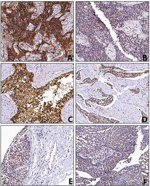
Univariate and multivariate Cox regression analysis for progression free survival according to common clinical-pathological variables, p53 expression, and TATI expression
LNI, Lymph node invasion; LVI, Lymphovascular invasion;
Immunohistochemical images of the combined expression of TATI and p53 in bladder cancer tissues (20 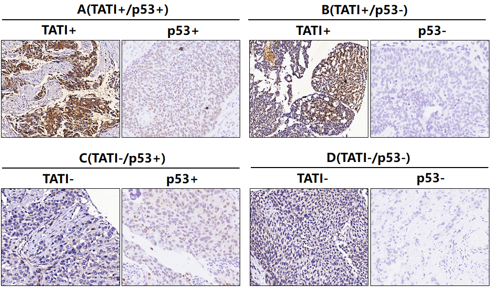
Association of TATI and p53 expression with clinical-pathological features
TATI expression was loss in 49.1% (54/110) of the tumor tissues from the patients undergoing RC. The association between TATI expression and clinical-pathological features was displayed in Table 1. We found that loss of TATI expression was significantly associated with advanced tumor stage (
Association of the combined expression of TATI and p53 with clinical outcomes
The average follow-up period was 36.8 months (range 2.0–88.0). According to Kaplan-Meier analysis, significant differences regarding PFS and OS were found in patients with TATI-positive vs. TATI-negative specimens (Log-rank test,
Relative risks of disease progression among different expression patterns according to TATI expression and p53 expression
Relative risks of disease progression among different expression patterns according to TATI expression and p53 expression
HR, hazard ratio; CI, confidence interval; A, TATI
Kaplan-Meier analysis of PFS and OS in bladder cancer. A: PFS rates according to TATI expression, B: OS rates according to TATI expression, C: PFS rates according to p53 expression, D: OS rates according to p53 expression, E: PFS rates according to the combined expression of TATI and p53, F: OS rates according to the combined expression of TATI and p53.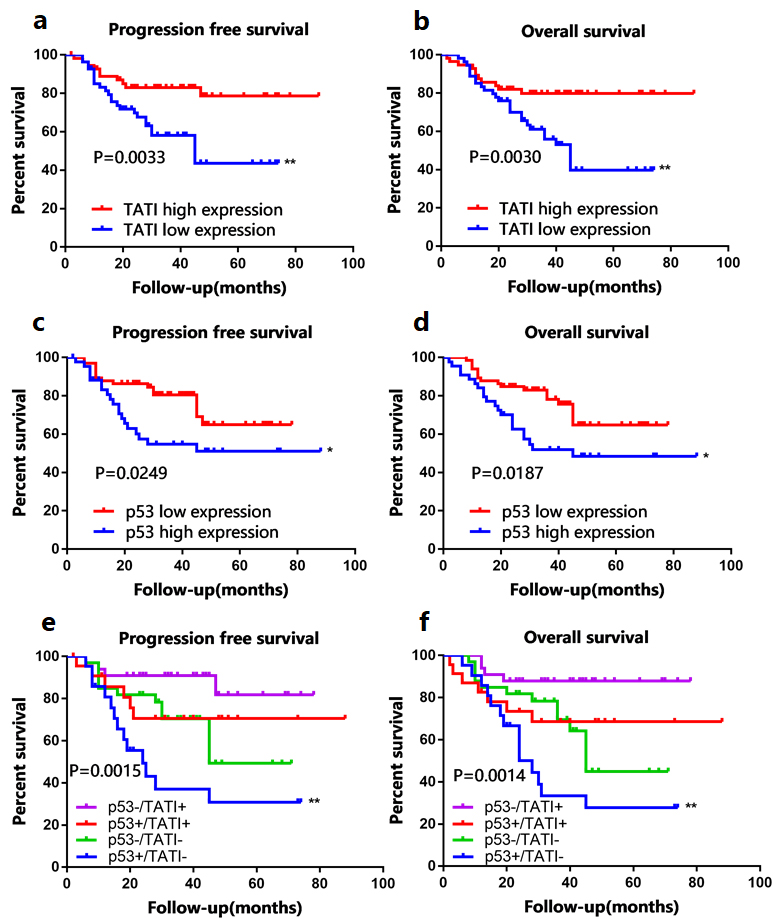
The results of univariate and multivariate Cox regression analyses for PFS are shown in Table 2. In the univariate analysis, tumor number, tumor stage, tumor grade, lymph-node invasion, TATI expression level, and p53 expression level were significantly associated with disease progression. Multivariate Cox regression analysis demonstrated that tumor number (hazard ratio [HR]: 2.20, 95% confidence interval [CI]: 1.89–2.57), tumor stage (HR: 1.55, 95% CI: 1.23–1.96), tumor grade (HR: 2.15, 95% CI: 1.51–3.07), TATI expression level (HR: 0.26, 95% CI: 0.21–0.31), and p53 expression level (HR: 1.87, 95% CI: 1.60–2.19) were independent prognostic factors for PFS. TATI-positive expression was an independent protective factor for patients with bladder cancer undergoing radical cystectomy.
We further compared the prognostic value of the different conditions for PFS, especially when classifying protein expression patterns into 4 subgroups and controlling for TATI and p53 expression separately, as shown in Table 3. In multivariate Cox regression analysis, compared to that of the TATI
Of note, the prognostic significance of TATI expression on PFS in the p53-negative expression group was much stronger than that found in the p53-positive group (HR: 0.05 vs. 0.37). When comparing the prognostic significance of p53 expression according to TATI expression, the TATI-positive group resulted advanced (HR 16.44 vs. 1.84). In other words, TATI-positive expression could significantly increase the prognostic value of p53 expression on PFS of patients with bladder cancer undergoing RC.
Discussion
Despite of rapid improvements on minimally invasive surgery, adjuvant chemotherapy, and targeted treatments for bladder cancer, mortality in this specific type of cancer has only decreased 5%, compared with a 20–40% relative risk reduction in prostate, breast, lung, and colon carcinoma observed over the last 15 years [15]. One of the most important reasons is that clinical-pathological features, such as those established by the European Organization for Research and Treatment of Cancer [EORTC] and the Spanish Urological Club for Oncological Treatment [CUETO] scoring systems, have not been able to provide enough prognostic information for patients with bladder cancer after receiving RC, making difficult for clinicians to select optimal treatment regimens timely [16, 17]. On the other hand, several studies had focused on the identification of molecular biomarkers associated with tumor recurrence, progression, metastasis, and resistance to chemotherapy based on the application of gene microarrays and next-generation sequencing. The prognostic value of many of these biomarkers has been demonstrated, having the ability to classify patients according to recurrence or progression risks more accurately than the clinical-pathological models [18, 19, 20, 21, 22, 23]. However, the clinical use of these promising biomarkers is still in an exploratory stage because of the poor reproducibility found within studies for each particular biomarker. Such shortcomings might be compensated to some extent by using combined expression models of two or more biomarkers in prognostic judgment.
In this study, tumor number, tumor stage, and tumor grade were found to have independent prognostic significance for PFS, determined by multivariate analysis. These results are in accordance with previously published reports [24, 25, 26]. Of note, altered TATI expression in tissues has been found to be associated with adverse clinical outcomes in a variety of malignancies, such as prostate [27], ovarian [6], and colorectal cancer [8]. In bladder cancer, several studies have demonstrated the association of decreased TATI expression with biologically-aggressive tumor features. Therefore, this parameter seems to play a vital role in tumor pathogenesis and progression and may serve as a good biomarker for tumor staging, although no prognostic benefit for patients receiving RC has been recognized [11, 28, 29]. In our study, we identified a correlation between loss of TATI expression and increased stage and grade of the tumor. Interestingly, the univariable Kaplan-Meier analysis revealed a relationship between loss of TATI expression and unfavorable prognosis, suggesting that this marker could serve as an independent prognostic factor for PFS, when adjusted for standard clinical-pathological features. These results seem to disagree with previous reports, in which the correlation between decreased TATI expression and increased tumor grade/advanced tumor was less significant than that shown in our study. Therefore, the association and prognostic value of TATI expression for the clinical outcomes of patients with bladder cancer undergoing RC should be revisited.
The prognostic value of p53 status for progression-free, disease-free, and disease-specific survival has been assessed in both non-MIBC and MIBC [30, 31, 32, 33]. Although p53 expression does not provide effective information for predicting the clinical outcomes of patients with MIBC undergoing RC compared to those with lower-stage organ-confined tumors, available evidences support that the expression of p53 increase gradually with progression from normal urothelium to non-MIBC, and then to MIBC [34]. Therefore, used in combined-expression models that include other markers related to proliferation, apoptosis, and immunity, mutant p53 expression could compensate the heterogenic basis of single biomarkers on prognosis adjustments [35]. For example, Seo et al. [36] determined that the simultaneous overexpression of p53 and Ki-67 did not predict PFS (HR: 1.16; 95% CI: 0.21–6.20,
In conclusion, our study indicated that decreased expression of TATI and overexpression of p53 were independent predictive factors for progression of bladder cancer in patients who had undergone RC. In addition, combined TATI expression patterns could enhance the prognostic value of p53 overexpression on progression.
Footnotes
Acknowledgments
The present study was supported by Ph.D. Innovation Fund of the Second Military Medical University and the present study was supported by Ph.D. Innovation Fund of the Second Military Medical University and the National Nature Science Foundation of China (grant no. 8150101083).
Conflict of interest
The authors declare they have no competing interests.