Cancer stem cells (CSCs) have the ability to dictate tumor initiation, recurrence, and metastasis. Here, we examined the expression of a21 in laryngeal cancer tissues and further determined the effect of 21 on the migratory ability and tumorigenicity of laryngeal cancer cells. Immunofluorescence staining revealed that 21 was positive in 13 (13/16, 81.25%) cases in laryngeal squamous cell carcinoma (LSCC) tissues, 7 (7/16, 43.75%) cases in paracancerous tissues and only 2 (2/16, 12.5%) cases in normal tumor tissues. Our quantitative RT-PCR assays further showed that 21 LSCC cells expressed significantly higher levels of stem cell-associated genes and drug efflux and resistance genes versus21 cells. Sphere-forming assays demonstrated higher sphere-forming efficiency in the 21versus21 subpopulation. Our Matrigel assays showed that 21 cells exhibited significantly greater invasive and migratory ability than 21 cells. Furthermore, the percentage of purified 21 in TU686 and TU212 cells treated cisplatin or paclitaxel was significantly higher than that of the control group. Tumor xenograft assays revealed that the tumorigenicity of 21 cells was much higher than 21 cells. In conclusion, a 21 subpopulation with CSC-like property was present in laryngeal cancer and possessed high self-renewal activity and was sufficient for tumor growth, differentiation, migration, invasion, and chemotherapeutic resistance. They could represent a promising therapeutic target for LSCC.
Laryngeal cancer is the second most common head and neck malignancy. Globally, approximately 110,000 to 130,000 new cases are diagnosed each year [1]. In China, the crude incidence rate of laryngeal cancer was 1.86/100,000 between 2008 and 2012 and the crude mortality was 1.01/100,000 [2]. Mainstay treatments for laryngeal cancer include radiotherapy, chemotherapy and surgical resection; however, management of advanced laryngeal cancer is complex and a consensus therapeutic strategy has not yet emerged.
Cancer stem cells (CSCs) are implicated in tumor initiation and differentiation, and are of high self-renewal properties, and can drive the tumorigenic process. The ability of CSCs to drive cancer initiation and progression also make them prime targets for treatments [4]. Evidence suggests that a minor population of CSCs in laryngeal cancer is extremely tumorigenic and possesses the potential to differentiate into cells that are responsible for tumor propagation and relapse [4]. Laryngeal CSCs are characterized by the expression of aldehyde dehydrogenase isoform 1 (ALDH1) [5], CD133 [6], and CD44 [7, 8]. However, laryngeal CSCs appear to differ considerably and may contribute to the heterogeneity of laryngeal cancer [9]. The calcium channel 21 subunit serves an important role in regulating calcium oscillation amplitude [10] and has been reported in hepatocellular carcinoma (HCC) [11]. This subpopulation of HCC cells possesses stem cell-like properties. In comparison to the non-purified cells, the invasiveness ability, self-renewal ability and the expression level of genes related to stem cells were increased in 21 HCC cells [11, 12]. Knocking down the 21 gene in combination with doxorubicin treatment compromises HCC oncogenesis [11]. Recently, Yu et al. have demonstrated that non-small cell lung cancer cells expressing 21 demonstrated CSC-like properties, and may contribute to chemoresistance [13].
Currently, no study is available on the role of 21 in laryngeal cancer. In the present study, we examined the expression of a 21 in laryngeal cancer tissues and further determined the effect of 21 on the migratory ability and tumorigenicity of laryngeal cancer cells.
Materials and methods
General design
The current study included 3 major parts. In the first part that involved clinical samples, 21 expression was compared between primary lesions versus non-cancerous adjacent tissues collected from 16 LSCC patients receiving larygectomy and no neoadjuvant therapy. The second part of the study was conducted using 2 representative laryngeal squamous cell carcinoma (LSCC) cell lines: TU212 (highly malignant) versus TU686 (less malignant). Briefly, 21 cells were purified using flow cytometry from cultured cells. The following features were compared between 21 and 21 cells: 1) representative CSC markers (BMI1, SOX2, EPCAM, OCT4, CNTTB, KLF4, NANO, ABCG2 and MDR1); 2) sphere formation; 3) differentiation; 4) migration/invasion; 5) sensitivity to cisplatin/paclitaxel. These features were also examined after 21 knockdown using shRNAs in 21 cells. In the final step of xenograft experiments with NOD/SCID mice, the ability of 21 versus 21 TU686 cells to form tumor and subsequent growth was compared using limited dilution.
Tissue specimen acquisition
Archived surgically resected laryngeal squamous cell carcinoma (LSCC) tissue specimens were obtained from 16 treatment-naive male patients and snap-frozen in liquid nitrogen. The study protocol was approved by the institute ethics committee of Beijing Friendship Hospital, Capital Medical University (no. 2017-P2-187-01) and written informed consent was obtained from all the study subjects.
Immunofluorescence staining
Frozen tissues were sectioned with acryostat and fixed with methanol for 30 seconds. After blocking with 5% nonfat milk in PBS, slides were incubated with 21 monoclonal antibody (dilutions 1:100; Catalog no. MA3-921, ThermoFisher Scientific) overnight at 4C, followed by incubation with FITC-conjugated goat-anti-mouse IgG. In negative control, the antibody was omitted. Nuclei were counterstained with 4,6-diamidino-2-phenylindole dihydrochloride (DAPI; Polysciences, Warrington, PA, USA). The slides were mounted in 90% glycerol/PBS containing 2.5% 1,4-diazabicyclo (2,2,2) octane and examined with a Leica SP5 confocal microscope (Leica, Wetzlar, Germany).
Cells
Human LSCC cell lines TU212 and TU686 were obtained from Shanghai Huiying Biological Technology (Shanghai, China), and were cultured in RPMI 1640 medium (Invitrogen, Grand Island, NY, USA) supplemented with 10% fetal bovine serum (FBS), 100 U/mL penicillin, and 100 mg/mL streptomycin (Invitrogen) at 37C in a humidified atmosphere with 5% CO.
Primer sequences for quantitative PCR in the study
Gene
Sense
Anti-sense
BMI1
5-AGCAGCAATGACTGTGATGC-3
5-CAGTCTCAGGTATCAACCAG-3
SOX2
5-ACATGAACGGCTGGAGCAAC-3
5-AGGAAGAGGTAACCACAGGG-3
EPCAM
5-CTGAATTCTCAATGCAGGGTC-3
5-CCCATCTCCTTTATCTCAGCC-3
OCT-4
5-TGGGCTCGAGAAGGATGTGGTC-3
5-AGTTTGAATGCATGGGAGAGC-3
CTNNB
5-TGATGGAGTTGGACATGGCC-3
5-CTCATACAGGACTTGGGAGG-3
KLF4
5-AAGCCAAAGAGGGGAAGAC-3
5-CATCTGAGCGGGCGAATTTC-3
NANOG
5-TGCCTCACACGGAGACTGTC-3
5-TGCTATTCTTCGGCCAGTTG-3
ABCG2
5-GGAGGCCTTGGGATACTTTGAA-3
5-GAGCTATAGAGGCCTGGGGATTAC-3
MDR-1
5-GCCTGGCAGCTGGAAGACAAATAC-3
5-ATGGCCAAAATCACAAGGGTTAGC-3
GAPDH
5-GACCCCTTCATTGACCTCAAC-3
5-CTTCTCCATGGTGGTGAAGA-3
Flow cytometry
For isolation of 21 cells, TU212 and TU686 cells were rinsed in phosphate buffered saline (PBS), and dissociated with 0.25% trypsin (Thermo Fisher Scientific, Waltham, USA). The cells were stained with FITC-conjugated 21 monoclonal antibody (dilutions: 1:100; Catalog No. MA3-921, ThermoFisher Scientific) for 30 min at 4C. Then, 21 cells were sorted using a Facscan flow cytometer (Becton Dickinson, Mountain View, CA, USA). Furthermore, TU686 cells were treated with cisplatin (6 mol/L) or paclitaxel (0.2 mol/L) for 72 hours. The percentage of 21 cells was then detected by flow cytometry. The results were calculated with the software FlowJo (Tree Star Inc., Ashland, Oregon) and FACSCanto II (BDBiosciences).
Quantitative reverse transcription (qRT)-PCR
Total RNA was extracted from 21 and 21 cells with Trizol reagent (Invitrogen). The mRNA levels of BMI-1, SOX2, EpCAM, OCT4, CTNNB, KLF4, NANOG, ABCG2 and MDR1 were determined by quantitative reverse transcription PCR (qRT-PCR) and normalized against -actin. qRT-PCR was performed on an ABI7500 PCR machine using the SYBR Green PCR Master Mix (Toyobo Co. Ltd., Osaka, Japan). The primer sequences are shown in Table 1 [14, 15, 16, 17, 18, 19, 20, 21, 22]. Expression differences were calculated by the 2 method [23]. -actin was used as an endogenous reference.
Plasmids
Retroviral-based plasmids containing a short hairpin RNA (shRNA) against human 21 were purchased from Origene (Rockville, MD, USA). The U6 promoter-driven shRNA expression cassettes were transferred to the lentiviral shuttle vector plenti6 (Invitrogen). Lentiviral packaging, infection, and selection of blasticidin-resistant cell pools were performed as previously described [24]. The sequences of human 21-specific 29-mer shRNAs were as follows: shRNA53, 5’-ACTCAACTGGACAAGTGCCTTAGATGAAG-3’ and shRNA56, 5’-AGATGCAAGAAGACCTTGTCA CACTGGCA-3’. Scrambled shRNA was used as a negative control.
Sphere formation assays
Sphere-formation assays were carried out as previously published [10]. Briefly, 21 and 21 cells were cultured in DMEM supplemented with 1% methylcellulose (Sigma, St. Louis, MO, USA), B27 (Invitrogen), 20 ng/mL basic fibroblast growth factor (bFGF) (Peprotech, Rocky Hill, NJ, USA) and 20 ng/mL epidermal growth factor (EGF) (Peprotech) using a 96-well plate with ultralow attachment. The culture medium was replenished with 200 L supplementary media every other day. Spheroids ( 100 m) were counted using a microscope (Zeiss, Oberkochen, Germany) after 4 weeks. In addition, passage two 21 cells and 21 cells treated with shRNA53 and shRNA56 were used for sphere-formation assays where indicated.
Cell differentiation assays
21 cells were seeded in a Petri dish containing 10 mL RPMI 1640 and 10% FBS and cultured at 37C in a humidified incubator with 5% CO. After 1 week of culture, the percentage of 21 cells was calculated by flow cytometry.
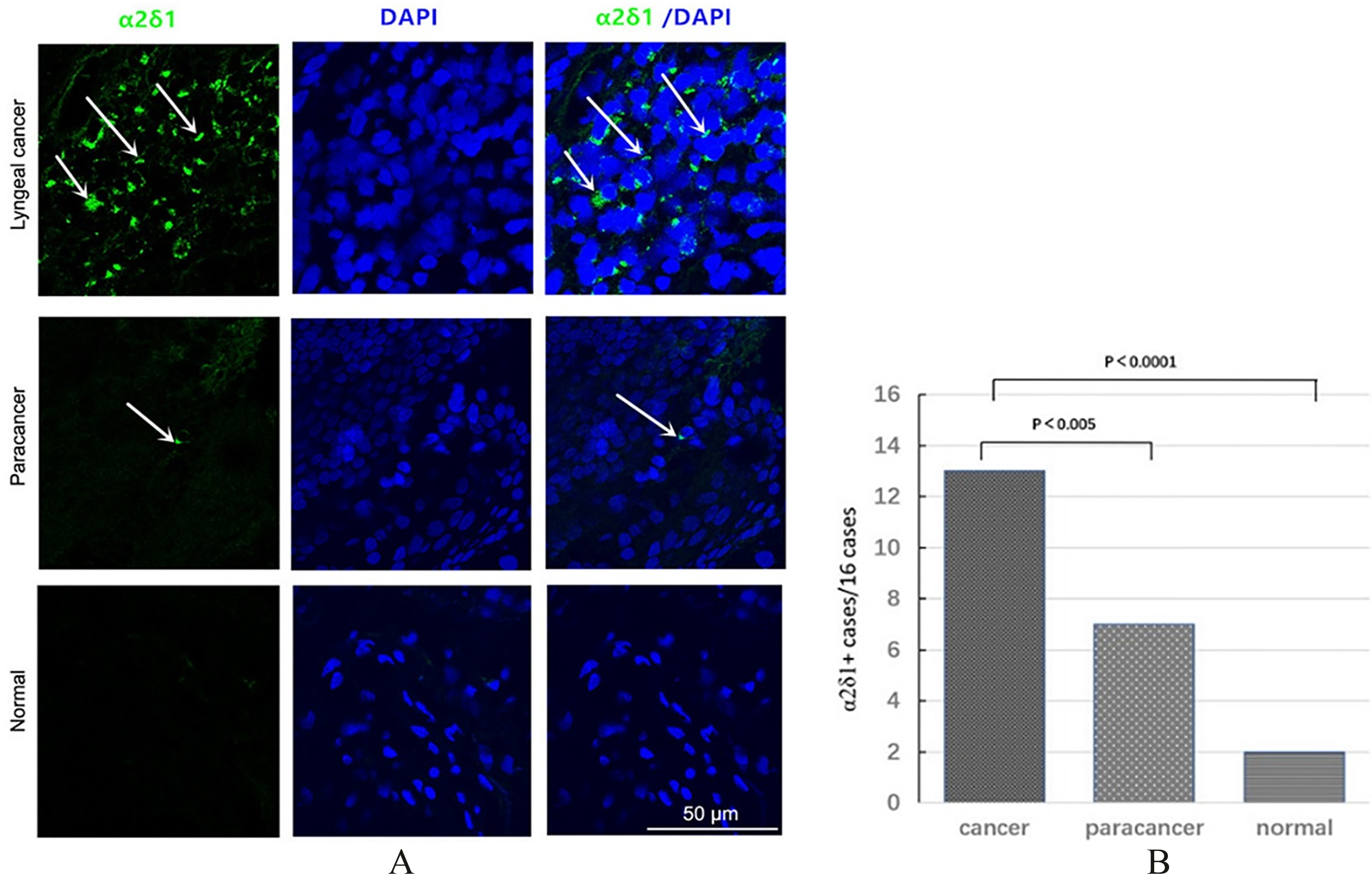
Expression of 21 in primary LSCC tissues. (A) Representative images of immunofluorescence expression of 21 in laryngeal tissue, paracancerous tissue and normal adjacent tissue in a patient with T3N0M0 laryngeal cancer are shown. 21 (green) was stained with specific monoclonal antibody and nuclei (blue) were stained with DAPI. Arrows indicate 21-positive cells. (B) Summary of 21 expression in LSCC tissues, paracancerous tissues and normal adjacent tissues.
Matrigel assays
For detection of migration and invasion of 21 cells, 5 10 cells were added onto a porous membrane (pore size, 8 m; BD Biosciences) that was coated with 2 mg/mL Matrigel. After 48-hour incubation at 37C, cells were washed three times with PBS, and fixed with 4% neutral formaldehyde prior to routine hematoxylin staining for 5 min. The number of cells that invaded through the membrane (migration) or Matrigel (invasion) was counted in 10 representative fields at 20 magnification. Images were acquired and analyzed using SPOT imaging software (Nikon).
Tumor xenograft assays
Four to 6 week-old female non-obese diabetic/severe combined immunodeficient (NOD/SCID) female mice were purchased from Huafu Kang Experimental Animal Co., Ltd (Beijing, China), and maintained in a SPF facility. All animal experiments were performed in accordance with the National Institutes of Health Guide for the Care and Use of Laboratory Animals. The protocols were approved by the Animal Care and Use Committee at Peking University Cancer Hospital.
Cells were suspended in 50 mL in a 1:1 mixture of RPMI 1640 and Matrigel (BD Biosciences) and 10 and 10 cells were injected into the right and left flank of each mouse, respectively. Tumor formation was monitored weekly. Twenty weeks after inoculation, all mice were euthanized with an overdose of anesthesia(20% urethane). Tumor volume was determined using the formula 0.5, where and represent the largest and the smallest diameter, respectively.
Statistical analysis
Data was analyzed using SPSS 17.0 software (IBM, Armonk, New York, NY, USA). Student’s test, test and one-way analysis of variance (ANOVA) were used to analyze the differences. Tumorigenic cell frequency was calculated based on extreme limiting dilution analysis using the web tool at http://bioinf.wehi.edu.au/ software/elda/. 0.05 (2-sided) was considered statistically significant.
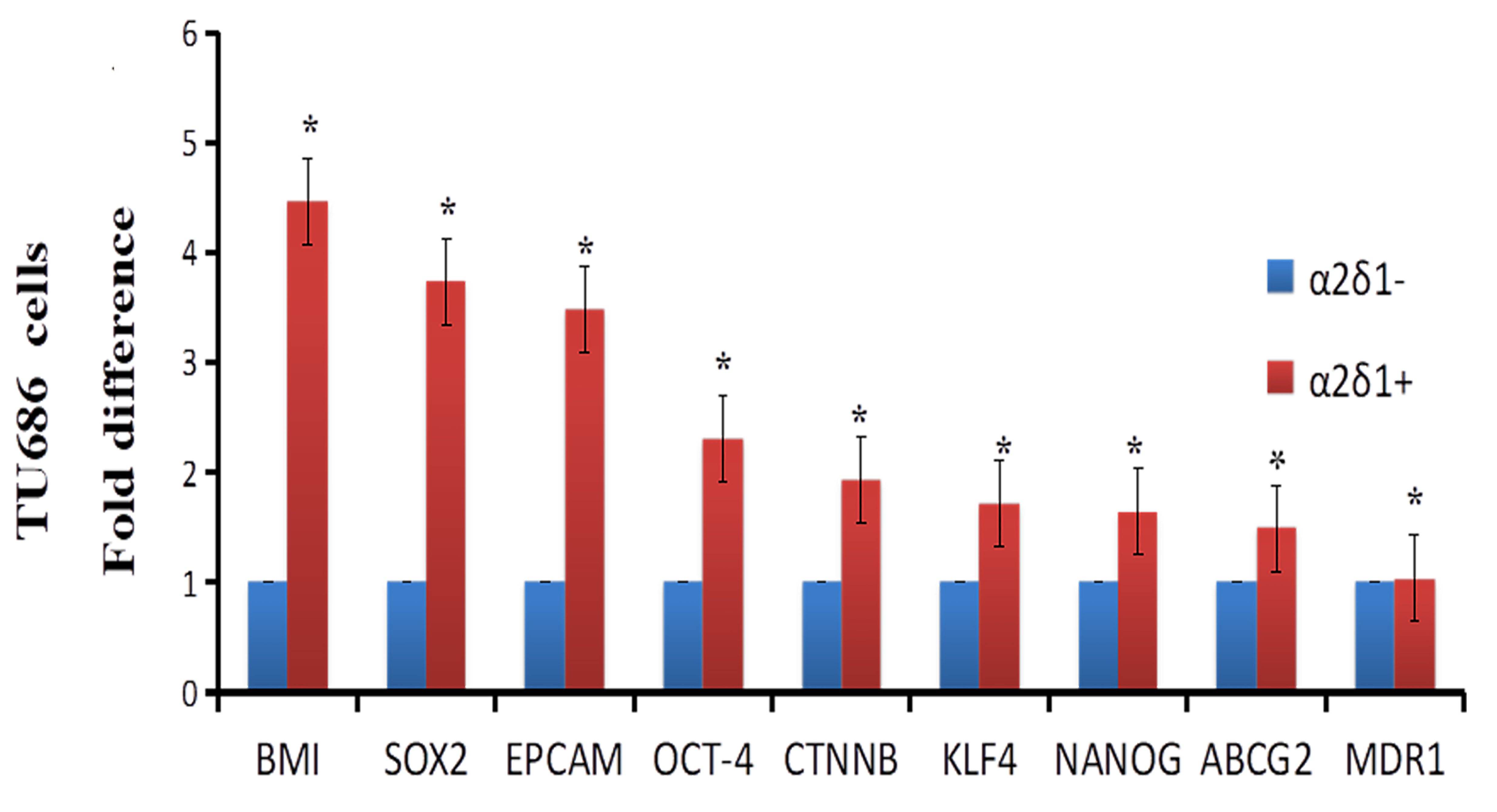
Expression of stem cell and drug efflux and resistance genes in 21versus21 subpopulation of TU686 cells. qRT-PCR analysis of the expression of stem cell markers and drug-resistance-related genes in purified 21 and 21 subpopulations of TU686 cells. Data are presented as fold difference over 21 cells for each gene. Compared with 21 cells, 21 cells consistently expressed 1 to 4.5 fold higher levels of BMI1, SOX2, EPCAM, OCT4, CNTTBKLF4, NANOG, ABCG2 and MDR1. *Error bars indicate SD.
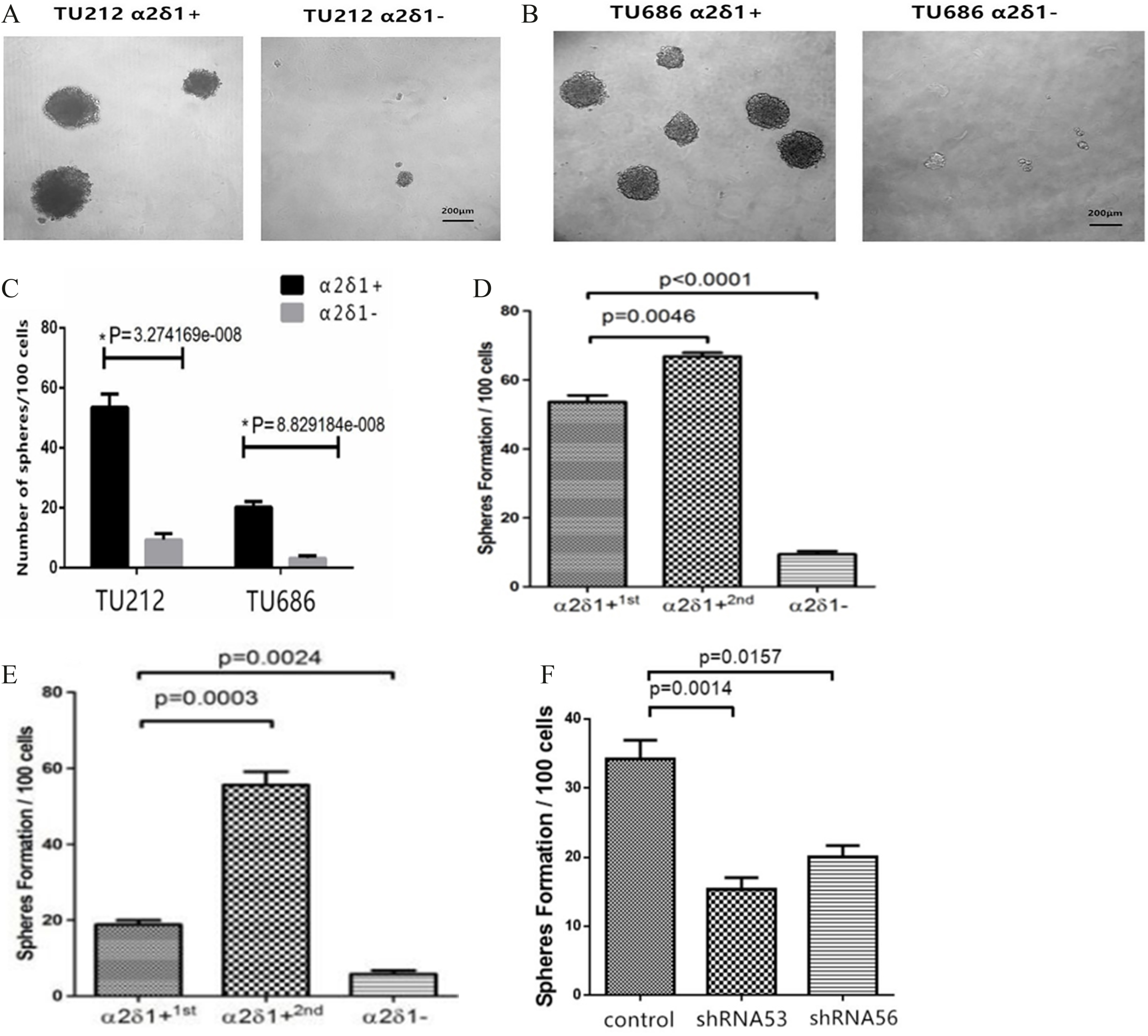
Sphere-forming efficiency of 21 and 21 cells. (A, B) The sphere-forming ability of 21 and 21 cells was examined by sphere formation assays as detailed in Methods. (C) Histograms show the spheroid forming efficiency of FACS-sorted 21 and 21fractions from indicated sources. The ability of the spheres formed by 21 TU 212 cells (D) and TU 686 cells (E) to form secondary spheroids is also shown (21/2 passage). (F) The sphere forming ability of purified 21 TU212 cells was reduced when 21cells were incubated with indicated lentivirus for 4 hours, and were plated at 100 cells per well ( 5). One hundred cells per well were plated( 5). Spheroids ( 100 m) were counted under a stereomicroscope. Statistical analysis was carried out using data on sphere formation in 100 cells (C, F). Data are represented as mean SD.
Results
21 is predominantly expressed in LSCC tissues
Immunofluorescence staining revealed abundant 21 cells in LSCC tissues, but hardly any 21 cells in normal laryngeal tissues and occasional 21 cells in paracancerous tissues (Fig. 1A). Furthermore, 21 was positive in 13 (13/16, 81.25%) cases in LSCC tissues, 7 (7/16, 43.75%) cases in paracancerous tissues and only 2 (2/16, 12.5%) cases in normal tumor tissues (Fig. 1B).
Stem cells-associated genes and drug efflux and resistance genes are significantly upregulated in 21 TU686 cells
We isolated 21 and 21 cells in TU686 cells via FACS. Our qRT-PCR assays showed that 21 cells expressed significantly higher levels of stem cell-associated genes versus21 cells: EpCAM (3.5 fold), OCT4 (2.3 fold), CNTTB1 (2.0 fold), KLF4 (1.7 fold), NANO (1.6 fold), and ABCG2 (1.5 fold) (all 0.05). In addition, the 21subpopulation expressed significantly higher levels of the drug efflux transporter gene BMI1 (4.5 fold) and the multidrug resistance gene SOX2 (3.8 fold) compared to 21 cells ( 0.05) (Fig. 2).
Self-renewal property of 21 cells
Sphere formation assays further showed that 21 cells formed a significantly higher number of spheres than 21 cells ( 0.05) (Fig. 3A–C). Furthermore, passage 2 21 cells had significantly increased sphere-forming ability versus passage 1 21 cells (Fig. 3D and E). Moreover, knocking down 21 by 21-specific shRNAs significantly inhibited the sphere forming ability of 21 cells (Fig. 3F).
Differentiation properties of 21cells
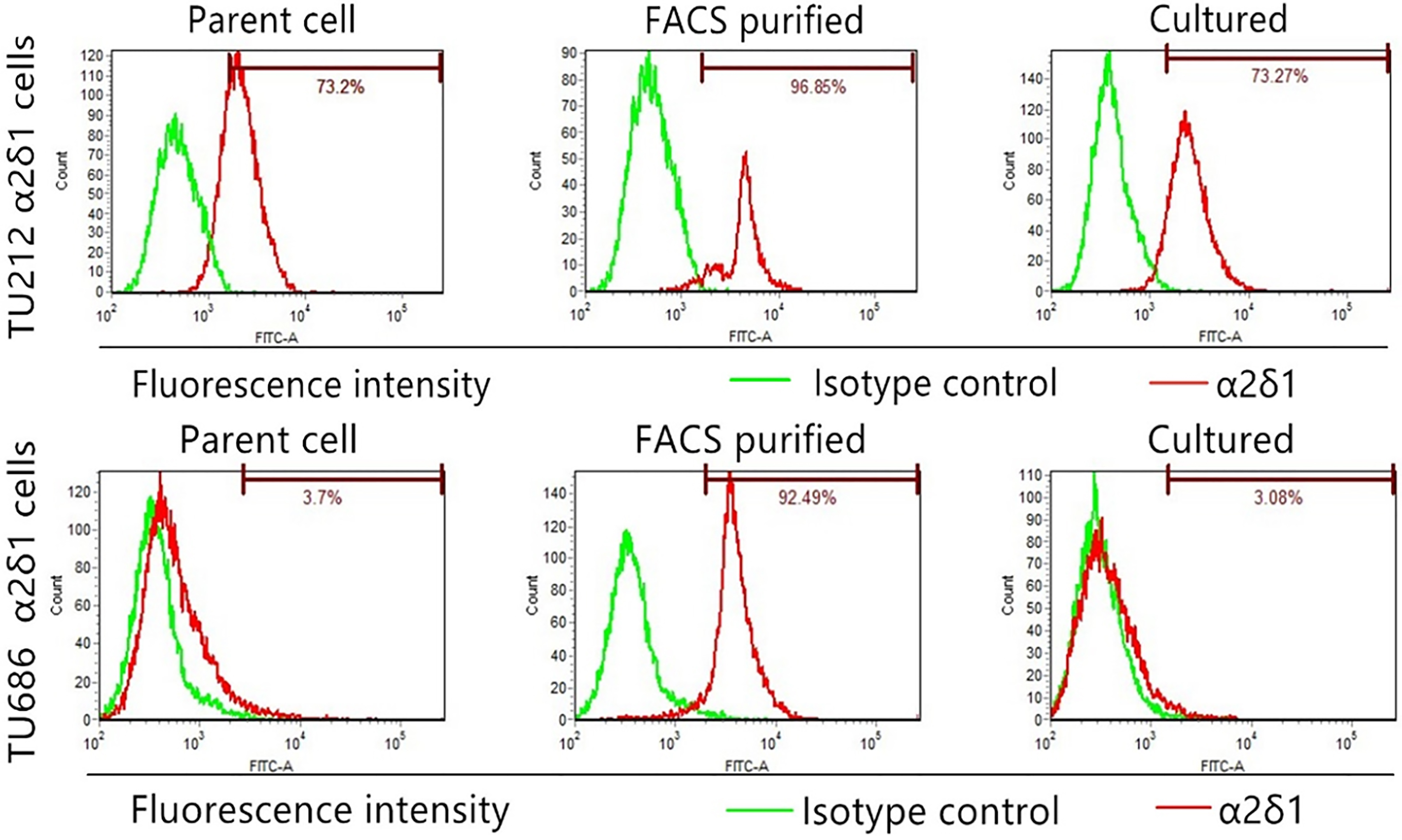
After purified TU212 21 cells were cultured for 1 week, the percentage of 21 cells decreased from 96.85% to 73.27%; the percentage of 21 TU686 cells decreased from 92.49% to 3.08% (Fig. 4), suggesting that 21 cells differentiated into 21 cells.
Migratory and invasive potential of 21cells
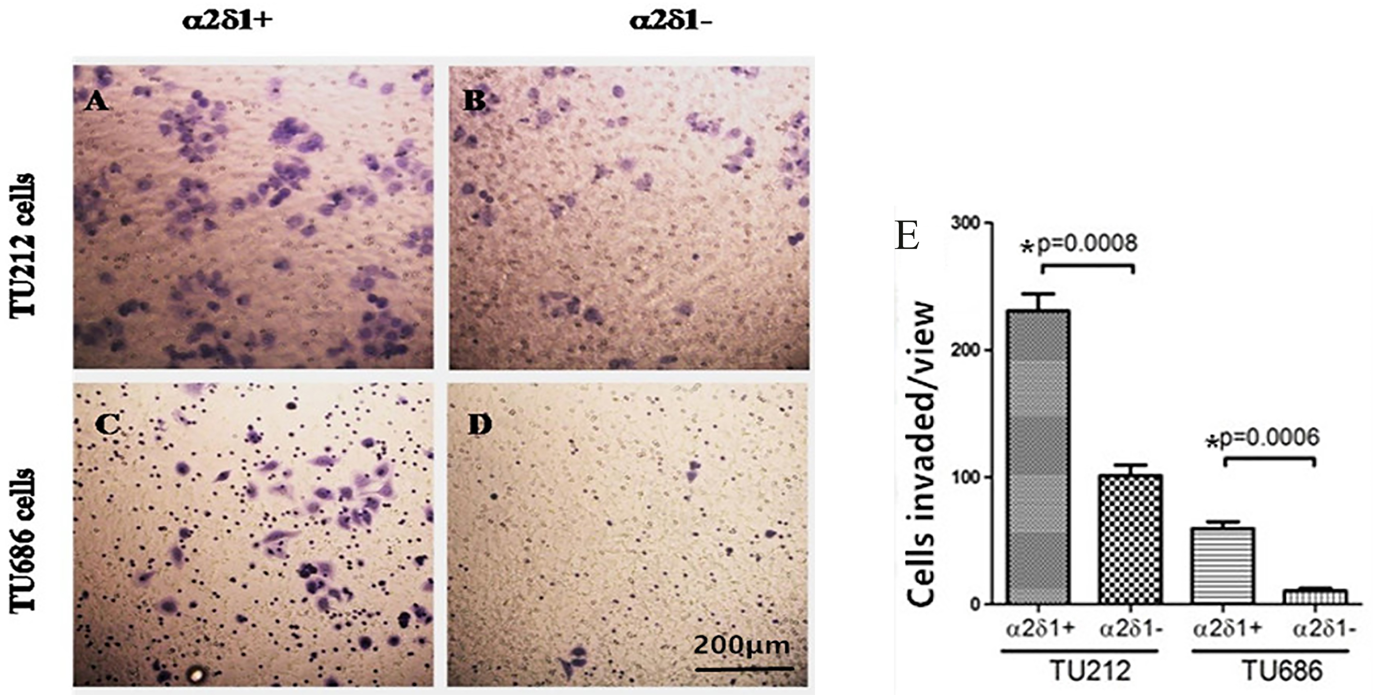
Our Matrigel assays showed that 21 cells exhibited significantly greater invasive ability than 21 cells (Fig. 5A–D). Furthermore, in comparison to 21 cells, 21 cells showed significantly greater migratory ability (Fig. 5E).
Chemoresistance of 21 cells
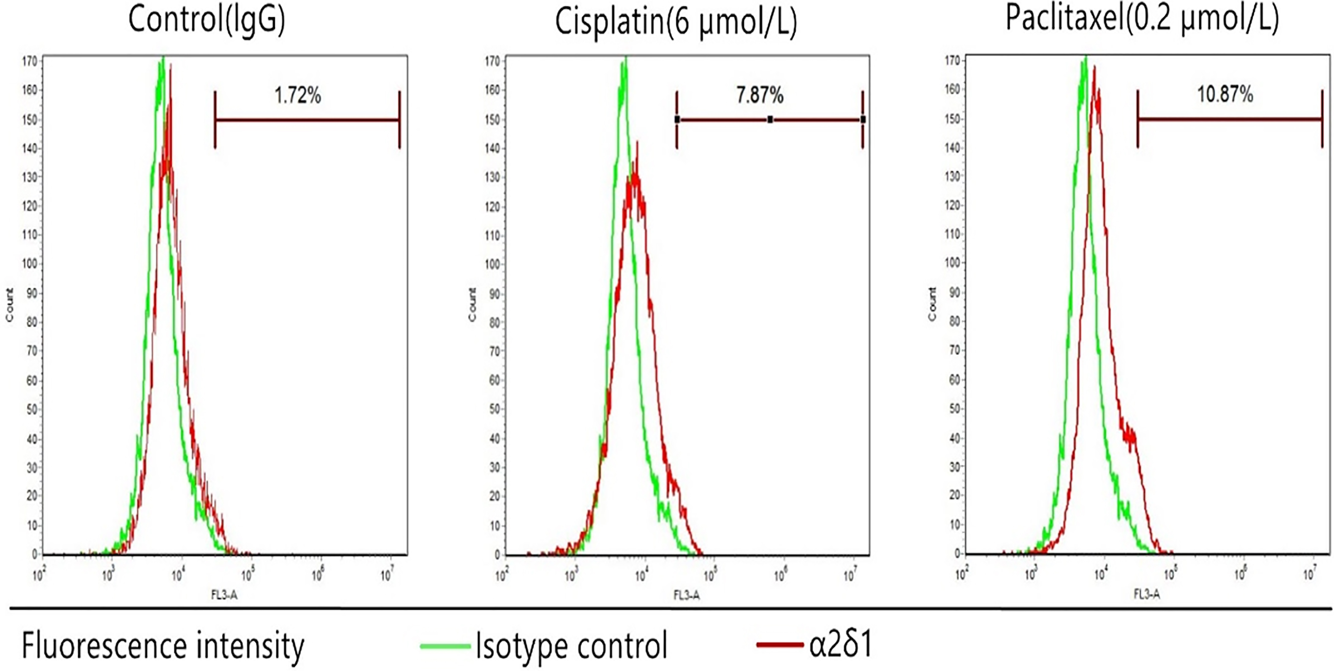
We treated TU686 cells with cisplatin (6 mol/L) or paclitaxel (0.2 mol/L) for 72 hours. Our flow cytometric analysis showed the percentage of purified 21 cells in TU686 cells treated cisplatin or paclitaxel was significantly higher than that of the control group (cisplatin: 7.87% vs. control: 1.72%; paclitaxel:10.87% vs. control: 1.72%) (Fig. 6).
Tumorigenesis of 21cells
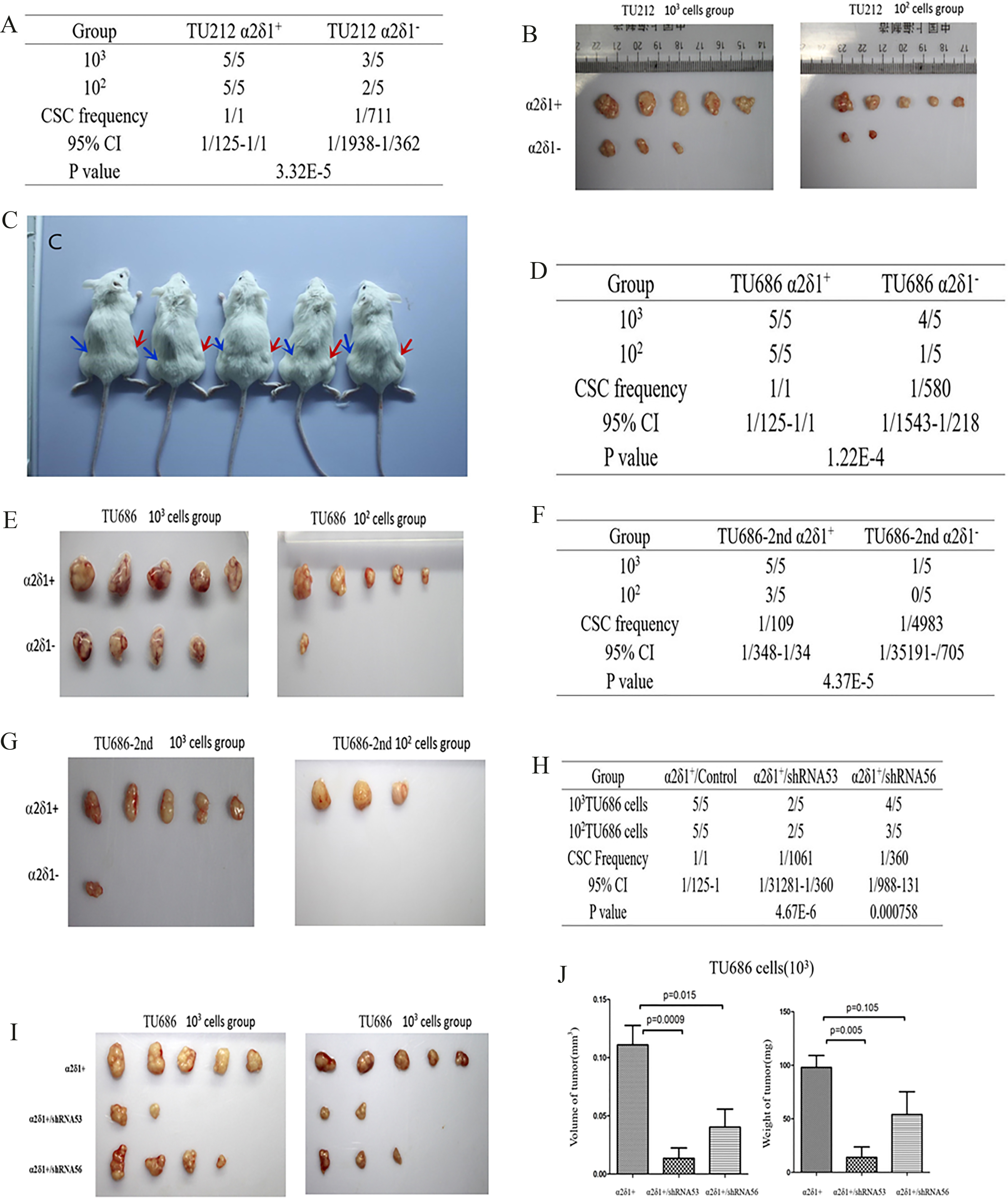
As few as 10-10 purified 21 cells from the TU212 and TU686 cell lines initiated tumor formation in almost all inoculated mice (Fig. 7A–E). By contrast, 21 cells were completely non-tumorigenic or formed tiny nodules only occasionally (Fig. 7A–E). Passage 2 21 and 21 cells further confirmed that 21cells were consistently more tumorigenic than 21 cells (Fig. 7F and G). As a matter of fact, mice inoculated with passage 2 21 cells showed no evident tumor formation at 12 weeks (Fig. 7F and G). Furthermore, shRNA53 and shRNA56 21 knockdown significantly suppressed the of tumor xenografts from 21 TU686 cells (Fig. 7H–J).
Differentiation properties of 21 TU212 and TU686 cells. Flow cytometry shows the percentage of 21 cells in TU212 and TU686 cells, FACS-purified 21 cells and purified 21 cells cultured in 10% serum-containing medium for 1 week (cultured).
Migration and invasive potential of 21 cells. Sorted 21 and 21 cells were assayed for their invasive ability on Matrigel using a Boyden chamber assay. The invasive and migratory potential of 21 cells (A, C) and 21 cells (B, D) were detected. Statistical results of invasive cells were calculated (E). Data represent the mean SD of three independent experiments. *Student’s -test.
Chemoresistance ofTU686 cells. The percentage of sorted 21 TU686 cells after exposure to cisplatin (6 mol/L) or paclitaxel (0.2 mol/L) for 72 hours.
Tumorigenesis of 21vs.21subpopulation. (A) TU212 cells were transplanted subcutaneously in NOD/SCID mice as indicated. (B, C) Representative photographs showing dissected tumors by FACS-purified 21 (red arrows) and 21 (blue arrows) TU212 cells. (D) TU686 cells were inoculated subcutaneously in NOD/SCID mice as indicated. (E) Photographs showing dissected tumors formed by FACS-purified 21 and 21 TU686 cells. (F, G) Serial inoculation of 10 and 10 TU686 cells into NOD/SCID mice from tumors formed by 21 TU686 cells. (H, I) Tumorigenicity of TU686 21 cells FACS-purified from indicated sources after 21 shRNA53/shRNA56 knockdown. TU686 21 cells were incubated with indicated lentiviruses for 4 hours, and 10/10 cells per mouse were injected. The statistical results of tumorigenic formation in 5 mice were calculated. (J) The volume and weight of tumors by 21 cells, and 21 cells knockdown with shRNA53and shRNA56 were detected.
Discussion
21 is a voltage-gated calcium (Cav) channel subunit [25] that governs synaptic input integration, activation of calcium-dependent signaling cascades, neuronal migration, neurotransmitter, and hormone release [26]. Existing evidence shows that 21 is expressed in HCC [10]. Knocking down 21 induced apoptosis of tumor-initiating cells (TIC) and inhibited tumor formation and self-renewal capacities of HCC cells. In contrast, 21 overexpression increased sphere formation. These results indicated that 21 HCC cells possess stem cell-like properties. In the current study, we found that 21 was expressed in a subpopulation of laryngeal cancer TU212 and TU686 cells, and 21 LSCC cells possessed stem cell-like properties.
Immunofluorescence staining of 16 LSCC specimens showed that 21 was significantly more frequently expressed in laryngeal cancer tissue than that in paracancerous tissues and normal tissues. This result is consistent with previous findings in other malignancies [10, 13]. Next, we used qPCR to detect CSC-associated genes and drug resistance genes. The results showed high level of expression of stem cell-associated genes, including BMI-1 [4], SOX2 [27], EpCAM [28], OCT4 [29], CTNNB1 [30], and ABCG2 [31, 32, 33] in 21 TU686 cells. These data together showed that a CSC subpopulation in laryngeal cancer is marked by a surface phenotype 21. Only 21 cells for this marker showed stem-cell like characteristics, including the ability to differentiate and for self-renewal, and the capability of resisting standard chemotherapy.
Next, we showed that TU212 and TU686 cells also contain a 21 subpopulation. Purified 21 cells had the capability to multiply as spheroids in growth factor-supplemented, serum-free medium under non-adherent conditions. After culturing for 4 weeks, cancer spheres of undifferentiated 21cells were obtained. In contrast, the sphere-forming ability of 21 cells were much weaker than 21cells under the same conditions. Cell spheres deriving from single cells that were acquired from dissociated 21 cells showed nearly limitless growth capability and could be expanded in subsequent serial propagations. Therefore, we speculate that 21 may play a crucial role in the sphere formation ability of TU212 and TU686 cells. This is consistent with a previous study [10], which found that 21 Cav channel overexpression resulted in increased sphere formation in HCC Hep-11 and Hep-12 cells. Self-renewal ability is one of the characteristics of CSC; these results indicate that 21 cells in TU212 and TU686 cells may represent CSCs.
We next evaluated the differentiation and migration/invasion potential of the 21subpopulation in TU212 and TU686 cells. The results showed that 21 cells could differentiate into 21 cells and 21 cells while 21 cells did not have this property. 21 cells also had greater differentiation and migration/invasion potential than 21cells. The ability to differentiate and migrate/invade is also a feature of CSCs.
Consistent with a previous study on 21 cells in lung cancer [13], treatment with cisplatin and paclitaxel increased the percentage of 21cells in TU686 cells in our experiments, suggesting that laryngeal cancer cells expressing 21 are resistant to chemotherapeutic agents. According to previous report [13], we believe that cisplatin/paclitaxel may cause 21cells to undergo apoptosis or necrosis, while 21 cells are insensitive to chemotherapeutic drugs and can survive the tumoricidal effect of cisplatin/paclitaxel. Therefore, the ratio of 21 cells to all viable cells increases, and the effect of enriching CSC is achieved. In future studies, we will verify the above speculation and grading the concentration of chemotherapeutic drugs to see if the change in the ratio of 21 cells is time- and dose-dependent in a range of drug concentrations. Available evidence indicates that 21 cells are resistant to cisplatin/paclitaxel. Resistance to radiotherapy and chemotherapy is one of the important biological characteristics of CSCs.
Previous studies have confirmed that 21 plays a crucial role in regulating the amplitude of CSC calcium oscillations [10]. Usually, spontaneous calcium oscillations in cells are caused by excitable tissues such as muscles, nerve tissues, embryonic stem cells, and immature dendritic cells. In most cases, their regulatory mechanisms and biological functions have not been elucidated. Calcium oscillations, a major form of calcium signaling, can promote the expression of specific genes, which is related to the magnitude and duration of calcium transients. It is possible to bind to the enhancer site and initiate transcription by keeping the transcription factor in the nucleus at a sufficiently high level. We hypothesize that 21 may be involved in the “amplitude-encoding” signal in LSCC, thereby maintaining the stem cell characteristics of LSCC.
Upon in vivo tumor xenograft assays, 100 purified 21 cells were sufficient for tumor formation. In contrast, 21 cells either were completely non-tumorigenic or formed tiny nodules only occasionally. In vivo serial inoculation assays with re-sorted cells from tumors formed by implanted 21 cells confirmed the tumorigenesis potential of 21 but not 21 cells. Knocking down 21 in purified 21 cells using shRNA reduced sphere formation and decreased tumorigenic cells in TU686 cells. These data indicate that 21 has an important influence on sphere formation or tumorigenicity of LSCC both in vivo and in vitro, and has the potential to become a LSCC CSC marker. Nevertheless, the role of 21 in the onset, progression and metastasis of LSCC remains to be further studied.
Conclusion
In summary, the results from the current study indicated the presence of a 21 subpopulation with CSC-like property in laryngeal cancer. The specific mechanism of CSC-like effects exerted by 21 in laryngeal cancer requires further study.
Footnotes
Acknowledgments
This research was supported by the Scientific Research Common Program of Beijing Municipal Commission of Education (#KM201510025028) and Research Foundation of Beijing Friendship Hospital, Capital Medical University (#yydszx2015-02, #yyqdkt 2014-23).
Conflict of interest
The authors declare that they do not have any potential conflict of interests in relation to the contents of this manuscript.
References
1.
SiegelR.WardE.BrawleyO. and JemalA., Cancer statistics, 2011: The impact of eliminating socioeconomic and racial disparities on premature cancer deaths, CA: A Cancer Journal for Clinicians61 (2011), 212–236.
2.
LiuY.ZhaoQ.DingG.ZhuY.LiW.ChenW. and ChinJ., Incidence and mortality of laryngeal cancer in China, 2008–2012, Cancer Res30 (2018), 299–306.
3.
DuL.LiH.ZhuC.ZhengR.ZhangS. and ChenW., Incidence and mortality of laryngeal cancer in China, 2011, Chinese J Cancer Res27 (2015), 52–58.
4.
YuD.LiuY.YangJ.JinC.ZhaoX.ChengJ.LiuX. and QiX., Clinical implications of BMI-1 in cancer stem cells of laryngeal carcinoma, Cell Biochem Biophys71 (2015), 262–269.
5.
MartínM.HinojarA.CerezoL.GarcíaJ.LopezM.PradaJ.MarínA. and GamalloC., Aldehyde dehydrogenase isoform 1 (ALDH1) expression as a predictor of radiosensitivity in laryngeal cancer, Clinical and Translational Oncology18 (2016), 825–830.
6.
WangJ.WuY.GaoW.LiF.BoY.ZhuM.FuR.LiuQ.WenS. and WangB., Identification and characterization of CD133+ CD44+ cancer stem cells from human laryngeal squamous cell carcinoma cell lines, J Cancer8 (2017), 497–506.
7.
de JongM.C.PramanaJ.van der WalJ.E.LackoM.Peutz-KootstraC.J.de JongJ.M.TakesR.P.KaandersJ.H.van der LaanB.F.WachtersJ.JansenJ.C.RaschC.R.van VelthuysenM.L.GrénmanR.HoebersF.J.SchuuringE.van den BrekelM.W. and BeggA.C., CD44 expression predicts local recurrence after radiotherapy in larynx cancer, Clin Cancer Res16 (2010), 5329–5338.
8.
BaumannM. and KrauseM., CD44: A cancer stem cell-related biomarker with predictive potential for radiotherapy, Clin Cancer Res16 (2010), 5091–5093.
9.
SuerI.KaratasO.F.YuceturkB.YilmazM.GuvenG.BugeO.CansizH. and OzenM., Characterization of stem-like cells directly isolated from freshly resected laryngeal squamous cell carcinoma specimens, Curr Stem Cell Res Ther9 (2014), 347–353.
10.
ZhaoW.WangL.HanH.JinK.LinN.GuoT.ChenY.ChengH.LuF.FangW.WangY.XingB. and ZhangZ., 1B50-1, a mAb raised against recurrent tumor cells, targets liver tumor-initiating cells by binding to the calcium channel α2δ1 subunit, Cancer Cell23 (2013), 541–556.
11.
SainzB. and HeeschenC., Standing out from the crowd: Cancer stem cells in hepatocellular carcinoma, Cancer Cell23 (2013), 431–433.
12.
ZhangY.ZhaoW.HanH.LiS.ChenD. and ZhangZ., MicroRNA-31 suppresses the self-renewal capability of α2δ1+ liver tumor-initiating cells by targeting ISL1, Oncotarget8 (2017), 87647–87657.
13.
YuJ.WangS.ZhaoW.DuanJ.WangZ.ChenH.TianY.WangD.ZhaoJ.AnT.BaiH.WuM. and WangJ., Mechanistic exploration of cancer stem cell marker voltage-dependent calcium channel α2δ1 subunit-mediated chemotherapy resistance in small-cell lung cancer, Clin Cancer Res24 (2018), 2148–2158.
14.
ZareiS.BayatA.A.HadaviR.MahmoudiA.R.TavangarB.VojganiY.Jeddi-TehraniM. and AmirghofranZ., Production and characterization of a peptide-based monoclonal antibody against CD44 variant 6, Monoclonal Antibodies in Immunodiagnosis and Immunotherapy34 (2015), 36–43.
15.
ZhaoC.MaZ.G.MouS.L.YangY.X.ZhangY.H. and YaoW.C., Targeting effect of microRNA on CD133 and its impact analysis on proliferation and invasion of glioma cells, Genetics and Molecular Research16 (2017), 1–9.
16.
LiS.K.M.SmithD.K.LeungW.Y.CheungA.M.S.LamE.W.F.DimriG.P. and YaoK.M., FoxM1c counteracts oxidative stress-induced senescence and stimulates bmi-1 expression, J Biol Chem283 (2008), 16545–16553.
17.
KleinS.DvornikJ.L.YarrabothulaA.R. and SchanielC., A Marfan syndrome human induced pluripotent stem cell line with a heterozygous FBN1 c.4082G > A mutation, ISMMSi002-B, for disease modeling, Stem Cell Res23 (2017), 73–76.
18.
EastburnD.J.SciambiA. and AbateA.R., Picoinjection enables digital detection of RNA with droplet rt-PCR, Plos One8 (2013), e62961.
19.
KimR. and NamJ., OCT4 expression enhances features of cancer stem cells in a mouse model of breast cancer, Lab Anim Res27 (2011), 147.
20.
DurandJ.LampronA.MazzucoT.L.ChapmanA. and BourdeauI., Characterization of differential gene expression in adrenocortical tumors harboring β-catenin (CTNNB1) mutations, J Clin Endocrinol Metab96 (2011), E1206–E1211.
21.
EvseenkoD.A.PaxtonJ.W. and KeelanJ.A., The xenobiotic transporter ABCG2 plays a novel role in differentiation of trophoblast-like BeWo cells, Placenta28 (2007), S116–S120.
22.
MarchesiF.MontiP.LeoneB.E.ZerbiA.VecchiA.PiemontiL.MantovaniA. and AllavenaP., Increased survival, proliferation, and migration in metastatic human pancreatic tumor cells expressing functional CXCR4, Cancer Res64 (2004), 8420–8427.
23.
PfafflM.W., A new mathematical model for relative quantification in real-time RT-PCR, Nucleic Acids Res29 (2001), e45.
24.
LanL.HanH.ZuoH.ChenZ.DuY.ZhaoW.GuJ. and ZhangZ., Upregulation of myosin Va by Snail is involved in cancer cell migration and metastasis, Int J Cancer126 (2010), 53–64.
25.
WuJ.YanZ.LiZ.QianX.LuS.DongM.ZhouQ. and YanN., Structure of the voltage-gated calcium channel Cav1.1 at 3.6 Å resolution, Nature537 (2016), 191–196.
26.
Van PetegemF.ClarkK.A.ChatelainF.C. and MinorD.L., Structure of a complex between a voltage-gated calcium channel β-subunit and an α-subunit domain, Nature429 (2004), 671–675.
27.
HuiL.ZhangJ.DingX.GuoX. and JiangX., Matrix stiffness regulates the proliferation, stemness and chemoresistance of laryngeal squamous cancer cells, Int J Oncol50 (2017), 1439–1447.
28.
MurakamiN.MoriT.YoshimotoS.ItoY.KobayashiK.KenH.KitaguchiM.SekiiS.TakahashiK.YoshioK.InabaK.MorotaM.SumiM. and ItamiJ., Expression of EpCAM and prognosis in early-stage glottic cancer treated by radiotherapy, Laryngoscope124 (2014), e431–436.
29.
El DeebN.M.F. and AbdelzaherE., Stem cell markers OCT4 and nestin in laryngeal squamous cell carcinoma and their relation to survivin expression, Pathol Res Pract210 (2014), 751–758.
30.
MachinP.CatasusL.PonsC.MuñozJ.Matias-GuiuX. and PratJ., CTNNB1 mutations and β-catenin expression in endometrial carcinomas, Hum Pathol33 (2002), 206–212.
31.
ShenB.LiD.DongP. and GaoS., Expression of ABC transporters is an unfavorable prognostic factor in laryngeal squamous cell carcinoma, Ann Otol Rhinol Laryngol120 (2011), 820–827.
32.
QiX.YuD.JiaB.JinC.LiuX.ZhaoX. and ZhangG., Targeting CD133+ laryngeal carcinoma cells with chemotherapeutic drugs and siRNA against ABCG2 mediated by thermo/pH-sensitive mesoporous silica nanoparticles, Tumor Biol37 (2016), 2209–2217.
33.
PuY.J.YanL.WeiZ.DanY.Lian-JiW.ShunJ.C., Chemoresistance of CD133+ cancer stem cells in laryngeal caronoma, Chin Med J (Engl)124 (2011), 1055–1060.