
Abstract
BACKGROUND:
Colorectal cancer (CRC) is a common type of cancer around the world. Detection of microRNA (miRNA) aberration in blood samples is a novel approach for CRC screening.
OBJECTIVE:
The purpose of this study was to explore the serum miR-497 expression pattern in CRC and examine its potential usefulness as a biomarker for CRC diagnosis and prognosis.
METHODS:
Serum miR-497 expression was evaluated by quantitative reverse transcription polymerase chain reaction (qRT-PCR) in 110 patients with CRC, 35 cases with colorectal adenoma, 54 cases with colorectal polyps, and 70 healthy individuals.
RESULTS:
The expression level of miR-497 was significantly downregulated in CRC tissues compared to the normal tissues based on the data from three independent studies GSE68204, GSE41655 and GSE35834. Compared to healthy controls, the serum miR-497 level was significantly decreased in patient with CRC or benign lesion (colorectal adenoma and polyps). Serum miR-497 level was dramatically increased in the post-operative blood samples from early stage CRC patients. Receiver-operating characteristic (ROC) analysis showed that serum miR-497 had a high sensitivity and specificity for discriminating CRC or precancerous colorectal lesion from normal controls. Moreover, low serum miR-497 expression was closely correlated with aggressive clinical features and shorter overall survival (OS). Kaplan-Meier analyses also revealed that OS was strongly associated with lymph node invasion, TNM stage and histological grade. Furthermore, univariate and multivariate analysis showed serum miR-497 was an independent prognostic factor for CRC.
CONCLUSIONS:
Collectively, serum miR-497 may serve as a promising biomarker for diagnosis and prognosis of CRC.
Introduction
Colorectal cancer (CRC) is one of most commonly diagnosed cancer with a high mortality rate. Each year, more than 1.2 million cases are diagnosed with CRC and more than 0.6 million cases die from this malignancy worldwide [1, 2]. In China, CRC is the fifth or fourth most frequent malignancy in female or male population respectively [3]. Unfortunately, a significant proportion of new cases are diagnosed at an advanced stage or present with distant metastasis, leading to the dismal prognosis and poor survival rate [4, 5, 6]. Therefore, identification of novel biomarkers for early detection and improvement of prognosis prediction is of great importance.
MicroRNAs (miRNAs) are a class of short, small non-coding RNAs with an average length of 22 nucleotides that regulate target messenger RNAs (mRNAs), leading to mRNA degradation or translational suppression [7, 8]. Dysregulation of miRNAs was closely associated with tumor initiation, progression and cellular process, including cell growth, proliferation, migration and apoptosis [9, 10]. Thus far, a number of miRNAs have been reported to function as oncogenes or tumor-suppressors, and their expression patterns are significantly altered in many cancers, including CRC. MiRNAs are extremely stable in the circulating system, which enable them to be excellent biomarkers for many human diseases including CRC [11]. For instance, the expression level of miR-1290 was significantly increased in the serum samples of CRC patients. In addition, upregulation of serum miR-1290 was significantly associated with unfavorable clinical outcome of CRC [12].
Several studies have explored the role of miR-497 in CRC. In a study carried out by Xu et al, the expression level of miR-497 was significantly decreased in CRC tissues compared to the normal tissues. Upregulation of miR-497 suppressed the proliferation, migration, invasion of colorectal cancer cells by degrading insulin receptor substrate 1 (IRS1) [13]. Zhang et al. reported that miR-497 was downregulated in CRC cells and overexpression of miR-497 inhibited the invasive capacity of cancer cells through regulating Fos-related antigen-1 (Fra-1) [14]. Similarly, miR-497 was found to play a tumor suppressive role in CRC by targeting insulin-like growth factor 1 receptor (IGF1-R) [15]. Wang et al. showed that miR-497 expression level was significantly lower in CRC tissues than in normal controls, and ectopic expression of miR-497 suppressed the oncogenic behaviors of colorectal cancer cells and enhance cancer cell sensitivity to chemotherapeutic agent by directly regulating kinase suppressor of ras 1 (KSR1), which was a known oncogene [16]. These findings suggested miR-497 might function as a tumor suppressor in CRC. However, the potential diagnosis and prognosis value of serum miR-497 in CRC remained poorly known. The aim of current study was to detect serum miR-497 level in patients with CRC, and then assessed its potential clinical significance.
Materials and methods
Patients and sample preparation
This study was approved by the Ethics Committee of Yantai Hospital of Traditional Chinese Medicine. Informed consent was obtained from all the participants. Blood samples were collected from 110 patients with CRC, 35 cases with colorectal adenoma, 54 cases with colorectal polyps, and 70 healthy individuals. The patients with colorectal adenoma or colorectal polyps were categorized into benign lesion group. None of CRC subjects had undergone any therapy, such as radiotherapy, chemotherapy or immunotherapy. The clinical staging and histological grade of CRC was classified based on the TNM staging system of the American Joint Committee on Cancer (AJCC)/Union for International Cancer Control (UICC). The mean age of the patients was 58.52 years, with 46 patients less than 55 years old. Forty-three patients were females. Eighty-five patients had tumors located in colon while the remaining 25 cases were located at rectum. Sixty-five patients had tumor size smaller than 4 cm, and 62 had no lymph node metastasis. When classified into tumor stage, 58 patients were at the early stage and 52 patients were at the advanced stage. For the histological grade, 69 patients had well or moderate differentiated cancer. The median follow-up period was 41.1 months (range
RNA isolation and quantitative reverse transcription-polymerase chain reaction (qRT-PCR)
Total RNA was extracted from serum samples using miRNeasy RNA isolation Kits (Qiagen, Valencia, CA, USA) following the manufacturer’s instructions, and the RNA concentration was measured with a NanoDrop 2000 spectrophotometer (Thermo Fisher Scientific, Wilmington, DE, USA). Reverse transcription was performed using the TaqMan MicroRNA Reverse Transcription Kit (Applied Biosystems, San Diego, CA, USA). Subsequently, quantitative PCR of miR-497 was performed in triplicate using the 7500 real-time PCR system (Thermo Fisher Scientific, Waltham, MA, USA). Cel-miR-39 served as the reference control, and the relative expression levels of serum miR-497 were calculated using the 2
The expression level of tissue miR-497 was significantly higher in benign lesion, CRC and metastatic CRC compared to the normal controls (A–C). Serum miR-497 level was significantly lower in patients with CRC or patients with benign lesion compared to the healthy controls (***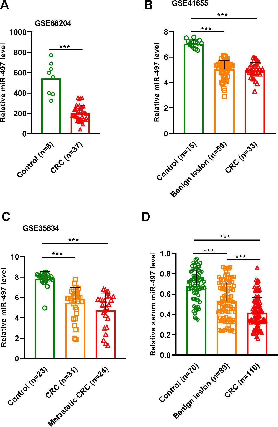
Statistical analyses were performed using MedCalc 16.7 (MedCalc, Ostend, Belgium) and GraphPad 7.0 (GraphPad Software Inc., La Jolla, CA, USA). As the expression level of serum miR-497 in CRC patients didn’t obey the normal distribution, the median value of serum miR-497 level was used as the cut-off point to separate the CRC patients into high serum miR-497 expression group and low serum miR-497 expression group. Differences of serum miR-497 expression among groups were calculated using the Mann-Whitney U test or Kruskal-Wallis test. The Chi-square test was used to examine the correlations between serum miR-497 expression and clinical variables. The receiver operating characteristic (ROC) curve and area under the ROC curve (AUC) were established for identifying CRC, precancerous colorectal lesions from healthy controls. Overall survival was defined as the time interval from the initial diagnosis to death from any cause or to the last follow-up in censored patients. Survival time was calculated in months and the maximum follow-up time was 60.0 months. Overall survival analyses were performed with Kaplan-Meier curves, and differences in survival of subgroups were compared by log-rank test. Cox’s proportional hazard regression test was used to analyze univariate and multivariate hazard ratios for overall survival (OS). Proportional hazards assumption was investigated by examining the scaled Schoenfeld residuals.
Results
Serum miR-497 level was downregulated in CRC and precancerous colorectal lesion
ROC curve analysis the diagnostic value of serum miR-497 for discriminating CRC from healthy controls (A) or benign lesion from healthy controls (B).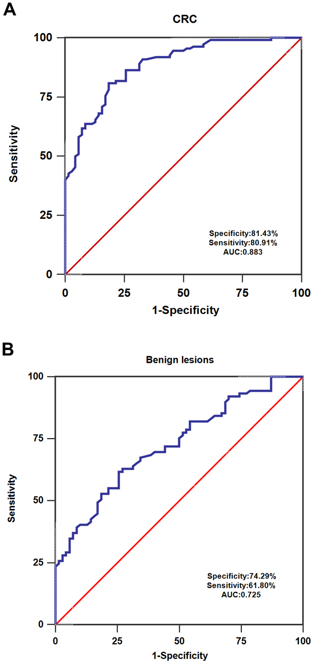
The expression pattern of tissue miR-497 was first evaluated using the three independent studies from GSE. The results of GSE68204 showed that the expression level of miR-497 was significantly downregulated in CRC tissues compared to the normal mucosa (***
The expression level of serum miR-497 was greatly elevated in CRC patients at the early stage following treatment (***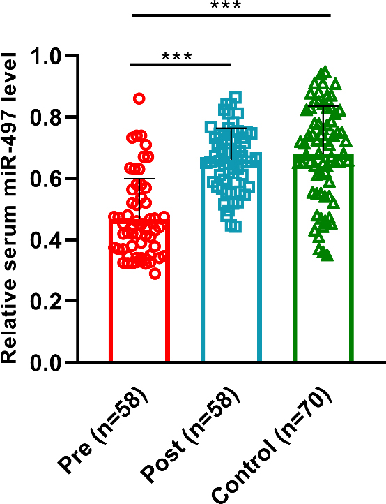
Of all CRC subjects, 58 cases were at the early clinical stage (stage I/II). We obtained the blood samples from early stage CRC patients one month after treatment, and measured the serum miR-497 expression levels. As illustrated in Fig. 3, serum levels of miR-497 in post-operative samples were significantly higher than those in pre-operative samples, whereas no significant difference was noted between post-operative samples and healthy controls.
Correlations between the serum miR-497 expression and clinical variables in 110 patients with CRC
Correlations between the serum miR-497 expression and clinical variables in 110 patients with CRC
The association between serum miR-497 level (A), histological grade (B), lymph node metastasis (C), TNM stage (D) and the overall survival of CRC.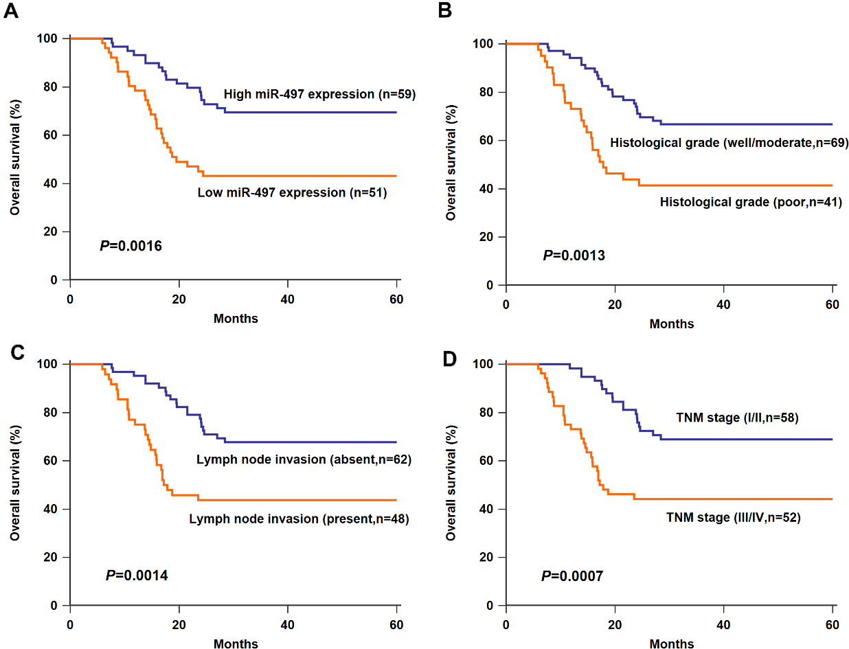
Univariate and multivariate analysis to identify factors that predict overall survival of patients with CRC
According to the median expression levels of serum miR-497, all CRC cases were classified into either the high serum miR-497 expression group (
Prognostic significance of serum miR-497
To further evaluate the association between serum miR-497 level with the prognosis of CRC, Kaplan–Meier survival analysis was performed. Patients with lower serum miR-497 levels had statistically significantly worse OS in comparison with those with serum miR-497 levels (Fig. 4A,
Then, univariate analysis revealed that serum miR-497 (HR 4.65; 95% CI, 1.54–7.93;
Discussion
Since the tumor often spread to distant organs at the time of diagnosis, late detection of CRC is one of the leading causes for the high mortality of this malignancy. Therefore, identification of novel biomarkers is crucial for improving the prognosis of CRC. In this study, we demonstrated that the expression level of miR-497 was significantly downregulated in CRC tissues compared to the normal tissues based on the data from three independent studies, which was consistent with the previous findings [13, 14, 15, 16]. Then the serum miR-497 level was found to be significantly lower in patients with CRC or precancerous colorectal lesion than in healthy controls. Serum miR-497 level was dramatically increased in the post-operative blood samples from CRC patients at the early stage. ROC analysis revealed that serum miR-497 could well differentiate CRC or precancerous colorectal lesion from normal control. In addition, low serum miR-497 expression was significantly correlated with aggressive clinical variables and shorter OS. The Kaplan-Meier analyses also revealed that OS was closely correlated with lymph node invasion, TNM stage and histological grade. Additionally, serum miR-497 was strongly associated with OS, and identified as independent prognostic factors for CRC. These data suggested that serum miR-497 might be a promising biomarker for the diagnosis and prognosis prediction CRC.
One possible reason for the decreased serum miR-497 in patients with CRC is that the cancer cells secret less miR-497 into the circulating system. The contents of tumor exosomes include protein and RNA species, which carry an imprint of the parent cells. Cancer cells secret much more exosomes than the normal cells. Therefore, it is also possible that less miR-497 is packaged into the tumor cells derived exosome, leading to the downregulation of serum miR-497 in patients with CRC. Both colorectal adenoma and colorectal polyps are the precancerous lesions for CRC. The expression level of serum miR-497 was significantly downregulated in patients with colorectal adenoma or colorectal polyps compared to the normal controls, indicating that serum miR-497 might be useful for detecting CRC even at the precancerous stage. Although serum miR-497 level was not powerful for discriminating the patients with precancerous colorectal lesions from patients with CRC, this is the disadvantage of most biomarkers. Therefore, combining the serum miR-497 and other known circulating biomarkers as well as clinical information might contribute to differentiate the CRC from precancerous colorectal lesions.
Besides CRC, miR-497 downregulation had also been observed in many human cancers. For instance, miR-497 was downregulated in bladder cancer tissues and reduced miR-497 was closely associated with worse clinical variables [17]. In non-small cell lung cancer, miR-497 expression was significantly decreased both in tumor tissues and cell lines. In vitro and in vivo evidence showed that miR-497 overexpression greatly inhibited cancer cell proliferation, colony formation, tumor growth and angiogenesis by negatively regulating plasmacytoma variant translocation [18], yes-associated protein [19] or hepatomaderived growth factor [20]. Similarly, downregulation of serum miR-497 was also reported in prostate cancer, and restoration of miR-497 decreased cell proliferation, migration, invasion or induced apoptosis of prostate cancer cells [21, 22, 23]. Serum miR-497 levels were significantly reduced in patients with osteosarcoma and could be used to differentiate osteosarcoma cases from healthy controls. In addition, miR-497 upregulation attenuated malignant behaviors of cancer cells and enhanced the sensitivity to cisplatin in vitro as well as inhibited tumor growth in vivo [24, 25, 26]. Furthermore, miR-497 expression was markedly reduced in hepatocellular carcinoma (HCC) tissues and cell lines. Enforced miR-497 expression suppressed the proliferation, colony formation and tumor growth of HCC. Checkpoint kinase 1, VEGFA and AEG-1 were identified the downstream targets of miR-497 [27, 28, 29]. Additionally, accumulating evidence have shown that ectopic expression of miR-497 remarkably reduced the proliferation and metastatic potential of cervical cancer cells by targeting RAF-1 [30] or IGF-1R [31]. Abnormal expression of miR-497 was correlated with worse prognosis and aggressive clinical features of renal cancer [32], glioma [33] and pancreatic cancer [34], indicating miR-497 could be used as a promising prognostic marker. MiR-497 also acted as a tumor suppressor in many cancer types, such as nasopharyngeal carcinoma [35], breast cancer [36, 37], multiple myeloma [38]. On the contrary, miR-497 was remarkably upregulated in oral squamous cell carcinoma (OSCC) tissues in comparison with the adjacent normal controls. In vitro analysis showed that miR-497 overexpression promoted cell migration and vice versa, indicating miR-497 functioned as an oncogene in OSCC [39]. Collectively, miR-497 might act as either an oncogene or a tumor suppressor in human malignancies with a cancer-type dependent manner. Further research is needed to explore the underlying molecular mechanisms.
One limitation of our study was the relatively small sample size. Further studies with larger patient cohort are needed to validate our findings. Deregulation of serum miR-497 has also been reported in other human cancer such as osteosarcoma, cervical cancer and malignant astrocytomas [24, 40, 41]. Therefore, examining the serum miR-497 level alone is not enough detect CRC at the early stage or predict the prognosis of CRC. Several studies have also demonstrated that abnormal expression of other circulating miRNAs was associated with the unfavorable clinical outcome of CRC [12, 42]. Therefore, combining multiple serum biomarkers and clinical data might be an effective strategy to diagnose and predict the prognosis of CRC with high accuracy. Finally, the molecular mechanisms accounting for the tumor suppressive role of miR-497 in CRC is poorly known. How aberrant expression of miR-497 affecting the malignant behaviors of colorectal cancer cells needs further exploration.
Conclusions
In conclusion, low serum miR-497 expression was observed in CRC patients and strongly associated with worse clinical variables, as well as shorter survival time. In addition, serum miR-497 was confirmed to be an independent prognostic predictor for CRC. Taken together, serum miR-497 might serve as a promising biomarker for CRC diagnosis and prognosis.
Footnotes
Conflict of interest
None.