
Abstract
OBJECTIVES:
To investigate TCF4 expression in epithelial ovarian cancer, and to explore its correlation with clinicopathological parameters and clinical prognosis of epithelial ovarian cancer.
METHODS:
From 2009 to 2017, 188 cases of paraffin-embedded epithelial ovarian cancer tissues and 41 paratumor ovarian tissues which had been confirmed at the memorial hospital of Sun Yat-sen University were collected in this study, and the expression of TCF4 was performed by immunohistochemistry using a polyclonal antibody specific for TCF4.
RESULTS:
The expression of TCF4 protein was associated with disease progression free survival and overall survival in epithelial ovarian cancer patients; and TCF4 overexpression was associated with age, FIGO stage, lymph node metastasis, intraperitoneal metastasis, intestinal metastasis, vital status, intraperitoneal recurrence, and serum CA153. Moreover, in a multivariate Cox regression analysis TCF4 overexpression was an indeed independent prognostic factor in epithelial ovarian cancer.
CONCLUSIONS:
TCF4 may play an oncogenic role in epithelial ovarian cancer, and TCF4 is a useful independent prognostic biomarker of epithelial ovarian cancer, and it may provide a candidate target therapy treatment in future.
Introduction
Ovarian cancer is the first leading cause of cancer death in female genital malignant tumors. Approximately 238,700 women are annually diagnosed with this disease worldwide, and about 151,900 associated deaths [1]. The prognosis of epithelial ovarian cancer patients remains poor, which is because of the lack of specific symptoms and the absence of effective early diagnostic method [2, 3], so emphasizing the development of new diagnostic and treatment strategies is very ugent. Recently serum biomarkers such as serum CA125 and HE4 have been used to monitor the diagnose, treatment effect, progress and prognosis of epithelial ovarian cancer, and also have been used to detect the recurrence of disease after cytoreductive surgery or chemotherapy. However, these biomarkers are neither extremely sensitive nor particularly specific. So it is important to develop novel diagnostic and treatment strategies.
The Wnt/
In this study, we aimed to investigate the expression, clinicopathological parameters, and clinical significance of TCF4 in 188 cases patients with paraffin-embedded epithelial ovaria cancer tissues.
Materials and methods
Paraffin-embedded ovarian cancer tissue samples
From 2009 to 2017, a total of 188 cases of paraffin-embedded epithelial ovarian cancer tissues and 41 paratumor ovarian tissues which had been confirmed pathologically at the memorial hospital of Sun Yat-sen University were collected in this study. Survival time was calculated from the operation date until 14 April 2018 when any remaining survivors were censored. This trial was obtained ethics approval from the memorial hospital of Sun Yat-sen University Ethics Committee.
Comparison and representative photomicrographs of ovarian cancer/paratumor tissues immunohistochemically stained for TCF4: A. Comparison of ovarian cancer and paratumor tissues immunohistochemically stained for TCF4; B. Representative photomicrographs of ovarian cancer and paratumor tissues immunohistochemically stained for TCF4 (400 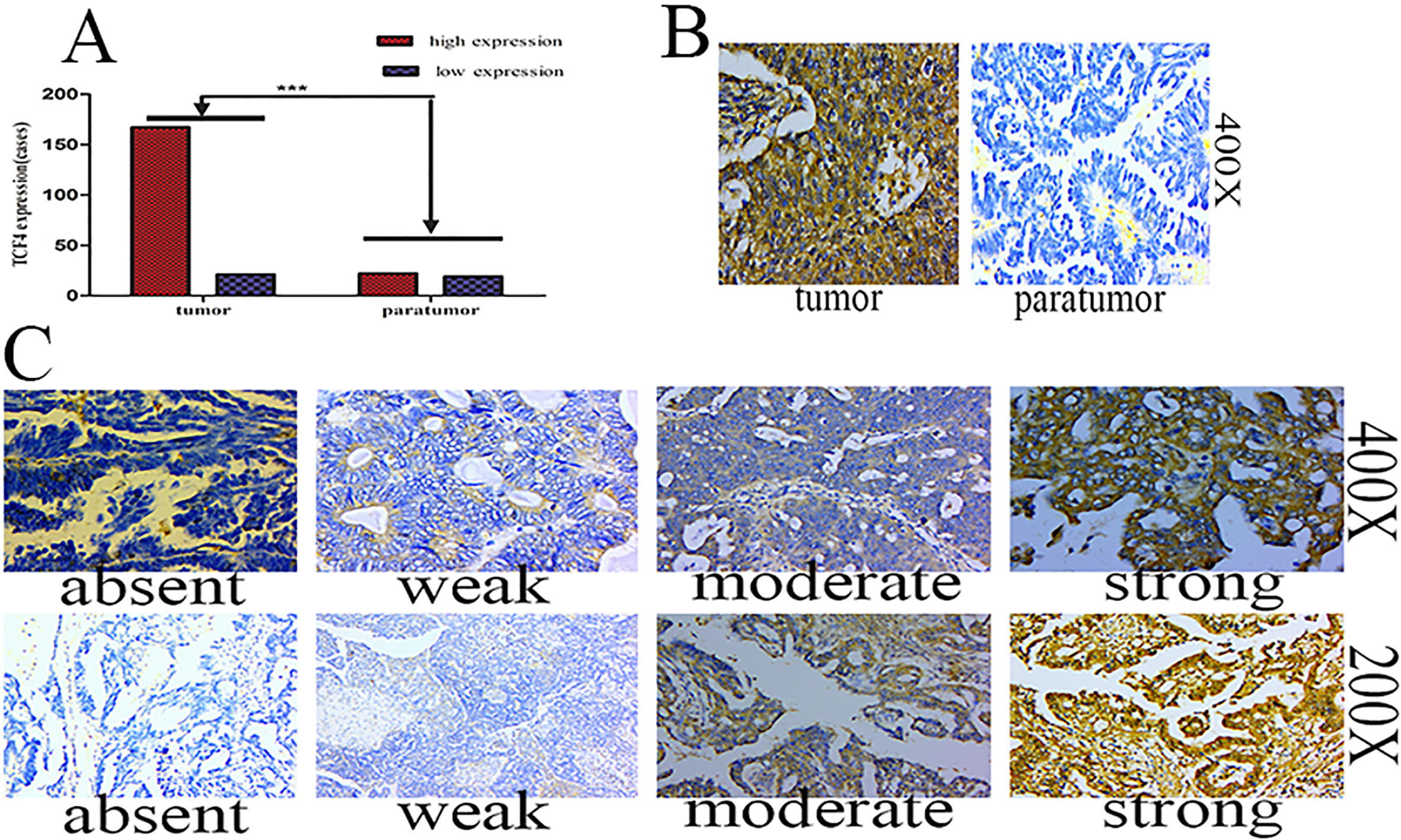
The TCF4 protein expression in epithelial ovarian cancer tissues and paratumor tissues were all detected by immunohistochemical technology. The step was following, 4
TCF4 protein overexpression is associated with disease progression free and overall survival in epithelial ovarian cancer patients.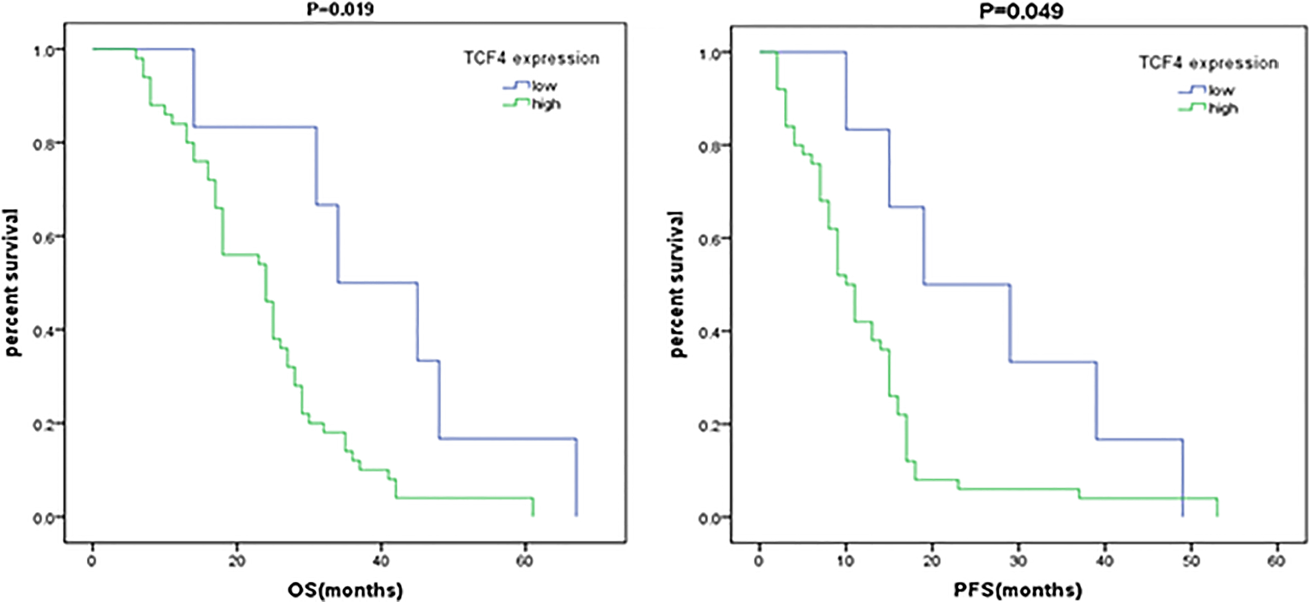
All of the statistical analyses were calculated with the statistical software package SPSS 21.0. The
Results
Comparison and representative photomicrographs of ovarian cancer/paratumor tissues immunohistochemically stained for TCF4
In 188 cases of ovarian cancer tissues stained with the TCF4 specific antibody, 21/188 (11.17%) had weak/absent staining (also called low expression) and 167/188 (88.83%) had strong/moderate staining (also called high expression), while in 41 ovarian paratumor tissues, 19/41 (46.34%) had weak/absent staining and 22/41 (53.66%) had strong/moderate staining, and there was significant difference between the two groups (
TCF4 protein overexpression was associated with disease progression free and overall survival in epithelial ovarian cancer patients
In order to determine whether TCF4 expression was associated with epithelial ovarian cancer patients survival, we stained 188 epithelial ovarian cancer cases and 41 paired ovarian paratumor tissues using immunohistochemistry with a rabbit polyclonal antibody specific for TCF4. Kaplan-Meier survival analysis demonstrated that there was a statistically significance on disease progression free survival between high and low expression of TCF4 (
TCF4 overexpression was correlated with the clinical features of ovarian cancer
Subsequently, we studied that TCF4 overexpression was correlated with the clinical parameters of epithelial ovarian cancer. The percentages of patients with stages I, II, III, and IV tumors were 14.36%, 11.17%, 58.51%, and 15.96%, respectively (Table 1).
Clinicopathological parameters and tumor expression of TCF4 in epithelial ovarian cancer
Clinicopathological parameters and tumor expression of TCF4 in epithelial ovarian cancer
In addtion, the results of the statistical analysis using the
Using
Univariate analysis of TCF4 expression in association with standard clinicopathological variables using the
-or Fisher’s Exact test
Univariate analysis of TCF4 expression in association with standard clinicopathological variables using the
Cox regression univariate and multivariate analyses of prognostic factors in epithelial ovarian cancer
Furthermore, we also assessed the prognostic value of TCF4 overexpression in epithelial ovarian cancer patients. In a univariate Cox analysis, TCF4 expression, intraperitoneal metastasis, postoperative chemotherapy, and hyperthermic intraperitoneal chemotherapy were significant prognostic factors (Table 3). In addition, multivariate Cox regression analysis further showed that TCF4 protein expression was an indeed independent prognostic factor of epithelial ovarian cancer (Table 3). Taken together, these results suggested that TCF4 expression was an independent prognostic predictor, and TCF4 may contribute to the prognosis of epithelial ovarian cancer, which suggested that TCF4 may be a useful independent prognostic marker in epithelial ovarian cancer. Moreover, In this multivariate model, intraperitoneal metastasis was no longer significant.
Discussions
As we all know, the Wnt/
Prior to our research, several clinical studies have reported that TCF4 is an indicator of poor prognosis or malignant potential in hepatocellular carcinomas [14], colon cancer [15], lung adenocarcinomas [16] and esophageal squamous cell carcinoma [13]. And interestingly, consistent with these reports, we found that TCF4 predicted the poor prognosis of patients with epithelial ovarian cancer. And our data for the first time suggested that TCF4 can be used to act as a candidate useful biomarker for predicting prognosis in patients with epithelial ovarian cancer. In this study, we found that patients with high expression of TCF4 had a poorer prognosis.
Moreover, Ishiguro et al. reported [13] that TCF4 expression was correlated with Tumor status, pathological stage, lymphatic invasion and blood vessel invasion, but did not correlate with age, gender or lymph node status. However, in the study overexpression of TCF4 in epithelial ovarian cancer patients was found to be significantly related with age, FIGO stage, lymph node metastasis, intraperitoneal metastasis, intestinal metastasis, vital status, intraperitoneal recurrence, and serum CA153. Thus, we concluded that patients with TCF4 overexpression were more likely to trend for older patients, which indicated that TCF4 detection should be performed in older people in order to diagnose earlier. Moreover, TCF4 overexpression were more in III/IV stages, and this showed that TCF4 overexpression were more likely to exhibit advanced disease, so we concluded that TCF4 expression increased with the occurrence and progress of ovarian cancer. In addition, we found that TCF4 overexpression were more likely to develop lymph node metastasis, intraperitoneal metastasis, and intestinal metastasis, which showed that TCF4 was correlated with ovarian cancer metastasis and migration. Moreover, we found that TCF4 overexpression were more likely to develop intraperitoneal recurrence, which suggested that TCF4 was associated with intraperitoneal recurrence of epithelial ovarian cancer. To the best of our knowledge, epithelial ovarian cancer metastasis and recurrence are the major risk factors of poor survival of patients. Patients with metastasis and recurrence usually have a worse prognosis. Thus, an early diagnosis of intraperitoneal metastasis and recurrence is important for the survival of ovarian cancer patients, but only a few tumor markers are currently used to predict recurrence and metastasis of ovarian cancer in the clinical work. So our data showed that TCF4 is meaningful as a biomarker or predictor to diagnose metastasis and recurrence of ovarian cancer. We concluded that TCF4 may directly or indirectly participate in the migration and invasion of epithelial ovarian cancer. But it need more in viro and in vitro experiments to identify its role in ovarian cancer.
And in a multivariate Cox regression TCF4 was still associated with survival, which showed that TCF4 is a useful indeed independent prognostic marker in ovarian cancer. Taken together, our research showed that TCF4 was associated with the prognosis of epithelial ovarian cancer for the first time.
Conclusions
TCF4 may play a candidate oncogenic role in epithelial ovarian cancer, and TCF4 is a useful independent prognostic marker in ovarian cancer, and it may provide a candidate target therapy treatment in future.
Footnotes
Acknowledgments
This work was supported by the Nature Science Fund of Guangdong Province (no.2016A030313536).
Conflict of interest
The authors declare no conflict of interest.