
Abstract
BACKGROUND:
The microRNA plays an important role in tumor progression. MiR-1236-3p acts as a tumor suppressor in various malignancies.
OBJECTIVE:
The aim of present study was to explore the expression of miR-1236-3p in gastric cancer (GC) and its correlation with clinicopathological features, and evaluate the feasibility of using it as a prognostic biomarker in GC.
METHODS:
Seventy-six pairs of tissue specimens were collected from GC patients. MiR-1236-3p expression level was detected by using qRT-PCR. The diagnostic value of miR-1236-3p was evaluated by receiver operating characteristic curve, and Kaplan-Meier method was used to analyze the overall survival. Prognosis analysis was performed using multivariate cox proportional hazards regression analysis.
RESULTS:
The expression of miR-1236-3p was significantly reduced in tumor tissues (
CONCLUSIONS:
The study showed that miR-1236-3p is downregulated in GC tissues, and low expression of miR-1236-3p is associated with a poor prognosis in GC. It may be a new diagnostic and prognostic biomarker for GC.
Introduction
Gastric cancer (GC) is the fourth most common cancer and the second major cause of cancer-related death in the world [1, 2]. Because of the lack of early diagnostic indicators, most of the patients were diagnosed with advanced GC. Although cancer diagnosis and treatment have made great improvement, advanced GC patients still always have poor prognosis mainly due to frequent metastasis and recurrence after surgery [3], effective therapeutic methods that can be used for them are limited [4, 5]. Therefore, it is imperative to identify novel biomarkers for its diagnosis and to develop anticancer targeted therapies.
MicroRNAs (miRNAs) are a class of small, non-coding, single stranded, regulatory RNA species, approximately 19–25 nucleotides in length, which can promote or inhibit the expression of gene through binding to the 3’-untranslated region of the target mRNA at the post-transcriptional level [6, 7, 8]. Accumulating evidences have proved that miRNAs have associated with various biological processes such as proliferation, differentiation, apoptosis, and metastasis [9, 10]. Furthermore, there is increasing evidence that miRNAs serve a crucial role in cancer development and progression. They can function as tumor suppressor genes or oncogenes in different types of cancer through the regulation of cellular proliferation, metastasis, and apoptosis [11, 12, 13]. Numerous miRNAs dysregulation has already been reported in GC occurrence and progression; this suggests that miRNAs could serve as a diagnostic marker or target of therapeutics for GC [14, 15].
Mir-1236-3p, an intronic miRNA, is located in the Chr6p21.33 and embedded within the intron of the NELFE gene. MiR-1236-3p has been identified as a tumor suppressor gene, and downregulation of miR-1236-3p has been specified in some cancers, including hepatocellular carcinoma, ovarian cancer, renal cell carcinoma, bladder cancer, lung cancer, and breast cancer [16, 17, 18, 19, 20]. However, its biological function in GC is remains unclear, few studies have investigated the association between miR-1236-3p and GC development. In the present study, we quantitatively compare the expression level of miR-1236-3p in gastric cancer tissues relative to their non-tumor counterparts. Moreover, the association of miR-1236-3p misregulation with clinicopathological features and prognostic value has been investigated. The aim of this study is to evaluate whether miR-1236-3p is capable of acting as a potential diagnostic and prognostic biomarker and therapeutic target for GC patients.
Primers of qRT-PCR
Primers of qRT-PCR
Clinical samples and cell lines
Seventy-six pairs of cancerous tissues and their paired adjacent non-tumor tissues were obtained from patients with gastric cancer who underwent surgery at the Cancer Research Institute of China Medical University. All specimens had been diagnosed with GC by histopathological confirmation. None of the patients underwent chemotherapy before the operation. Tissue samples were snap frozen in liquid nitrogen after surgical removal. The study protocol was approved by the Ethics Committee of China Medical University, and the study conforms with The Code of Ethics of the World Medical Association (Declaration of Helsinki). All human gastric cancer cell lines, MKN45, MGC803, HGC27, SGC7901, and the human normal gastric epithelial cell line (GES-1) were purchased from Shanghai Institute of Cell Biology, Chinese Academy of Sciences (Shanghai, China).
RNA isolation and RT-qPCR
Total RNA was extracted using TaKaRa MiniBEST Universal RNA Extraction Kit (Takara, Dalian, China). The cDNA was reverse-transcribed using Hairpin-it
Receiver operating characteristic (ROC) curve analysis
The relative levels of miR-1236-3p expression in gastric cancer and non-tumorous tissues were recorded. ROC curves were generated, with the horizontal axis as specificity and the vertical axis as sensitivity, using GraphPad Prism 6 software. According to the drawn ROC curves, the diagnosis cut-off points and their specificity and sensitivity were analyzed and calculated. The diagnostic value of miR-1236-3p in GC was evaluated, and presented by the area under the curve (AUC).
Statistical analysis
Statistical evaluation was performed using SPSS 19.0 software and GraphPad Prism 6 software. Data were shown as mean
MiR-1236-3p was downregulated in GC tissues and cells. (A) The expression levels of miR-1236-3p in 76 pairs of human GC tissues and adjacent normal tissues measured by qRT-PCR. (B) The expression levels of miR-1236-3p in GC cells and GES-1 cells. *, 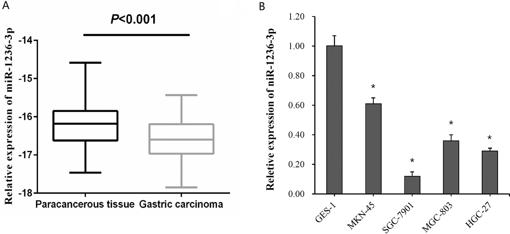
Associations between miR-1236-3p level and clinicopathological parameters in patients with GC
The
The respective area under receiver operating characteristic curves of miR-1236-3p in GC.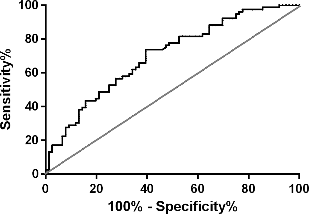
Univariate and multivariate Cox regression analyses for overall survival
MiR-1236-3p expression was downregulated in GC tissues and cells
The relative expression level of miR-1236-3p was analyzed in 76 paired clinical GC and adjacent non-tumor tissues using qRT-PCR to consider whether miR-1236-3p has a role in gastric tumorigenesis. As shown in Fig. 1A, the expression of miR-1236-3p was significantly downregulated in GC tissues when compared with adjacent normal tissues (
Association between the clinicopathological features and expression levels of miR-1236-3p in human GC
To elucidate the underlying function of miR1236-3p in the development of GC, We analyzed the associations between the miR-1236-3p expression level and the clinicopathological parameters of the patients with GC. The patients were divided into two groups. The cancer tissues with higher expression of miR-1236-3p than their adjacent normal tissues were selected as the high group, while those with less expression of miR-1236-3p than their adjacent normal tissues were selected as the low group. As shown in Table 2, the results indicated that a low expression level of miR-1236-3p was correlated with high TNM stage (
Kaplan-Meier survival curve analysis of miR-1236-3p expression levels.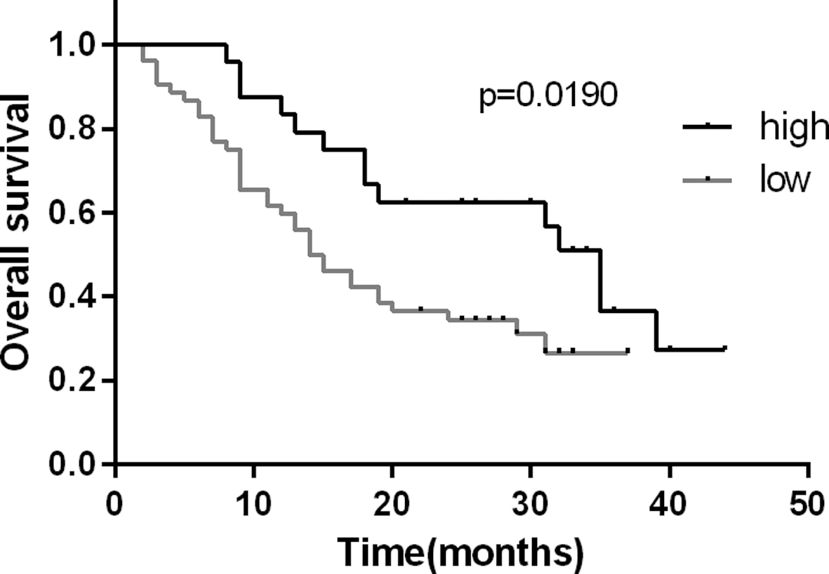
ROC curves were employed to analyze and assess the diagnostic value of miR-1236-3p in GC (Fig. 2). Comparing tumor tissues with adjacent normal tissues, ROC curve analysis demonstrated that the AUC of miR-1236-3p was 0.7016 (95% confidence interval: 0.6195–0.7837), with a diagnostic threshold of-16.28, specificity and sensitivity were 60.53% and 73.68%, respectively.
Significance of miR-1236-3p expression in GC prognosis
To further investigate the prognostic value of miR-1236-3p expression, we used Kaplan-Meier method and log-rank test. As shown in Fig. 3, survival analysis indicated that overall survival of GC patients with high miR-1236-3p levels were significantly higher than those of patients with low miR-1236-3p levels (in the log-rank test,
Discussion
Despite considerable improvement in cancer diagnosis and treatment, patients with advanced GC have poor prognosis primarily due to frequent tumor metastasis and tumor recurrence after surgery [3]. Patients with GC were often diagnosed at a late stage and had a low survival rate. Therefore, clarification of the molecular mechanism of GC is crucial for developing effective intervention and therapeutic strategies. However, identification of molecular biomarkers with clinical value is still a great challenge.
Many biomarkers like carcinoembryonic antigen (CEA), carbohydrate antigen (CA) series (CA19-9, CA72-4) and p53 gene have been reported to have association with diagnosis and prognosis in GC, but they are lack of specificity and sensitivity [21, 24]. In the samples of GC patients, Liu et al. [22] compared the AUC values of CEA and CA19-9 which were only 0.503 and 0.600, respectively. However, in our study, miR-1236-3p in GC patients had significantly lower expression than in the control group, the AUC was 0.7016 (95% CI: 0.6195–0.7837). The results show that miR-1236-3p has some advantages as a tumor marker. Zhang et al. [23] analysed the diagnostic accuracy of p53 gene for detection of GC, the AUC value was 0.70. It’s close to our results. But the association between p53 overexpression with lymph nodes metastasis and shorter survival is still controversial; therefore, p53 cannot be considered a trustworthy prognostic biomarker [24].
MiRNAs have gained much attention due to its important role in carcinogenesis. As the biomarkers of diagnosis, tissue miRNAs are most widely used in scientific research. A large number of miRNAs have been found in GC, but their underlying molecular mechanism in GC development is still poorly understood [25, 26].
Previous studies have shown that decreased expression of miR-1236-3p was observed in many malignancies. For instance, Gao et al. [16] showed that miR-1236-3p could suppress the proliferation, migration and invasion capacity of hepatocellular cancer cells by targeting the 3’ UTR of AFP mRNA and down-regulating its expression. Wang et al. [17] found that miR-1236-3p overexpression suppressed the migration and invasion of ovarian cancer cells by targeting EMT-inducer ZEB1. Wang et al. [19] observed that overexpression of miR-1236-3p inhibited the proliferation, migration and invasion of bladder cancer cells by upregulating p21. They also identify that miR-1236-3p directly interacted with p21 promoter. Butkytė et al. [27] showed that miR-1236-3p expression was down-regulated in GC cell line KATOIII. All these findings indicated that miR-1236-3p may serve as a tumor suppressor in various cancers. However, the clinical significance of miR-1236-3p in GC tissue has not been investigated.
In the current study, miR-1236-3p expression in GC tissues was significantly lower than that in normal tissues. MiR-1236-3p served as a tumor suppressor, which was similar to the other cancers. We further found that the level of miR-1236-3p in GC was strongly correlated with TNM stage, Lymph node Metastasis, and differentiated degree. GC patients with low miR-1236-3p expression had poorer overall survival. Compared with above mentioned biomarker p53 gene, miR-1236-3p had better credibility as a biomarker. The AUC value was 0.7016 and the sensitivity was obtained about 73.68%, which indicates potency of miR-1236-3p for differentiating the healthy group from patient group. Furthermore, univariate and multivariate survival analysis indicated that miR-1236-3p was an independent prognostic factor for overall survival of GC prognosis and could be used as a potential prognostic biomarker. According to the authors’ knowledge, this is the first study to investigate the impact of miR-1236-3p expression on diagnosis and prognosis of GC patients using a large number of clinical samples.
The present study did have limitations. There may have been selection bias in operation when the inclusion criteria were formulated. We noticed that there were no significant differences between miR-1236-3p expression and TNM invasion, age, and gender of the GC patients under study. This could be owing to the limited sample size, the effect of the expression pattern of tumorous cells on healthy adjacent cells, and the selection of tumorous samples from patients.
In order to better understand the role of miR-1236-3p in GC, we still need to do further mechanism research. Like many other microRNAs, miR-1236-3p may also affect the progression of GC by acting on downstream target genes. This study confirmed the expression of miR-1236-3p in GC tissues and cells. In the following experiments, we will further study the effects of miR-1236-3p on the biological function of GC cells, look for and verify the downstream target genes. At the same time, we will explore the effects of miR-1236-3p on epithelial-mesenchymal transition and related signaling pathways.
In conclusion, the present results demonstrated that miR-1236-3p expression levels were downregulated in GC patients. Down-regulation of miR-1236-3p is associated with the tumor stage, lymph node metastasis and differentiated degree. Therefore, miR-1236-3p may be a potential biomarker for diagnosing GC, as well as predicting the prognosis, evaluating the curative effects, and monitoring the recurrence of GC patients. It also may be a potential therapeutic target for gastric cancer. The exact efficiency of miR-1236-3p as the tumor marker remains to be confirmed by further confirmation.
Footnotes
Acknowledgments
This work was supported by Liaoning Province Science and Technology Plan Project (No. 2013225021) and the Natural Science Foundation of Liaoning Pro- vince (No. 201602817).