
Abstract
This article has been retracted, and the online PDF replaced with this retraction notice.
Introduction
Osteosarcoma is a primary malignant bone tumor commonly observed in children or teenagers, and occupies about 5% of total childhood tumors [1]. Osteosarcoma is commonly occurred in epiphysis end of long bones such as femora and humeral, and has characteristics such as high malignancy and invasiveness [2]. With abundant blood flow at epiphysis, osteosarcoma cell is predisposed to have distal metastasis via blood circulation, leading to treatment difficulty and unfavorable prognosis, bringing heavy burdens for public health [3]. Recent study has revealed certain anti-tumor effect of sclareol in inhibiting tumor growth such as osteosarcoma, breast cancer, gastric cancer and colorectal carcinoma [4, 5]. Sclareol, also named as sclarcol, is a di-tert-alcohol firstly extracted from sage clary plants in 1982 and later in other plants [6]. Study has found its LD
Materials and methods
Reagents and equipment
DMEM medium, fetal bovine serum (FBS) and trypsin were purchased from Gibco (US). DMSO, sclareol and PI dyes were produced by Sigma (US). CCK8 test kit was obtained from Toyobo (Japan). Culture dish was a product of Corning (US). CO
Cell line and culture
MG63 osteosarcoma cells were purchased from Shanghai Cell Biology Institute, Chinese Academy of Sciences. Cells were kept in DMEM medium containing 10% FBS, and were kept in a humidified chamber with 5% CO
CCK8 assay
Cells at log-phase were collected by centrifugation. After re-suspension, cells were inoculated in 96-well plate (1
Effect of sclareol on MG63 cell proliferation.
MG63 cells were treated with DMSO (control) or sclareol. After 48 hours, the medium was removed, following by the digestion by 0.25% trypsin. Cells were rinsed in DMEM to prepare single cell suspensions, which were centrifuged at 1000 g for 5 min. PBS was then added to rinse cells again for further centrifugation. Supernatants were removed with adding 0.2 mL blocking buffer. The mixture was then incubated at room temperature for 15 min. 0.5 mL buffer solution was then added, followed by centrifugation. 0.2 mL annexin V dye was added, mixed and incubated for 10 min. After rinsing and washing by centrifugation, PI dye was added. The mixture was then loaded for flow cytometry to detect apoptosis.
Sclareol induced MG63 cell apoptosis. A to D, flow cytometry for apoptosis in MG63 cells treated with DMSO (A), 2 
Cells were digested in 0.25% trypsin, and were rinsed to prepare single cell suspension, which was centrifuged at 1000 g for 5 min. DMEM medium was then added to prepare single cell suspension (1
Western blot analysis of the expression of cytochrome c, Bax and Bcl-2 in cells after treatment with different concentrations of sclareol.
Meanwhile, cells were mixed with 10
RIPA lysis buffer was added into cells for extraction of total proteins, which was quantified by BCA assay. 40
Statistical analysis
SPSS 20.0 software was used to process all collected data. Measurement data were presented as mean
Sclerol’s effect on mitochondrial membrane potential. Flow cytometry was performed for measuring mitochondrial membrane potential in MG63 cells treated with DMSO (A), 2 
Sclerol’s effect on mitochondrial membrane potential. Confocal microscopy analysis was performed for measuring mitochondrial membrane potential in MG63 cells treated with DMSO, 2 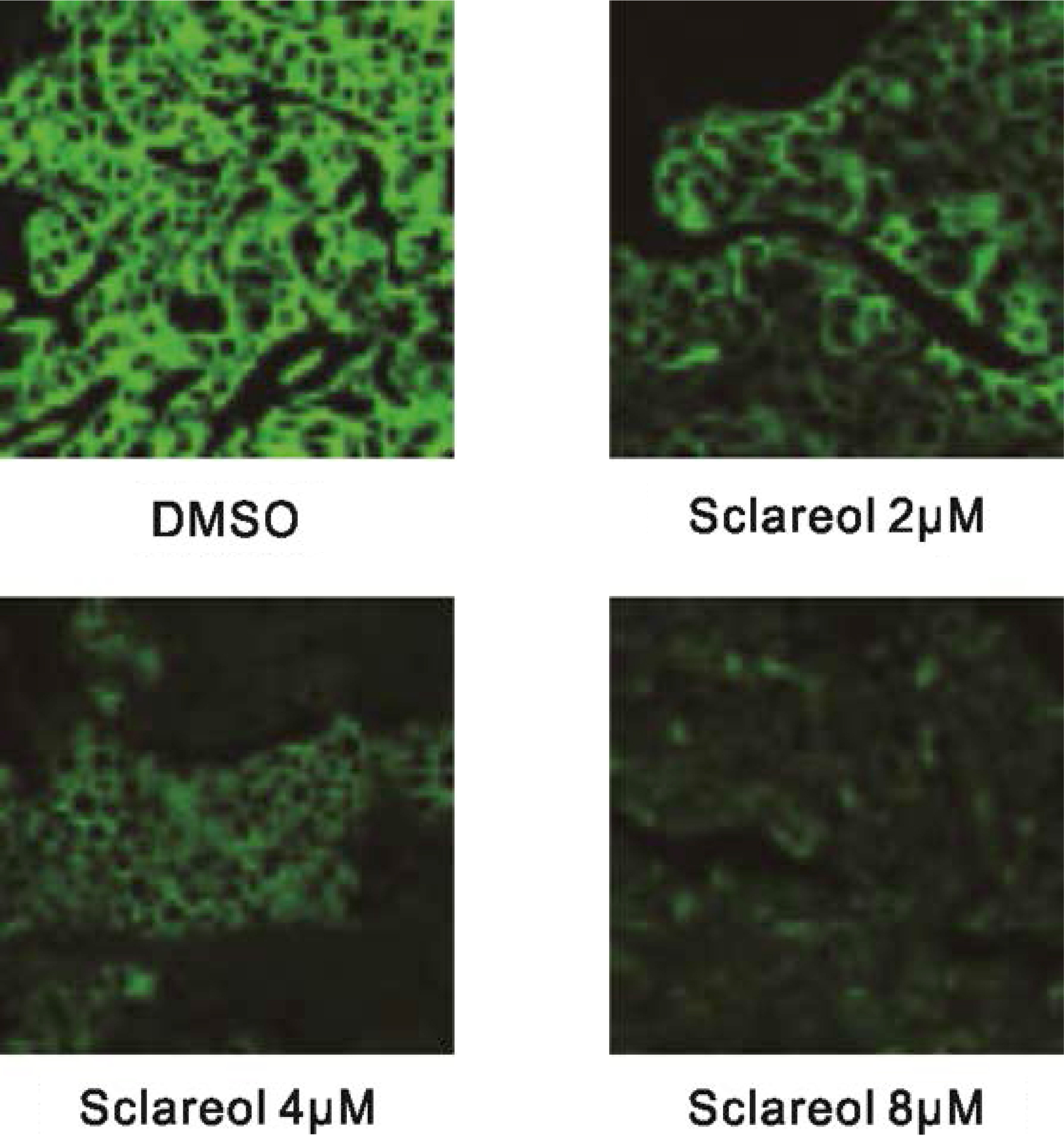
Effect of sclareol on MG63 cell proliferation
MG63 osteosarcoma cells were divided into control, DMSO and experimental group, which received no treatment, DMSO treatment and gradient concentration of sclareol (0.5
Sclareol induced MG63 cell apoptosis
To study the mechanism of sclareol on inhibiting MG63 cell proliferation, we further investigated the effect of sclareol on MG63 cell apoptosis. We divided cells into DMSO group and treatment group, which was further sub-divided into 2.0
Effect of sclareol on mitochondrial membrane potential (
)
To evaluate the effect of sclareol on mitochondrial membrane potential, flow cytometry was performed using JC-1 reagent. Twelve hours after treatment with different concentrations of sclareol (2.0
Discussion
Sclareol is a di-tert-alcohol firstly extracted from sage clary plants, which are mainly distributed in China and Mediterranean regions [8]. Traditionally it is used to produce perfumes and additives, and also as materials for drug development [9]. Previous reports indicated the anti-tumor activity of sclareol as it showed inhibitory effects on osteosarcoma, breast cancer, gastric carcinoma and colorectal carcinoma, but leaving the exact mechanisms largely unknown [15]. To study the anti-tumor activity of sclareol, we selected MG63 osteosarcoma cell as the model, in which the effect of sclareol on cell viability was observed using CCK8 assay. All cells were divided into blank control, solvent control and experimental group. Our results showed decreased proliferation ability of cells after sclareol treatment. Within certain dose range, there was a negative correlation between sclareol concentration and cell viability. Under the same concentration of drug, the proliferation ability of MG63 cell was decreased with elongated treatment period. To further study the mechanism underlying sclareol inhibition on osteosarcoma cells, we divided cells into DMSO and sclareol group, in which flow cytometry was used to test cell apoptosis. Our results showed significantly elevated apoptotic cell ratio in treatment group comparing to DMSO treated cells (
However, the mechanism underlying apoptosis of MG63 cells induced by sclareol is still unknown. Previous literature showed certain role of mitochondria in cell apoptosis, whose membrane potential has close relationship with cell apoptosis [16]. As an organelle with double lipid membrane, mitochondrion is the main location for producing energy of cell aerobic respiration. It also participates in signal transduction and cell apoptosis, with regulatory role on cell growth and cell cycle [17]. Recent study has shown the decrease of mitochondrial membrane potential may change permeability of membrane and induce further apoptosis [18]. Therefore cell apoptosis can be observed by measuring the membrane potential of mitochondria. To further illustrate the molecular mechanism underlying sclareol-induced apoptosis, we divided MG63 cells into both DMSO and sclareol treated group, in which the effect of sclareol on membrane potential of cells was assessed by flow cytometry using JC-1 reagent. Results showed significantly depressed mitochondrial membrane potential of sclareol-treated MG63 cells compared to DMSO-treated ones (
In summary, sclareol can decrease mitochondrial membrane potential of MG63 osteosarcoma cells and alter mitochondrial biological functions, thus inducing cell apoptosis and inhibiting osteosarcoma cell growth and proliferation.
Footnotes
Conflict of interest
None.