
Abstract
BACKGROUND AND OBJECTIVE:
Propofol, an intravenous anesthetic agent, has been found to inhibit growth of breast cancer cells. However, the mechanisms underlying the antitumor are not known. A recent report has found that propofol could significantly downregulate miR-24 expression in the human malignant cancers. In breast cancer cells, overexpression of miR-24 promotes cell proliferation and inhibits cell apoptosis by downregulation of p27. The miR-24 has been reported to be overexpressed in breast cancer and breast cancer cell lines. In the present study, we hypothesized that propofol induces apoptosis of breast cancer cells by miR-24/p27 signal pathway.
METHODS:
Breast cancer MDA-MB-435 cells were exposed to propofol (10
RESULTS:
MDA-MB-435 exposed to propofol showed a significant increase in apoptotic cells, followed by the downregulation of miR-24, upregulation of p27 expression and cleaved caspase-3 expression. Targeting p27 inhibits propofol-induced cell apoptosis; miR-24 overexpression decreased propofol-induced cell apoptosis, cleaved caspase-3 and p27 expression.
CONCLUSIONS:
Propofol induce
Keywords
Introduction
Propofol is widely used in all kinds of surgeries due to its short effect and rapid recovery. Apart from its multiple anesthetic advantages, propofol exerts a number of non-anesthetic effects [1]. In HL-60 human promyelocytic leukemia cells, propofol was shown to inhibit growth and induce the formation of apoptotic bodies, increase DNA fragmentation and laddering, activate caspase-3, caspase-6, caspase-8 and caspase-9, and induce the cytosolic release of cytochrome
MicroRNAs (miRNAs) are emerging as robust players of gene regulation. Numerous miRNAs have been implicated in a variety of cellular processes including differentiation, cell proliferation, apoptosis, embryonic development, stress response, stem cell renewal and metabolism [7, 8, 9, 10, 11]. miRNAs have also been implicated to play important roles in inducing apoptosis [12, 13, 14]. However, the effect of miRs on anesthetic-induced apoptosis in breast cancer cells has yet to be studied.
Recent studies have shown that miR-24 is upregulated in highly differentiated CD8
Materials and methods
Cell culture
MDA-MB-435 cells (ATCC, Shanghai, China) were cultured in DMEM supplemented with 10% FCS, 2 mM L-Glutamine, 100 Units/ml Penicillin and100
Propofol exposure
Pure propofol (2, 6-diisopropylphenol) was obtained from AstraZeneca, and the intralipid (20% emulsion, phospholipid-stabilized soybean oil) was obtained from Sigma-Aldrich. MDA-MB-435 cells were cultured in 96-well plates (2
qRT-PCR assay
Total miRNA was isolated through the mirVana miRNA isolation kit (Ambion). Quantitative expression studies of miR24 transcripts were performed using TaqMan miRNA Assays (Applied Biosystem). The cDNA was obtained using the High Capacity RNA-to-cDNA Kit (Applied Biosystem). cDNA was amplified using specific primer for miR-24 (Applied Biosystem). qRT-PCR was performed using an Applied Biosystems 7900HT Fast RT-PCR system. The expression level of miR-24 was calculated according to the 2
Western blot analysis
The primary antibodies used were mouse anti-p27, rabbit anti-cleaved caspase-3, and mouse anti-GAPDH. The horseradish peroxidase-conjugated secondary antibody (1:5000; Santa Cruz Biotechnology) and chemiluminescence Supersignal (Pierce) was used to detect protein. Protein loading was normalized for GAPDH signal. Band intensity was quantified versus a control sample (considered as 1).
Propofol increases cell apoptosis in the propofol-treated cells. A, Imaging of cell apoptosis determined by TUNEL assay. Nuclei of apoptosis was identified as TUNEL positive (green fluorescent) cell. Scale bar represents 50 um (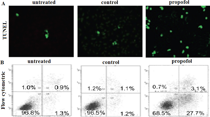
The cell arrays were fixed with 3.7% formaldehyde for 20 min at room temperature. After two PBS washes, the permeabilization was performed with 0.1% Triton X-100 in 0.1% sodium citrate for 2 min at room temperature. After washing with PBS, the appropriate volume of freshly prepared TUNEL reaction mixture (In Situ Cell Death Detection Kit, Fluorescein, Roche) was added to the slides and the slides were incubated in a humid atmosphere for 60 min at 37
Flow cytometric analysis of cell apoptosis
Apoptosis assay was performed using Vybrant Apo- ptosis Assay kit (Invitrogen) according to manufacturer’s instructions. Both apoptotic cells (PI- Annexin V
Statistical analysis
The data were expressed as the means
Results
Propofol induces cell death of MDA-MB-435 cells
The TUNEL-positive cells was 1.04%
Downregulation of miR-24 is required for the apoptotic activity of propofol. MDA-MB-435 cells were transiently transfected with miR-24 mimics or control miR or/and treated with 10 uM propofol for 6 h. A, miR-24 expression was detected by qRT-PCR assay; B, Imaging of cell apoptosis determined by TUNEL assay. Nuclei of apoptosis was identified as TUNEL positive (green fluorescent); C, Cells apoptosis induced by propofol was analyzed using the annexin V (FITC)/PI binding assay by flow cytometry (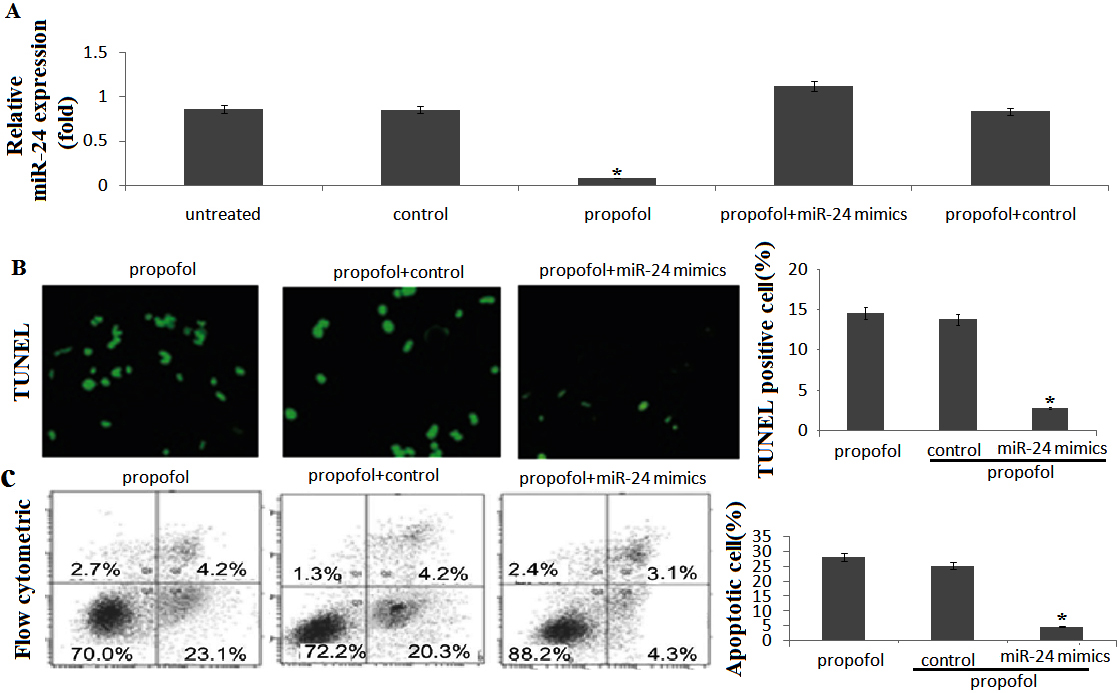
Propofol directly mediates miR-24/P27 induction. Cells were transfected with miR-24 mimics and/or treated with 10 uM propofol for 6 h. P27, caspase-3 and cleaved-caspase-3 was detected by western blot assay.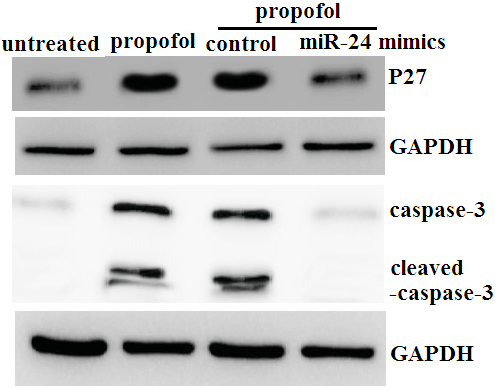
P27 mediates propofol-induced apoptosis. Cells were transfected with P27 siRNA and/or treated with 10 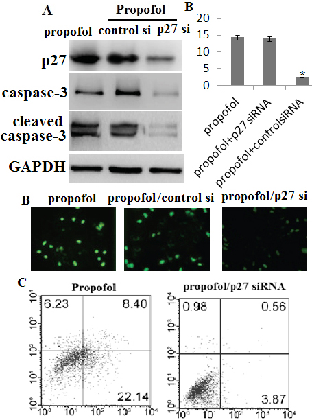
We first investigated the effects of propofol on miR-24 expression of MDA-MB-435 cells. Exposure to 10 uM propofol for 6 hours in MDA-MB-435 cells led to rapid miR-24 downregulation (Fig. 2A). However, transient miR-24 mimics transfection reveresd propofol-induced miR-24 downregulation (Fig. 2A) and reduced apoptosis following propofol treatment (Fig. 2B and C). Taken together, these data suggest that propofol decreased miR-24 expression, resulting in the induction of apoptosis in the MDA-MB-435 cells.
MiR-24-dependent induction of p27 by propofol
We then determined whether propofol-induced miR-24-dependent p27 expression. Exposure to 10 uM propofol for 6 hours in led to P27 upregulation (Fig. 3). However, transient miR-24 mimics transfection aggravated propofol-induced P27 upregulation (Fig. 3), suggesting that p27 was miR-24 negatively dependent regulation.
Caspases family plays an important role to regulate cell apoptosis, the mechanisms of which involved in the upstream initiators and downstream effectors. For example, caspase-3 is a main caspase effector. Therefore, we detected the effect of propofol on activity of caspase-3. As seen in Fig. 3, propofol significantly increased the caspase-3 activity by western blot assay. However, transient miR-24 mimics transfection inhibited propofol-induced caspase-3 upregulation and activation (Fig. 3).
P27 mediates propofol-induced apoptosis
We then investigate the role of P27 in propofol-induced apoptosis. Exposure to 10
Discussion
Previous studies on proof have mainly focused on its clinical effects and side effects [25, 26, 27] and some investigations have indicated that propofol could induce neurotoxicity [28]. A recent study showed that propofol can induce cancer cell apoptosis [29, 30, 31]. Studies have shown that propofol could induce tumor cell apoptosis by downregulation of miRs, such as miR-34a-5p [32], miR-4295 [33], miR-222 [34], miR-574–3p [35] and miR-9 [36]. However, the detailed mechanism about how miRs induces apoptosis is not clear. Studies have shown that miRs induce tumor cell apoptosis via regulation of p27 expression [37, 38, 39]. Lynch et al. has reported that miR-24 has a tumor suppressor role in prostate cancer and also targets p27 and p16 in prostate cancer cells [40]. However, miR-24 inhibits apoptosis in human breast cancer by targeting p27 [41, 42, 43]. miR-24 was found to be significantly decreased in the human embryonic stem cell-derived neurons induced by propofol and significantly induced cell death [44, 45]. Therefore, we explored whether propofol could control miR-24/P27 pathway to induce apoptosis in breast cancer cells.
In our study, miR-24 was significantly downregulated in MDA-MB-435 cells when exposure to 10 uM propofol for 6 hours followed by increasing cell death. However, overexpression of miR-24 by miR-24 mimics transfection in MDA-MB-435 cells significantly decreased toxic effects of propofol, suggesting that miR-24 plays an important role in resistance of MDA-MB-435 cells cell death.
miRNAs-mediated control is one of the posttranscriptional mechanisms responsible for p27 downregulation in cancer cells. p27 posttranslational regulation is mediated by multiple phosphoacceptor sites, controlling both nucleo-cytoplasmic shuttling and stability of the protein [46]. Reduced expression of p27 is frequently observed in various cancers [47, 48, 49] and correlates with poor prognosis [50, 51]. From these studies, it is clear that p27 is a promising target for tumor therapies. In fact, it is reported that overexpression of p27 suppresses tumor growth in various cancers [49, 52]. The role of miR-24 in regulating the expression of p27 and regulation of cell survival is well-established [41, 42, 43, 44, 45].
In the present study, we found that miR-24 was significantly downregulated and p27 and cleaved caspase-3 were significantly upregulated following exposure to 10
Some limitations are avaliable in the current study. First of all, we only used one cell line MDA-MB-435 at present. More breast cancer cell lines will be included in our following study. Secondly, MDA-MB-435 cells were treated with 10
In conclusion, propofol induces cell death in breast cancer cells via inactivation of miR-24 and activation of p27 signal pathway. miR-24 in propofol-induced cell apoptosis could possibly pave the way for new research into possible strategie for treatment of breast cancer.
Footnotes
Conflict of interest
No.