
Abstract
FTIR spectroscopy is an analytical technique widely applied for studying the vibrational fingerprint of organic compounds. In recent years, it has been applied to many biomedical fields because of its potential to detect the composition and molecular structure of various biological materials without the need of probe molecules. The coupling of IR spectrometers with visible microscopes has led to perform the imaging analysis of non-homogeneous samples, such as tissues and cells, in which the biochemical and spatial information are close related. In this review, we report the most significant applications of FTIR to the study of cells in different conditions (fixed, dried and living) with the aim to monitor their biochemical modifications, either induced or naturally occurring.
Introduction
Fourier Transform Infrared (FTIR) spectroscopy is a well-known technique widely applied for studying the vibrational fingerprint of organic compounds. It exploits a sensitive, non-invasive and non-damaging radiation, which causes vibrational transitions due to the interaction of matter with a broad band infrared source and carries a great amount of molecular information [4,50,60,69]. Within the mid-infrared range (MIR, 4000–400 cm−1), all molecules have specific vibrational frequencies corresponding to discrete vibrational energy levels, which characterize the infrared spectrum of each compound.
FTIR spectroscopy allows detecting the composition and molecular structure of biological materials without the need of probe molecules [11]. Its application to investigate biomedical questions took place more than 50 years ago, even if, owing to the lack of adequate technology, the potential of this technique was completely exploited only many years later [8,70]. Over the last 20 years, the coupling of IR spectrometers with visible microscopes has led to the successful use of this technique to perform imaging analysis, in which biochemical and spatial information of non-homogeneous biological samples, such as tissues and cells, are combined [23,36,41,46,49,55,72]. Fourier Transform Infrared Microspectroscopy (FTIRM) requires thin sections of sample (monolayer cell cultures or 5–10 μm thick tissues) on which IR maps can be acquired on previously selected areas. The spatial resolution is limited only by diffraction and this allows characterizing specific subcellular details, giving the possibility to mutually relate the vibrational local features with the morphology of the different compartments of the sample [1,52]. Owing to the large number of spectral data collected in a single map, specific software based on multivariate statistical analysis have been developed to highlight spectral similarities and differences in a simple readout format [36,64]. Hence, subtle changes caused by various biochemical processes, such as the occurrence of specific pathologies, benign and malignant ones, or by various cellular differentiation steps, can be detected [27,29,30,40,61,63]. Cells are dynamic systems, changing their macromolecular composition during the cell cycle. Moreover, the great majority of diseases usually begin inside a single cell, due to some misunderstanding of cellular processes. For these reasons, the study of single cells is important to investigate processes that cannot be otherwise well understood in heterogeneous cell samples [67]. In addition, the isolation and identification of single cells for biomolecular assays requires defined protocols and strict techniques [10]. The spectroscopic analysis of single cells can be considered an alternative interesting approach, even if not all the spectroscopic tools are suitable for this study. In fact, in some cases, i.e. ultraviolet light, the exposure to the electromagnetic radiation could cause stress factors resulting in apoptosis and necrosis processes [12 ,44].
The purpose of this review is to highlight the potential of FTIR technique for studying the macromolecular composition and eventually alteration of cell samples, moving from fixed to hydrated or even living ones [65,71,74]. Two different acquisition techniques (Attenuated Total Reflection ATR, and Transmission) will be described, each mode offering conveniences and challenges. The first experiments reported in literature were carried out on fixed or dried cells [9,24,33]. Unfortunately, these procedures caused alterations or artefacts in IR absorption spectra, which differ enough from those of cells in their natural aqueous state [47,53]. Hence, in recent years, a new methodology has been developed to analyze cells maintained in physiological solution [65]. The extensive literature on the processing of acquired spectral data will not be reviewed, unless relevant to the aim indicated above; to deepen this argument, readers refer to the Refs [1,49].
IR absorptions of biological samples
Biological samples contain macromolecules, such as nucleic acids, proteins, lipids and carbohydrates that have characteristic and well-defined IR vibrational modes. These bands can be used as markers for the biochemical response of cells and tissues to different treatments and pathologies [2,14,18]. In particular, the symmetric and asymmetric stretching vibrations of CH2 and CH3 groups, mainly contained in acyl chains of lipids, are found in the 3050–2800 cm−1 spectral region [39]; the =CH moiety of unsaturated chains shows a stretching vibration at ∼3010 cm−1; a further band ascribable to lipids, is that at ∼1745 cm−1, related to the C=O ester stretching of triglycerides [29]. Regarding proteins, the bands at ∼1650 cm−1 and ∼1540 cm−1 (respectively named Amide I and Amide II), attributable to the vibrations of peptide bonds (C=O and C–N stretching, and of N–H bending modes), are very sensitive to proteins secondary structure. Peaks at ∼1460 and ∼1400 cm−1 are generally due to the bending modes of CH2/3 groups present both in amino acid side chains and in fatty acids [39]. In the 1300–900 cm−1 spectral region, the absorptions resulting from carbohydrates as well as phosphates can be detected; in particular, the asymmetric and symmetric phosphodiester vibrations of nucleic acids are found at ∼1241 cm−1 and ∼1085 cm−1 [28]. The generic C–OH vibrational mode of carbohydrates is found at ∼1050 cm−1, while glycogen one falls at ∼1030 cm−1 [29]. RNA shows specific absorptions at ∼1120 cm−1 (ribose C–O stretching) and ∼998 cm−1 (uracil ring stretching), while DNA exhibits peaks at ∼1020 (deoxyribose C–O stretching) and ∼964 cm−1 (DNA backbone motions) [26].
ATR-FTIR spectroscopy
The presence of water can cause some problems in the spectral analysis of biological samples, due to its absorptions at ∼3285 cm−1, ∼2100 cm−1 and ∼1640 cm−1 that could overlap the bands of other components [37,56,57]. In this perspective, Attenuated Total Reflection Fourier Transform Infrared spectroscopy (ATR-FTIR) is considered the best approach for studying both hydrated and dried biological samples, such as cells and fluids. It overpasses this problem because of the full contact between the sample and the ATR element reduces the effective path length of IR light inside the sample itself, and hence the absorbance of water bands does not saturate the signal received by the detector.
During ATR measurements, samples are directly in contact with the ATR element (Ge, ZnSe, ZnS or diamond); the infrared light strikes the sample and enters the ATR element generating a critical angle. Due to internal reflection, the beam is reflected several times within the crystal creating an evanescent wave that extends beyond the ATR element. Because of the sample is in close contact with the ATR element, this evanescent wave loses energy at frequencies identical to the sample’s absorbance. The resultant beam is used to generate the absorption spectrum of the sample (Fig. 1). This allows the analysis of other components that are within the depth of penetration of the evanescence wave. To achieve reliable spectral data and to avoid artefacts resulting from light scattering or signal saturation, it needs a complete and homogeneous contact between the ATR element and the sample. In this way, the optical path length depends only on the geometry of the internal reflection element [31]. Regarding cell analysis, the chemical differences related to the different distribution of the cellular components may be detected because the region of the cell closer to the ATR element gives an higher absorbance than regions further away [21,34].
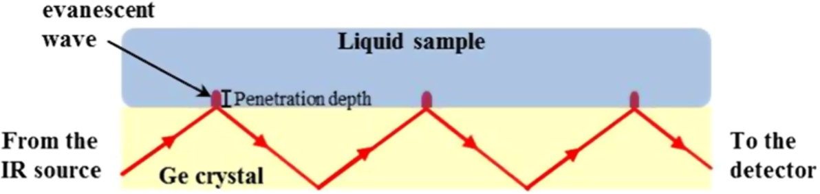
An illustration of the setup of an ATR element with a sample [this figure was originally published in [42]].
An interesting strategy to reduce the spectral contribution of water consists of replacing it with high concentrations of heavy water (D2O) in the preparation of media and buffers. The symmetric and asymmetric stretching modes of D2O appear at ∼2500 cm−1, where few chromophores of biological interest absorb; in addition, the bending mode of D2O at ∼1200 cm−1 stays away from the Amide I absorption (Fig. 2). The main drawback of D2O addition to cells is that their viability and survival are not guaranteed, since the substitution of H2O with D2O is generally stressful, although the cellular structure and chemical composition are retained. D2O is also widely used together with deuterated drugs, for studying drugs toxicity in human metabolism. An interesting study showed that D2O is more toxic to malignant animal cells than healthy ones [54].
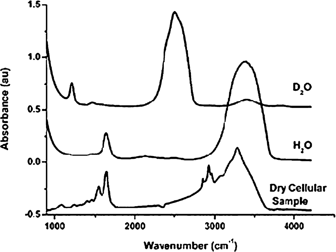
Absorption spectra of H2O and D2O compared with a typical cell spectrum. Comparison of the traces shows clearly that the bending mode of water around 1650 cm−1 overlaps with the Amide I absorption of cellular polypeptides in the same spectral region [this figure was originally published in [54]].
ATR-FTIR spectroscopy was applied to study different cellular topics [5,13,18,20,35]. An interesting application of this technique was the evaluation of the metastatic potential of cancer cells. Metastatic cells are known to have a higher motility and major fluidity of the plasma membrane compare to non-metastatic ones [16,68]. As already stated, their higher motility can be associated with the increase of cell’s membrane fluidity and of its hydration level, which may be consider a marker for the metastatic capacity of tumor cells [38,43,58,59]. Minnes et al. proposed an ATR-FTIR approach to detect the tumor stage of skin cancer cells, measuring the alterations of cell membrane due to hydration. They examined two murine melanoma cells, B16-F1 and B16-F10, with a common genetic background but a different metastatic level, and two human melanoma cells from the same patient, WM-115 and WM 266.4, which were respectively primary tumor cells and metastatic melanoma cells. IR measurements were carried out on live cell solutions placed on the Germanium ATR crystal and immediately acquired. During the experiment, cells’ concentration increased until the ATR element was fully covered by cells and the absorption spectrum reached a saturation value. The authors found that the absorbance of Amide II of proteins could be a reliable indicator for cell membrane hydration; in particular, a higher intensity of the band ratio 1540/1035 cm−1 (amide II/PO group of phospholipids) was detected in cells with a higher metastatic potential [42].
ATR-FTIR spectroscopy was also used to evaluate the cytotoxic effects of doxorubicin in situ in various immortalized human cell lines (i.e. HeLa, PC3, and Caco-2), and to provide information on their mechanisms of drug resistance [17]. Live cells were seeded with culture medium on the measuring surface of the ATR element until to a confluence > 90%. Measurements were carried out using a temperature controlled multi-bounce accessory through plate and a ZnS ATR element (Fig. 3). The path length produced from this accessory in living cells is 20–30 μm with a depth of penetration of 2–3 μm, which is smaller than the thickness of a living cell. This method let measure attached live cells with no significant contribution from the medium.
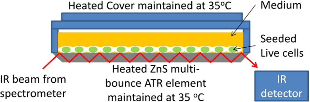
ATR element and cell culture setup for live cells’ FTIR measurement [this figure was originally published in [17]].
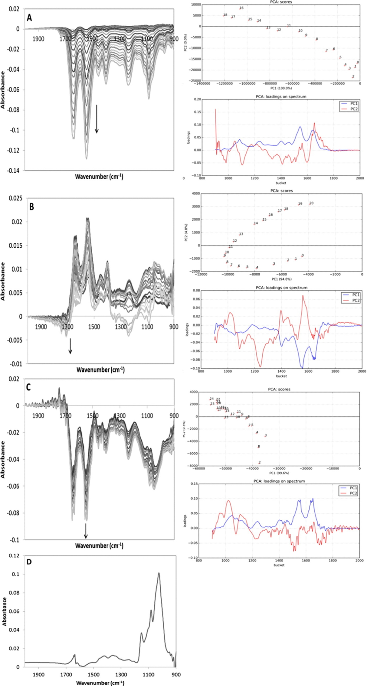
Difference spectra taken during 20 hours following the addition of 1 mM doxorubicin to (A) HeLa, (B) PC3 and (C) Caco-2 and their corresponding results from the PCA. Arrows point the sequence from 2 to 20 hours. (D) spectrum of glycogen [this figure was originally published in [17]].
Figure 4 shows the difference spectra respectively of HeLa, PC3, and Caco-2 cells after the application of 1 μM doxorubicin and the corresponding PCA analysis. HeLa cells resulted much more sensitive to drug effects than the other two cell lines and they showed a similar decrease of the absorbance of all the cellular components. PC3 cells showed positive Amide I and II bands, at 1645 and 1550 cm−1, when the drug was applied suggesting that the culture continued to grow in the first hours of the assay. This growth was followed by a proportional decrease of the bands at 1085 and 1240 cm−1, attributed to nucleic acids. This spectral performance could confirm that the amount of nucleic acids or triphosphate compounds with respect to protein content may be lower than adding doxorubicin. Caco-2 cells showed a smaller decrease, compared to HeLa, in all major peaks of the various cell components. However, a relatively large decrease in the absorbance band at 1024 cm−1, which led to the different shape of the spectra in the 1200–950 cm−1 region (relative to phosphate and carbohydrate), was found. Due to the reduction in absorbance in this region, spectral features resembled the spectrum of glycogen, generally accumulated in these cells as storage material to obtain energy (Fig. 4(D)). In summary, HeLa and PC3 cells, treated with low concentrations of doxorubicin (1 and 0.1 μM, respectively) showed a decrease in the nucleic acids amount together with an increase of protein ones. These findings could depend on alterations in DNA conformation caused by the drug or on a cell entry in a different phase of the cell cycle. With higher drug concentration, PC3 and Caco-2 cells showed a decrease in absorbance for all peaks of the principal cellular components suggesting a predominance of cell death.
Germanium is the most common element for ATR-FTIR spectroscopy, due to its low toxicity for most human cells. Nevertheless, Chan et al. noted that living cells let growth for more than 20 hours in contact with Ge could damage its surface [19]. They proposed a methodology to reduce this damage by coating germanium surface with a thin layer of biological materials that allow cell adhesion protecting the surface. Three different coating materials (gelatin, collagen and fibronectin) were deposited on Ge surface and let dry for 2 hours at 35°C. Then, HeLa and PC3 cell lines were let grow at 35°C overnight directly onto coated and uncoated Ge ATR plates. ATR-FTIR spectra of dried coatings were collected before and after adding culture medium. The spectrum of all coatings resulted higher in dried conditions with respect to when the medium was added, probably due to a partial possible dissolution of the coating material itself in the medium. Figure 5(a) shows that, during the first three hours after seeding cells on the Ge ATR element, the absorbance associated with cellular material increased, suggesting that the adhesion of cells on the ATR surface increases over time. After removal of the cells by trypsin, the Ge ATR surface without coating showed consistent damages due to a roughening of its surface, which increase the scattering loss of IR radiation by 40% (Fig. 5(b)). Conversely, coated Ge surface appeared smooth, above all when gelatin was used. The attach mechanism of living cells on Ge surface is not completely understood, but the same reduction of IR light was observed even when the cells were removed and the Ge surface cleaned, demonstrating that this permanent loss of throughput is related only to damages caused by cells on the surface. HeLa cells growth on coated Ge ATR element were also treated with doxorubicin, and the results confirmed that coatings did not affect the response of these cells to the drug and demonstrated the potential of ATR-FTIR to detect cellular changes in situ.
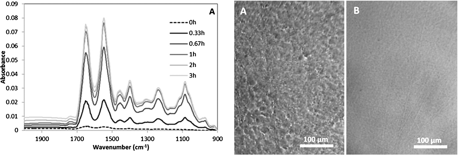
(a) FTIR absorbance spectra of cells in the first three hours after seeding in the ATR element. (b) Reflective visible images of a germanium ATR crystal surface, showing a roughness where the cells were attached to the crystal (left) and a smooth surface in the area not exposed in the cell culture (right) [this figure was originally published in [19]].
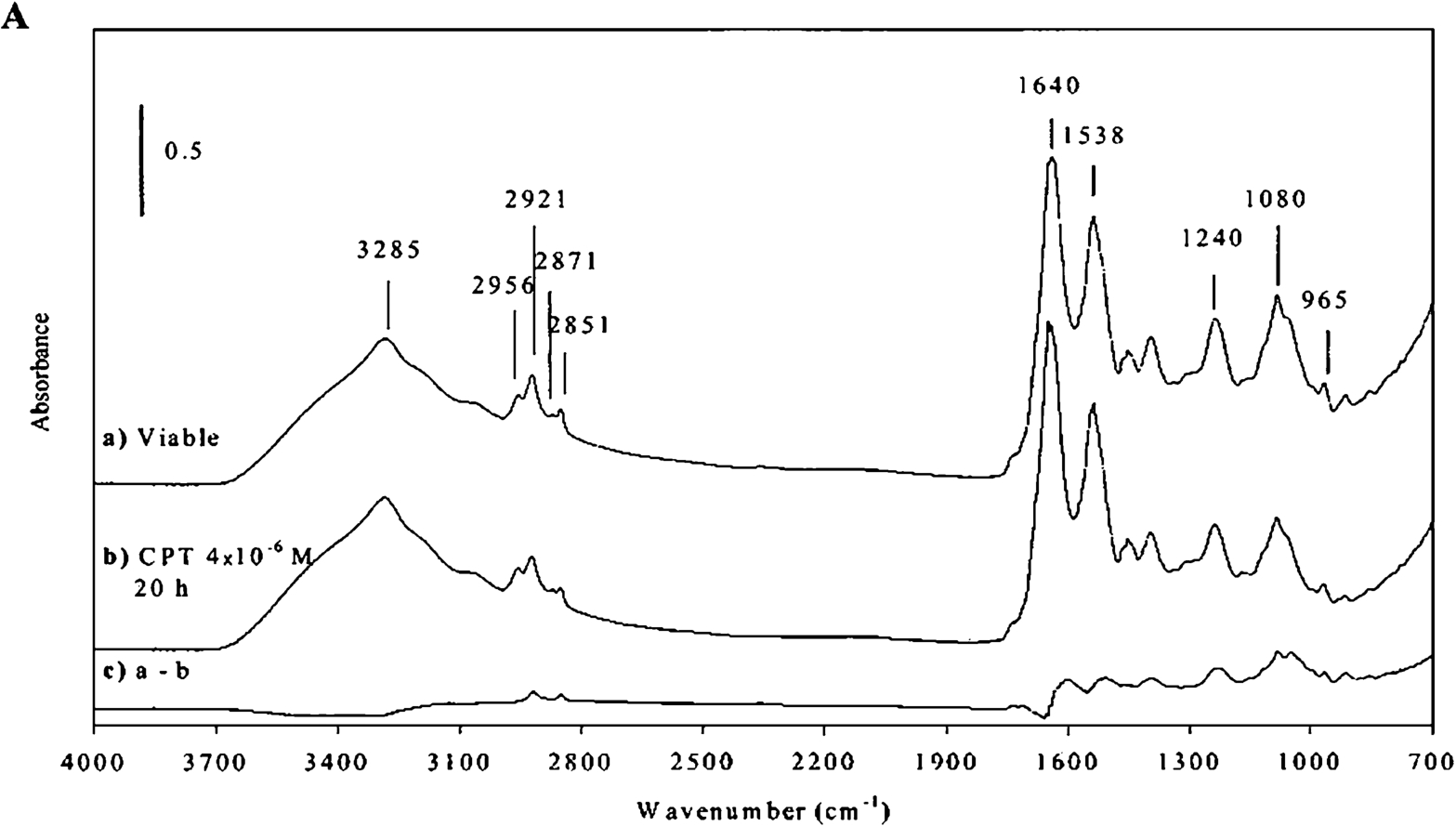
ATR-FTIR spectra of viable (spectrum (a)) and apoptotic (spectrum (b)) cells. Apoptosis was induced by treating the cells with camptothecin (CPT) for 20 hours. In order to emphasize the differences between spectrum (a) and spectrum (b), the difference spectrum (spectrum (c)) is also shown [this figure was originally published in [22]].
Gasparri et al. monitored apoptosis induction as a function of time on human HL60 leukemic cells incubated with camptothecin, a cytotoxic drug, by using ATR-FTIR analysis. In particular, cells were analysed as dehydrated bio-films on ZnSe crystal, obtained drying cell samples at room temperature for 15 min under airflow. During the apoptotic process, several spectral changes were detected and defined as diagnostic markers of apoptotic cells. The major differences between viable and apoptotic cells were ascribable mainly to nucleic acids, proteins and aliphatic chains (Fig. 6). The intensity of the region assigned to nucleic acids (1000–1140 cm−1) significantly decreased following apoptosis and it was considered an indicator of this process. The ratio between the band areas of Amide I (1599–1710 cm−1) and Amide II (1483–1595 cm−1) increased in apoptotic cells suggesting that changes in protein pattern occur during apoptosis. Finally, the region between 2800 and 3000 cm−1, relative to the absorption modes of aliphatic chains (in particular phospholipids and fatty acids) showed a slight decrease in the apoptotic cells compared with normal ones [22].
From the mid-1980s, the development of commercial visible-infrared microscopes let Fourier Transform Infrared Microspectroscopy (FTIRM) becoming a valuable tool for life science studies. The addition of a microscope as an accessory to conventional Fourier Transform Infrared spectrometers has led to the possibility of analyzing intact tissue sections and even single cells at cellular resolution.
FTIRM is a sensitive, non-invasive and non-radiation damaging technique, which allows correlate the morphological features of a sample with its vibrational patterns. The infrared radiation passes through the sample; the obtained spectrum is representative of the whole of the sampled volume and its intensity can depend on the size and the nature of the sample. FTIRM in transmission mode is useful for the spectral analysis of thin samples (<10 μm) such as cells and tissues and does not require too elaborate sample preparation procedures [48]. It is usually performed on dried or fixed biological samples, in order to avoid the strong absorbance of water in the cellular media.
Fixing processes could alter the fundamental cellular components, in particular nucleic acids and proteins, and modify the spectral profile. Some examples of FTIRM analysis, performed in transmission mode using different fixing protocols, will be discussed below.
Giorgini et al. exploited Fourier Transform Infrared Spectroscopy to study the aging effects of human female gametes and to identify their spectral biomarkers. The vibrational analysis was carried out in transmission mode, on 68 oocytes, distributed in two groups based on the donors’ age (group A: 30 ± 2 year old; B: 39 ± 2 years old). Each oocyte was deposed on a silicon support and air-dried for 30 minutes, without using fixatives. The comparative analysis of the representative spectra of the two experimental groups pinpointed some spectral evidences correlable with a decline in the quality of oocytes belonging to “old” women (B experimental group): an higher amount of peroxided fatty acyl chains with a more permeable plasma membrane; an increase in unordered structures suggesting an altered proteic pattern with an higher level of protein degradation; a variation in DNA conformation (Z-DNA) together with RNA degradation and an increase in the H-bond network; an increase in carbohydrates consumption, and the occurrence of a phosphorylative process [26].
FTIRM was also exploited to study the different degree differentiation in human dental pulp stem cells, with the aim of better understanding the molecular changes that occur during cell differentiation and to identify specific spectral markers of the different stages. Samples of undifferentiated stem cells were compared with preosteoblasts and osteoblasts. FTIRM measurements were carried out on cell samples previously fixed in 10% methanol at −20°C and air dried for 30 minutes. Spectral analysis carried out on the second derivative spectra of the three groups, showed changes mainly in phosphate stretching vibrations and Amide I, II and III bands; on differentiation, a modification of proteins secondary structure took place, in terms of a meaningful increase of helical structures with respect to β and random coil ones [27].
Gazi et al. performed an FTIR imaging analysis of PC-3 prostate cancer cell line previously fixed with formalin 4% in phosphate buffered saline (PBS). Formalin is a coagulative protein fixative, which causes the crosslinking of the primary and secondary amine groups of proteins and preserves lipids with the reaction between hydrated formalin and the double bonds of unsaturated alkyl chains. However, it must be emphasized that, because the formalin contains a significant amount of water, a post-fixation technique is necessary to remove this water component from the cells [24].
The spectral features of live U937 leukemic monocytes fixed with formalin or ethanol were compared with those of unfixed air-dried ones. Although ethanol showed to interact with membrane phospholipids, and formalin influenced their chemo-physical properties, lipids order and composition as well as proteins conformation were well preserved by both fixation protocols. Conversely, alcohol dehydration intensely affected both cellular macromolecular content and architecture [65].
Recently, the possibility to exploit FTIRM on live cells under physiological conditions can be considered a milestone to monitor the cell biochemical modifications, either induced or naturally occurring. The major advantage of working with living systems is the possibility to monitor the biochemical processes in real time and to resolve questions on the biochemical alterations induced by fixatives. A first approach to this topic was represented by the development of demountable liquid devices composed by two IR optical windows (CaF2, ZnSe or diamond), with plastic spacers, which allowed confine cells in compartments near their intrinsic volume, thus minimizing dilution effects and increasing detection sensitivity [25,62,73].
The first successful application of a microfluidic device with a precisely controlled path length was performed by Birarda et al. They analyzed the U937 monocyte cell line in vitro to establish its response to mechanical compression. The prototype device consisted of two CaF2 windows (25 × 12 × 2 mm) patterned with two larger wells for the accommodation of the cell suspension and/or pure buffer solution and by two smaller wells, used for air background acquisition. Different measurement chambers permitted to record air background as well as buffer and sample spectra without the necessity of disassembling the device [6].
Vaccari et al. proposed the fabrication protocol of the first example of a fully sealed transparent CaF2 microfluidic device for FTIRM analysis of live cells in transmission mode. Measurements were carried out using circular CaF2 devices composed by two concentric chambers, separated by a porous septum. Within the device, four regions water-free were lithographed for collecting air background. The lid was thermo mechanically sealed on the top of the bottom window in order to obtain a fully sealed microfluidic device [65]. It is noteworthy to evidence some problems arising from the fabrication protocol of CaF2 device, mainly related to the poor resist-substrate adhesion experienced on CaF2 surface, the effect of Ca2+ ions released by local dissolution of the substrate, and the quality of the sealing of the microfluidic device [7].
Based on these issues, Grenci et al. tried to solve these problems by introducing a thin layer of silicon on the CaF2 device in order to modify the surface energy and the wettability of the optical window [32]. Thanks to this improvement, the substrate showed a chemical behaviour similar to a Si surface and the thin layer (10–20 nm) acted as a contact-barrier between cells and CaF2. Moreover, the chemistry of Si may be exploited for surface functionalization with biomolecules, extending the applicability of infrared microspectroscopy to the study of the mechanisms of interaction between cells and nano-patterned substrates.
Further developments aimed at optimizing the fabrication protocol of microfluidic chip, were proposed testing new devices on BaF2 substrates modified with a 10–20 nm silicon layer [45]. Although BaF2 had an excellent IR transparency in a wider spectral window compared to CaF2, it was too soluble in water to perform experiments in physiological environment.
Currently, calcium fluoride is the most widely proposed substrate for the realization of microfluidic devices because it is an IR-transparent material with water solubility low enough for performing measurements in aqueous solutions.
An innovative approach to examine living cells was offered by Ebrahimifard et al. He presented an infrared absorption method based on deuterium oxide (D2O) as cell medium, in order to eliminate the high infrared absorption of H2O in the spectral range, where the symmetric and asymmetric stretching vibrations of CH2 and CH3 groups of lipids take place. The measurements were performed on two different living cell types, MDCK and Saccharomyces cerevisiae, washed in D2O-PBS before loading into the microfluidic chip to remove the H2O fraction. The designed IR sensor system was composed by a silicon nitride infrared light source, a monochromator, an InAs photodiode, an optical fibre and CaF2 lenses (Fig. 7). The IR spectra of both cell types showed different patterns, mostly in the 2800–3000 cm−1 region, probably due to differences in cell membrane composition [15].
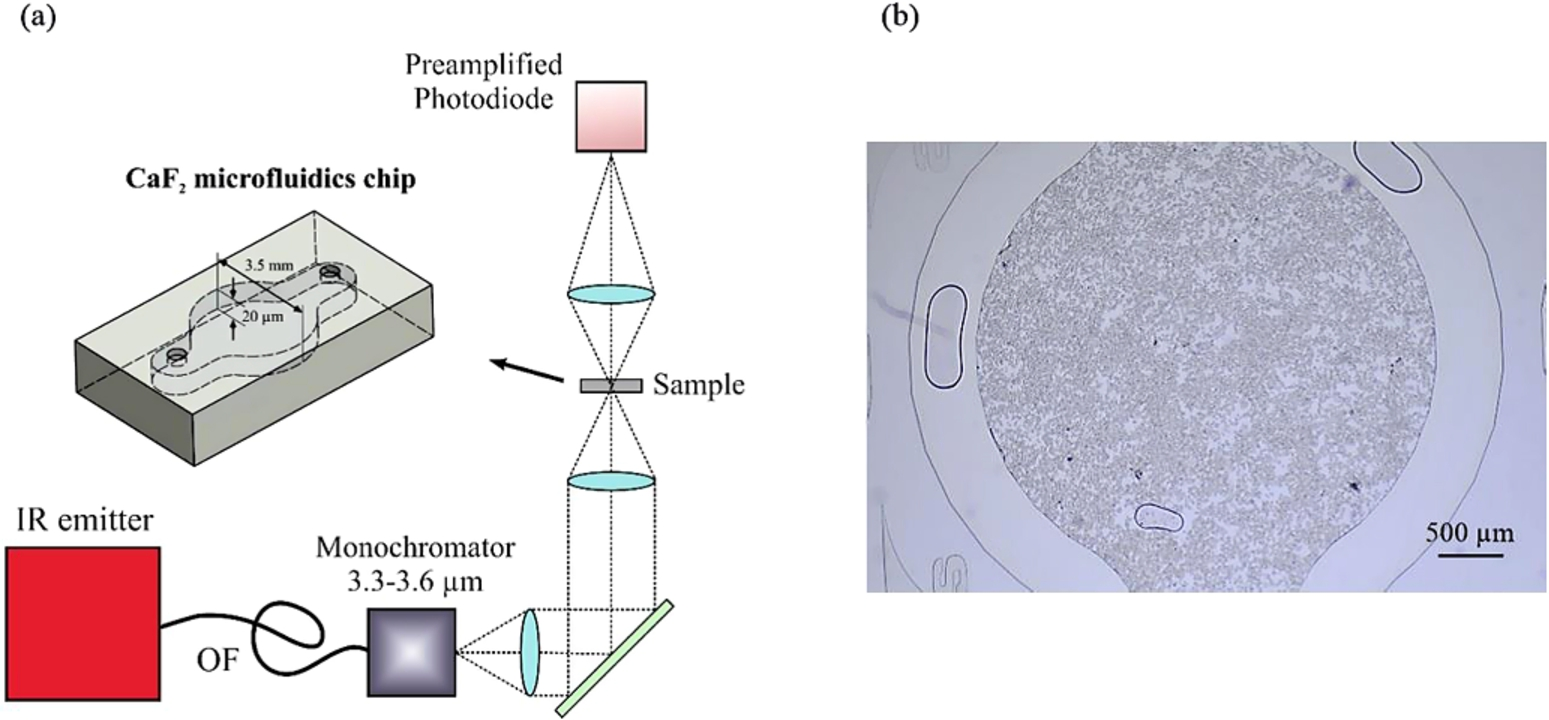
(a) Schematics of the IR sensor system setup and CaF2 chip; (b) living MDCK cells suspended in D2O-PBS loaded in a CaF2 chip [this figure was originally published in [15]].
The application of the microfluidic concept to IR transparent materials offers new potential for FTIR on living cells. The implementation of both IR and visible transparent 3D microfluidic devices allow the real time observation of the biochemical rearrangements undergone in living cells upon chemical and mechanical stimulations, limited only by the low throughput of IR microscopes operating at high spatial resolution, with apertures of few micrometers. The low brightness of IR conventional thermal sources together with the restriction of the beam size reduces the S/N ratio. The introduction of synchrotron light sources has provided an effective tool to overcome such limitations and to perform FTIR microspectroscopy on single cells and subcellular compartments in vivo, studying both structure and reactivity. Several studies demonstrated the usefulness of the high brilliance of the synchrotron radiation to examine single living cells at diffraction-limited spatial resolution and to identify the vibrational properties of biological components in single cells and in isolated cell nuclei [1,55,66].
Following the approach previously applied by several research groups [3,51,54] Liepic et al. compared the IR spectra of prostate cancer cells DU-145, derived from brain metastasis and treated with ionizing radiation, collected using a synchrotron radiation and a single point detector, with spectra collected using a conventional global source and a focal play array (FPA) detector. Due to the small cell sizes, the S/N ratio was lower when using the global source and FPA detector. Only the high brightness of the synchrotron radiation may allow investigate smaller regions with acceptable S/N ratio: SR-FTIR spectra were collected in transmission mode with a spectral resolution of 4 cm−1 in the region between 4000–600 cm−1. The spectrum of each cell was acquired with an aperture of 12 μm ×12 μm. The analysis of the 3000–2800 cm−1 spectral range, that provided information about the state-of-order of the hydrocarbon tails in lipids and acyl chains in general, pinpointed a greater bands width at 2852 and 2925 cm−1 (relative to CH2 symmetric and asymmetric stretching modes, respectively) in samples submitted to ionizing radiation compared to those not subjected to radiation [36].
Vaccari et al. analyzed living U937 monocytes, by using a CaF2 microfluidic device, with SR-FTIR, to monitor their biochemical response to both mechanical and chemical stimuli. In particular, by treating U937 monocytes with a synthetic peptide that promotes the monocyte extravasation for reaching the inflammation site, the synthesis of new adhesion proteins and the cell cytoskeleton rearrangement responsible for cell extravasation were observed. The response of U937 to mechanical deformation was also evaluated confining them in fluidic devices with different thickness, in order to achieve different conditions of deformation. Spectral analysis demonstrated that deeply deformed cells have a different cellular biochemistry compared to not deformed ones. In addition, it highlighted that the limit in fluidic device path length, imposed by the saturation of water bending band at ∼9 microns, was in turn limiting the type of cells can be sampled to the ones that have an height lower that the path length constrain [66].
Cells represent the fundamental biological unit from which the life of all living organisms depends. Knowledge of their morphology and above all their biochemical processes is extremely important in order to counteract the onset of cell anomalies or pathological conditions. Each cellular component shows a peculiar position in a cell IR absorption spectrum. The capability to extract specific informations from each spectrum is essential for drawing useful conclusions on the process of interest.
In this review, we have demonstrated that FTIR microspectroscopy is a valuable tool for studying single living cells and how this approach has evolved over time in order to get more and more reliable informations without isolating cells from their natural environment or subjecting them to conditions of stress. The development of IR microspectroscopy has allowed discovering the complexity of some cellular processes and we could start to wonder if these studies of biochemistry in action could open up new perspectives in cellular biochemistry.