
Abstract
BACKGROUND:
An electrodiagnostic evaluation is conducted to diagnose carpal tunnel syndrome (CTS) and evaluate its severity.
OBJECTIVE:
This study proposes a revised approach for classifying the severity of electrophysiological findings for patients with CTS.
METHODS:
This retrospective cross-sectional study included patients with CTS confirmed through electrodiagnostic evaluations. Based on the Stevens’ classification, the patients were divided into three groups (mild/moderate/severe). A new intermediate group was defined to identify patients with normal motor nerve conduction studies and abnormal electromyographic results. CTS pain was evaluated using a numeric rate scale. Physical examinations and sonographic evaluation were performed to detect anatomical abnormalities.
RESULTS:
Overall, 1,069 CTS hands of 850 CTS patients were included. The mean age was 57.9
CONCLUSION:
The intermediate CTS group showed clinical features that were intermediate to those of the moderate and severe CTS groups.
Introduction
Carpal tunnel syndrome (CTS) is a common condition characterized by compression of the median nerve of the wrist. The reported prevalence rates range from 1–5%, with a higher incidence in women than men [1, 2]. CTS results in various neurological symptoms, including sensory disturbances along the distribution of the median nerve in the fingers and weakness and atrophy of the thenar muscles [3]. Several methods have been used to diagnose and define the disease classification [4]. Commonly performed tests, such as the Tinel sign and Phalen test, aim to elicit characteristic symptoms; however, these tests, despite being easy to administer, often lack definitive diagnostic significance [5]. More objective diagnostic approaches have gained widespread use. These include electrodiagnostic testing and musculoskeletal ultrasound, which are known not only for their relatively high sensitivity and specificity in the diagnosis of CTS but also for their ability to classify the severity of CTS [6, 7].
The importance of clearly determining the severity of CTS lies in the fact that different treatment approaches are applied based on this parameter [8]. Various approaches are used to treat CTS. In cases with mild symptoms, rest and avoidance of excessive wrist movement are recommended, and a splint may be applied [9]. Non-steroidal anti-inflammatory drugs or steroids can be administered orally, and local steroid injections are used [10, 11]. If these conservative methods do not provide relief, surgical intervention may be considered to alleviate the increased pressure caused by the narrowed carpal tunnel [12]. The choice of treatment method is also determined by response to previous treatments.
Various classification criteria have been proposed to determine the severity of CTS; however, a standardized criterion is yet to be established. Severity classification methods based on electrodiagnostic tests have been developed [13, 14], among which the Stevens’ classification method has been widely used [15]. Based on this classification [15], the CTS is classified as mild when there is delayed distal sensory latency (DSL) or a decrease in the amplitude of the sensory nerve action potential (SNAP). It is classified as moderate in cases with delayed distal motor latency (DML) in the compound motor action potential (CMAP). Finally, for severe cases, the classification is determined by the absence of a response in SNAP, a decrease in the CMAP amplitude, or the observation of denervation potential in needle electromyography.
However, in clinical practice, there are occasional cases in which patients show normal DML and CMAP amplitude values but abnormal findings on needle electromyography (EMG), making it difficult to classify patients as moderate or severe. Therefore, we identified a relationship between sensory and motor nerve abnormalities in nerve conduction tests in patients with CTS. Nonetheless, there may be insufficient evidence of a precedent relationship between abnormal nerve conduction and the findings of electromyographic needle testing. Finally, we assumed that redefining the previous severity classification system was necessary.
In this study, we investigated the clinical characteristics of patients within the intermediate zone of the Stevens’ classification and compared them with patients who showed different levels of severity. By analyzing the clinical characteristics of these individuals and comparing them with those displaying clear-cut severity levels, we hope to shed light on an appropriate classification strategy. The overall objective of this study was to improve diagnostic accuracy and to guide clinicians in selecting the most suitable treatment approaches.
Materials and methods
Subjects and study design
Flowchart of inclusion of subjects in the study. CTS: carpal tunnel syndrome, EDX: electrodiagnosis, US: ultrasonography.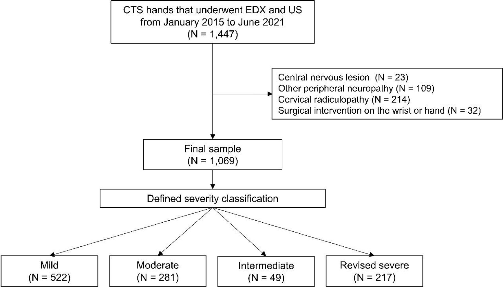
This retrospective cross-sectional study used the medical records of a single hospital between January 2015 and June 2021. This study focused on men and women 18 years and older who were diagnosed with CTS by electrodiagnostic and sonographic evaluations. Patients who had previously undergone surgery on the hand or wrist, including carpal tunnel release, were excluded, while those who had received general medical treatment for pain were included. Patients with concomitant central nervous system lesions, peripheral neuropathies in the upper limbs, or cervical radiculopathy were also excluded (Fig. 1). This study was approved by the Institutional Review Board of Pohang Stroke and Spine Hospital (approval number: PSSH0475-2022-HR-003-01), ensuring compliance with the Declaration of Helsinki. The informed consent requirement was waived due to the retrospective nature of this study and the anonymity of the data.
In the medical record, data collection from patients was conducted through interviews with specialists in physical medicine and rehabilitation. Clinical symptoms related to pain, such as numbness and tingling sensations, were evaluated using a numerical rating scale (NRS) to assess the severity of CTS pain. The duration of pain and the presence of nocturnal pain were determined based on patient reports. To evaluate thenar muscle weakness, the specialist explained movements involving the thenar muscle, such as turning and gripping a jar lid, and assessed whether the patient had experienced weakness in the corresponding muscle. In the case of thenar muscle atrophy, evaluation was carried out through a physical examination by the specialist.
Sonographic evaluation
Ultrasound examinations were conducted using an anterior approach to the wrist, enabling the assessment of the anatomical condition of the carpal tunnel. The sonographic evaluation occurred with the patient in a supine position, and the probe was oriented perpendicularly to prevent anisotropy through angulation. The examiner did not apply additional pressure when using the probe to avoid median nerve compression. The median nerve’s cross-sectional area (CSA) was measured when the scaphoid bone was medially visible and the trapezium bone was visible laterally [16]. The hypoechoic region closest to the hyperechoic nerve sheath was identified, and the largest diameter was measured three times, with the average value calculated.
Additionally, palmar bowing (PB) of the flexor retinaculum was measured at the trapezium and hook of the hamate level, indicating the extent to which it curves towards the palmar side [17]. A line connecting the trapezium and hook of the hamate was drawn, and the distance between this line and the upper portion of the flexor retinaculum was measured. If a subject was confirmed to have CTS in both hands, the evaluation was independently conducted for each hand. Sonographic assessments were performed by physicians with over ten years of experience using iU22 ultrasound machines (Philips, Bothell, WA, USA).
Electrodiagnostic evaluation
For SNAP, the recording electrodes were placed on the second digit. Electrical stimulation was applied to the wrist crease 14 cm from the active electrode. A DSL of 3.5 ms or less with an amplitude of 20
Defined severity classification
Comparison between groups classified through the Stevens’ classification and the revised method. SNAP: sensory nerve action potential, CMAP: compound motor action potential, EMG: electromyography.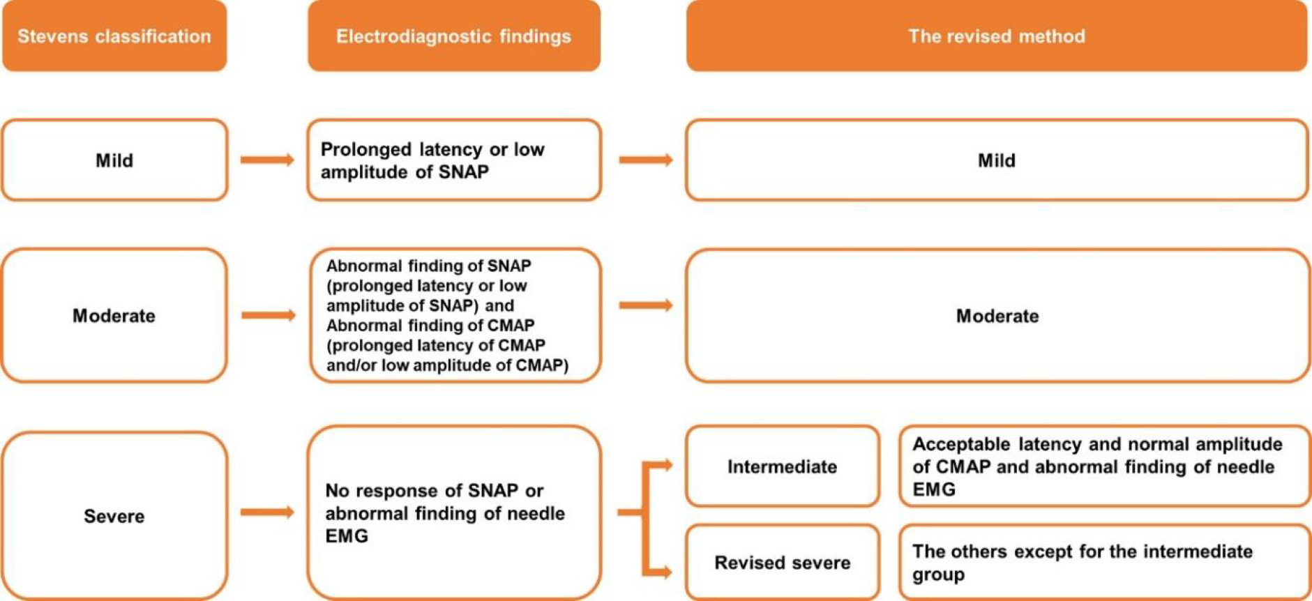
Based on the results of the electrodiagnostic test, patients were classified into three main groups according to the Stevens’ classification method: mild, moderate, and severe. The mild group was characterized by abnormal findings only in the SNAP. The moderate group exhibited delayed DML or decreased CMAP amplitude. Finally, the severe group was identified by the absence of a SNAP response or the presence of a denervation potential on needle electromyography. In this study, we retrospectively redefined an intermediate group that included cases with normal CMAP findings, but needle electromyography showed abnormal results. The revised severe group was confirmed as the severe group after excluding the intermediate cases (Fig. 2).
Under the assumption of the central limit theorem, continuous variables were assumed to follow a normal distribution and are presented as mean
Results
Demographics of the hands of the patients
Demographics of the hands of the patients
BMI: body mass index, CSA: cross-sectional area of the median nerve, PB: palmer bowing of the flexor retinaculum.
From January 2015 to June 2021, a total of 1,447 hands underwent electrodiagnostic and ultrasound examinations. Of these, 1,069 hands belonging to 850 patients were analyzed, with 378 hands excluded by the exclusion criteria (Fig. 1). The mean age of the total patient population was 57.9
Among the hands analyzed in this study, 266 were classified as severe according to the Stevens’ classification method. Of these, 49 hands were in the newly defined intermediate group. Based on our modified classification, the mild group included 522 hands (48.8%), the moderate group included 281 hands (26.3%), the intermediate group included 49 hands (4.6%), and the revised severe group included 217 hands (20.3%).
Comparative analysis of symptoms experienced by newly defined and existing patient groups of the Stevens’ classification
Comparative analysis between newly defined and existing patient groups in the Stevens’ classification
CSA: cross-sectional area of the median nerve, PB: palmer bowing of the flexor retinaculum, NRS: numeric rating scale of pain.
The proportion of hands exhibiting thumb thenar weakness and nocturnal pain symptoms increased progressively with disease severity, demonstrating a statistically significant trend (
In the present study, we redefined the CTS classification system based primarily on the Stevens’ classification system. By redefining the classification system, we provided a more refined and precise categorization of the severity of CTS. Our results revealed that by including patients who exhibited normal motor nerve conduction but abnormal needle electromyography findings, we could identify a subgroup with intermediate characteristics falling between the moderate and severe groups in the Stevens’ classification. This finding highlights the importance of considering additional parameters beyond motor nerve conduction in the classification and characterization of CTS. In addition, by recognizing this intermediate subgroup, we gained further insights into the spectrum of the disease and the diversity of presentations within the patient population. This may have important implications for the tailoring of treatment approaches and the optimization of clinical management strategies for patients in this intermediate category.
Electrodiagnostic testing is a valuable tool for the assessment of the neurophysiological characteristics of CTS, for which it is relatively well established compared to other peripheral neuropathies [23]. Electrodiagnostic testing also provides valuable information on the pathophysiology of CTS and enables the evaluation of nerve damage at various stages. In addition, the median nerve of the carpal tunnel is readily accessible for direct stimulation [24]. This allows the use of objective measures, such as distal latency and amplitude, and standardized testing techniques, which minimize variability between observers and improve the sensitivity and specificity of electrodiagnostic tests [25, 26]. In CTS, increased pressure within the carpal tunnel leads to changes in the microcirculation surrounding the nerves, leading to local hypoxia [27, 28]. The damage progresses sequentially from smaller-diameter nerve fibers, initially affecting autonomic nerve fibers, followed by sensory and motor nerve fibers [29, 30]. Peripheral nerve impairments can be identified using nerve conduction studies. When nerve damage reaches a certain degree, it begins to affect the muscle fibers controlled by the nerve and needle electromyography can then reveal signs of denervation [31]. Denervation potentials indicate compressive neuropathy that affects muscles, leading to additional symptoms such as muscle weakness and atrophy [32]. Therefore, for an accurate assessment of CTS, it is essential to consider both nerve conduction studies and needle electromyography findings. The severity classification of CTS is based on criteria such as SNAP, CMAP, and denervation potentials observed on needle electromyography. Most electrodiagnostic tests use these parameters to differentiate the severity of CTS. In addition to the Stevens’ classification method applied in our study, other classification systems, such as those of Bland et al. and Padua et al., have been proposed [13, 14]. However, the criteria used in these methods do not differ significantly from the Stevens’ classification, except for further subdivision of patients with normal distal sensory latency and amplitude of SNAP but delayed TCL, as well as subdivision within the severe group based on DML and the absence of CMAP findings. Furthermore, these classification methods involve at least five criteria, making them relatively complex and less commonly applicable in clinical practice than the Stevens’ classification method. Consequently, by understanding the sequential pattern of nerve involvement, clinicians can make better-informed decisions regarding diagnosis and management, considering the specific neurophysiological alterations observed in individual patients.
The ultrasound findings of our newly defined group showed characteristics similar to those of the moderate group of the Stevens’ classification. Ultrasound examination is a useful tool for objectively evaluating CTS and complements the information obtained from electrodiagnostic tests [33]. It is a non-invasive, painless procedure for patients, allowing direct visualization of the median nerve and facilitating both diagnostic purposes and therapeutic interventions, such as ultrasound-guided interventions [34]. Consequently, ultrasonography has gained increasing clinical significance and is regarded as a valuable tool that can replace or supplement traditional electromyography in the evaluation of CTS. Thus, because the intermediate group was anatomically closer to the moderate group than to the severe group, it has significant implications. Furthermore, the increasing use of ultrasound in clinical practice and its ability to assess parameters such as CSA and PB have demonstrated a correlation with severity classification based on the Stevens’ method, as previously reported by Kim et al. [35]. Thus, even if abnormal findings are observed on needle electromyography of the abductor pollicis brevis muscle and if the motor nerve conduction study is normal, it may be necessary to reconsider categorizing these patients as severe without further consideration of treatment decisions.
Analysis of the intermediate group provided information on the possibility that the worsening of anatomical indicators within the wrist preceded the exacerbation of symptoms reported by the patients. This suggests that symptom deterioration may occur because of neurophysiological changes not detected in motor nerve conduction studies [36]. Therefore, the evaluation of motor nerves in patients in the classification of CTS severity may require considerations beyond conventional measures, such as delayed DML or reduced amplitude of CMAP, and should include alternative approaches, such as comparing the latency of the second lumbrical and interosseous muscles during nerve conduction studies.
The characteristics of the intermediate group offer opportunities to explore novel clinical approaches for the treatment of CTS. The intermediate group anatomically presents similarities to the moderate group. However, these symptoms resemble those of the severe group, making them a subgroup in which surgical intervention may be considered more urgent than in the moderate group. Considering that the disease has not yet progressed anatomically to the extent observed in the severe group, early intervention, including surgery, for the intermediate group may lead to improved outcomes over surgery when patients are classified as the severe group [37]. Surgical intervention should be approached with caution as it can be considered the last resort for CTS patients. Given the adjustments made through this revised method, particularly in redefining the severe group, it can assist surgeons in making informed decisions about the optimal timing for surgery. This is especially relevant considering that the intermediate group exhibits differences in anatomical parameters compared to the original severe group, potentially exerting a significant influence on decision-making. Therefore, by initiating timely interventions, including surgical options, improved prognoses can be expected in the intermediate group compared to the severe group.
This study has several limitations. First, although the total number of patients included in the study was substantial, the number of patients in the intermediate group was relatively small. Second, since this was a retrospective study, we could not use a wider range of functional indicators beyond pain scores. Third, during the analysis of the ultrasound findings, we did not account for potential confounders such as age, sex, or body mass index, which may have influenced parameters such as CSA and PB. Fourth, we were unable to assess the association between our modified classification and treatment outcomes because of the lack of longitudinal follow-up data and retrospective study design. Further studies are required to investigate this aspect. Lastly, we did not directly present the anatomical and physiological findings associated with the electrodiagnostic examination. Further investigations incorporating surgical observations and histological findings may provide a more detailed understanding of this topic.
Conclusions
This study proposes a revised approach for CTS severity. By defining an intermediate group, we identified a group that exhibited characteristics between those of the moderate and severe groups. This study may improve the evaluation of the severity of CTS and the development of personalized treatment strategies. Future studies should focus on long-term follow-up with larger patient populations to further explore the prognostic implications and clinical applicability of our classification approach.
Author contributions
D.H.J. contributed to the investigation, visualization of data, and wrote the original draft. S.E.L contributed to the data resource and data curation. J.W.L and D.P contributed to the conceptualization and methodology of the study, conducted the data analysis, and edited the draft.
Data availability statement
All study data are contained in the article and supplementary material.
Ethical approval
The study was conducted according to the guidelines of the Declaration of Helsinki and was approved by the Institutional Review Board of Pohang Stroke and Spine Hospital (approval number: PSSH0475-2022-HR-003-01).
Funding
This research received no external funding.
Informed consent
Patient consent was waived because of the retrospective nature of this study and the anonymity of the dataset.
Supplementary data
The supplementary files are available to download from http://dx.doi.org/10.3233/BMR-230275.
Footnotes
Acknowledgments
The authors have no acknowledgments.
Conflict of interest
The authors declare no conflict of interest.