Breast cancer is a female malignancy that is a significant cause of mortality worldwide. Currently, investigations on natural ingredients as new candidates for chemopreventive agents and breast cancer chemotherapies are increasing. Propolis is a natural resinous material produced by honeybees that exhibit anticancer potential. Several studies have mentioned the major bioactive compounds of propolis, but their mechanism of action is not clearly understood.
OBJECTIVES:
The purpose of this review is to collect and summarize the evidence related to the effectiveness of propolis and its bioactive contents as candidates for breast cancer therapy and analyze the molecular mechanisms involved in their therapeutic pathways.
METHODS:
We reviewed 94 articles from journals and databases, extracted the results, and produced summaries and conclusions.
RESULTS:
Propolis and its bioactive ingredients show cytotoxic, anti-proliferative, pro-autophagic, anti-metastatic, and antioxidant activities, as well as synergistic effects with chemotherapy or radiotherapy in breast cancer. Its therapeutic activity involves various target molecules, including NF-κβ, Fas receptors, p53, TLR4, ANXA7, and voltage-gated Na+ channel (VGSC).
CONCLUSION:
The bioactive components of propolis and the target molecules involved need to be explored further to develop new breast cancer therapies and overcome the problem of chemoradiation resistance.
Breast cancer is the leading cause of female mortality related to malignancy worldwide. These malignant tumors have very heterogeneous characteristics, including their clinical development, prognosis, and underlying molecular mechanisms [1–3]. Based on their molecular characteristics, breast cancer is categorized as estrogen receptor (ER)-positive (e.g., MDA-MB134 and MCF-7 cell lines) and ER-negative (e.g., MDAMB453 and SKBR3 cells). ER-positive is the most common type of breast cancer, with an incidence rate of 50%–70%. Furthermore, based on the identified protein biomarker subtypes, breast cancer is categorized into triple-negative breast cancer (basal-like), luminal breast cancer, and HER-2 positive breast cancer [4–6]. This variation in molecular subtypes makes breast cancer difficult to treat. The current management of breast cancer is surgery, chemotherapy, radiotherapy, and even hormonal or targeted therapy in early cancerous lesions [7]. However, drug resistance and the severity of toxic side effects reduce the efficacy of the above therapeutic strategies. Therefore, many studies have explored the potential of natural ingredients in specifically inhibiting and killing cancer cells without damaging normal cells [1,8–10].
Propolis, a natural material in the form of resin produced by honeybees from various plants, is one of the many natural products that exhibit anticancer potential [11,12]. Propolis contains more than 300 active ingredients, including flavonoids and phenolic acid [13]. Many studies report that flavonoids demonstrate chemopreventive effects against carcinogenesis [14]. The unique feature of propolis that attracts many researchers’ attention is its selective targeting of cancer cells, which can be used as an alternative to conventional chemotherapeutic drugs [9,15–18]. The variety and color of propolis are influenced by its geographical area, season, and honeybee specificity [19,20]. The six most common types of propolis include poplar propolis, birch propolis, Brazilian green propolis, red propolis, pacific propolis, and Canarian propolis [21].
Caffeic acid phenethyl ester (CAPE) is an active ingredient in propolis that is frequently studied and appears to be a promising candidate as an anticancer drug. Specifically, several studies have proven it to be selectively cytotoxic against cancer cells based on dose, concentration, and the duration of exposure [1,22–24]. Kabala et al. reported that CAPE has a substantial cytotoxic effect on MDA-MB-231 cells as it reduced cell viability by 71.2%, 27.2%, 9.6%, and 5.6% at doses of 10, 25, 50, and 100 μM, respectively. Furthermore, CAPE administration at doses of 27.84 μM for 24 h and 15.84 μM for 48 h induced apoptosis in 50% of MDA-MB-231 cancer cells [1]. Wu et al. tested the effect of CAPE in vivo and demonstrated that tumor volumes in heterograft mouse models of MDA-MB-231 and MCF-7 cancer cells were reduced by 40%–60% after 3–4 weeks of exposure [23].
Several studies have concluded that propolis targets molecules that play key roles in apoptosis through the intrinsic pathway by activating caspase cascade mechanisms, releasing cytochrome C from the mitochondria into the cytosol, and acting as proapoptotic proteins. Watabe et al. showed that CAPE could suppress the activation of nuclear factor kappa-light-chain-enhancer B cells (NF-κB). NF-κB has the ability to prevent apoptosis, enhance proliferation, and promote angiogenesis. Interestingly, CAPE can trigger apoptotic pathways in cancer cells but not in normal WI-38 fibroblast cells [25].
The purpose of this review is to collect and summarize the evidence related to the effectiveness of propolis and its bioactive contents as candidates for breast cancer therapy and analyze the molecular mechanisms involved in their therapeutic pathway.
Propolis and its active ingredients
The chemical content in propolis varies greatly depending on its geographical origin, season, plants, and honeybee specificity. However, every variant of propolis contains flavonoids, terpenoids, steroids, and amino acids [2]. Many studies report that a more heterogeneous propolis content has stronger anticancer effects than those with only one active substance [26–28]. It was further reported that propolis shows a synergistic effect with standard chemotherapy drugs for breast cancer and radiotherapy [2,9,29–31].
Some active ingredients of propolis that have been successfully isolated and that exhibit anticancer effects include CAPE, caffeic acid, galangin, quercetin, nemorosone, apigenin, ferulic acid, and many more [8,32]. The mechanisms of action of the bioactive substances in propolis have been extensively studied and summarized in Table 1. Furthermore, Kuntz et al. showed that flavonoids, the most abundant ingredient in propolis, displayed anti-proliferative effects in vitro, but the relationship between its chemical structures and cellular bioactivity is not well understood [33].
Propolis has more than 300 active ingredients with various effects on cancer cells. The molecular mechanisms involved also vary.
Caffeic acid
Caffeic acid (CA) is one of the components in propolis that shows anticancer activity in vitro and animal models. CA is also reported to display anti-proliferative effects [43–46]. Kabała-Dzik et al. reported that CA showed a weak inhibitory effect on the MDA-MB-231 cell cycle. Furthermore, CA promotes apoptosis and inhibits proliferation by decreasing the viability of MDA-MB-231 and T47D cells in vitro [1,47]. Trials investigating MCF-7 cells showed moderate effects depending on the duration of exposure [8,47]. However, the conclusions regarding the cytotoxic and apoptotic effects of CA are still controversial and require further research.
CAPE
CAPE is a phenethyl ester derivative from CA found in propolis and is known as a bioactive ingredient. Among the compounds in propolis, CAPE is most frequently tested because of its dominant effects [48]. At present, evidence from several studies supports the potential role of CAPE as a chemopreventive agent or a new chemotherapy regimen, its proapoptotic activity, and its strong inhibitory effects on the cancer cell cycle [49–53]. CAPE is an anticancer agent that acts through diverse molecular mechanisms in several breast cancer cell pathways. Interestingly, CAPE shows selective effects on cancer cells and does not target normal cells [24,54–58]. Furthermore, CAPE inhibits lipid peroxidation and the effect of antioxidants if administered at a low dose [59,60].
Galangin
Galangin (3, 5, 7-trihydroxyflavone) is a yellowish crystal-like substance found in Serbian propolis [8,10]. This substance displays significant cytotoxic activity against MCF-7 cells by inhibiting the mitochondrial pathway and PI3K/Akt signaling [8,61]. Furthermore, Murray et al. reported that galangin inhibits the proliferation of Hs578T cells. Additionally, they showed that it was effective in combination with chemotherapy drugs that target the tumor cell cycle and overcame resistance to target-specific ER therapy [62].
Apigenin
Apigenin is a flavonoid substance found in fruits, vegetables, and propolis. Seyhan et al. reported that apigenin displays a dose-dependent cytotoxic effect that causes apoptosis in MCF-7 cells [8,34]. Tseng also observed that apigenin arrested the MDA-MB-231 cell cycle in the G2/M phase in vitro and in vivo. In addition, apigenin modulates epigenetic control in breast cancer cells [35].
Quercetin
The active substance quercetin is also one of the components of propolis that is often investigated. Quercetin belongs to the flavonoid group and is reported to be effective as a proapoptotic agent. It is also able to arrest the cell cycle in G0/G1 and DNA synthesis phases depending on the dose and duration of exposure in cancer cells [8,36,37]. The anti-proliferative effect of quercetin was also further investigated by Deng et al. and was shown to be effective in regulating survivin mRNA expression in MCF-7 cells. In addition, quercetin may induce the chemosensitivity of breast cancer cells by decreasing the expression of survivin genes [36,40].
Ferulic acid
Ferulic acid is a derivative of CA that is found in propolis. Researchers report that the effects of ferulic acid on MDA-MB-231 cells are conflicting [63,64]. Serafim et al. tested the effects of ferulic acid on MCF-7, HS578T, MDA-MB-231, and non-transformed human fibroblast cells and observed no cell growth activity [38]. By contrast, Seyhan et al. reported that after 24 h of exposure to cancer cells, ferulic acid had a mild apoptotic effect [8].
Nemorosone
Nemorosone, a polyisoprenylated benzophenone, is one of the main metabolites of brown Cuban propolis. Rubio et al. reported that nemorosone showed cytotoxic activity in several epithelial carcinomas with IC50 value ranging from 1.5–3.6 μg/ml [65]. Based on the 1,1-diphenyl-picrylhydrazyl (DPPH) assay, nemorosone acts as an active antioxidant compound [65]. Popolo A. et al. noted several mechanisms regarding nemorosone bioactivity on breast cancer cells, such as (i) selective response to ERα+ MCF-7 cells, (ii) arrest cell cycle in G1/G0 phase, (iii) reduce pERK1/2 and pAkt expression in estrogen signaling pathway [39]. Thus, nemorosone is stated to be a novel neoadjuvant on ERα+ breast cancer.
Myricetin
Myricetin, a substance in propolis in green to yellow crystals, is included in the subclass of flavones and flavonols. Myricetin shows anti-breast cancer effects on MDA-MB-231 cells when compared with other flavonoid substances [10]. Several studies report that myricetin displays a consistent cytotoxic effect compared with the other 23 flavonoid substances after 48 h of exposure in MDA-MB-231 cells [10,66].
Luteolin
Luteolin is another substance found in propolis in yellowish crystals and is classified in the flavonoid group. Together with myricetin, this substance shows cytotoxic activity after 72 h of administration at doses of 115.68 and 114.75 μM on MDA-MB-231 cells [10].
Pinocembrin
Pinocembrin is a dihydroxyflavanone found in propolis. This substance is reported as an anticancer, vasodilator, and neuroprotective agent [67]. Ozdal et al. noted that pinocembrin, together with other substances in Turkish propolis, showed cytotoxic effects on MDA-MB-231 cells through apoptotic activation and acted as an antioxidant [41].
Propolis therapeutic pathways in breast cancer cells
The development of cancer cells goes through multiple stages involving changes at cellular, genetic, and epigenetic levels. Cancer treatment generally aims to prevent, inhibit, or reverse this process. The current concept of breast cancer treatment involves targeting specific molecules in certain subgroups in the growth pathway and promoting the apoptosis of cancer cells to reduce heterogeneity [68]. The chemopreventive effect of a substance lies in its ability to inhibit absorption, deactivate carcinogens by inhibiting their binding to DNA, and repair DNA that has mutated [69–71]. The active substance in various herbs can act as a chemopreventive agent through the modulation of the intracellular pathways [72]. The regulation of programmed cell death is believed to be the key mechanism of potential anticancer agents [73].
Potential dosage of propolis bioactive substances in breast cancer cells
μM: micromolar; EXT: extract; DPPC: Dipalmitoylphosphatidylcholine; MCF-7, MDA-MB-231, SKBR-3, Hs578T, MDA-MB-468, UACC-3199, TD47D: various types of breast cancer cell pathways.
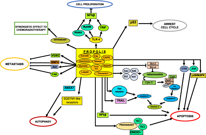
Many studies report that the anti-tumor effect of propolis on breast cancer cells works through several mechanisms [8,12,48,78–80]. The effectiveness of propolis depends on the dose and duration of exposure (Table 2). Generally, propolis can trigger apoptosis, inhibit cell cycle progression, reduce proliferation, induce autophagy, exhibit antioxidant effects, and inhibit metastases (Fig. 1).
Schematic diagram showing molecular pathways that play a role in propolis therapeutic activity against breast cancer cell pathways [2,9,12,16,76,85,89].
Apoptosis
Cytotoxicity is the most important effect of anticancer drugs. However, cytotoxic drugs are also a major issue in conventional chemotherapy because of their associated systemic side effects. Many studies report the natural components in propolis to show a selective effect against cancer cells without targeting normal cells. These bioactive compounds show effects against breast cancer cell cultures, ranging from potential to ineffective. The cytotoxic abilities of its bioactive substances remain inconsistent, according to some research reports [8]. However, there has also been convincing evidence related to the anticancer effects of the bioactive contents in propolis in vitro. The underlying molecular mechanisms also vary but generally involve apoptotic pathways.
There are many key apoptotic molecules related to the therapeutic mechanisms of propolis, including cytochrome C, protein caspases (3, 8, 9/10), p53, Bax, Bak, Fas receptor, TNF-related apoptosis-inducing ligand (TRAIL), nitrite oxide (NO), reactive oxygen species (ROS), and NF-κβ [16,81–86]. CAPE is a bioactive substance in propolis that induces apoptosis in breast cancer cells by modulating the intracellular pathways by triggering mitochondrial stress, resulting in the release of cytochrome C enzyme into the cytoplasm. With APAF-1, this protein complex then triggers a sequential reaction that activates caspase 9/10, converts procaspase three into caspase 3 to induce apoptosis. Interestingly, CAPE can increase the expression of p53, Bax, and tub proteins after 3 h of incubation and simultaneously decrease the expression of the anti-apoptotic protein Bcl-2 after 36 h of incubation [81]. Propolis can also activate apoptosis through the extracellular pathway mediated by Fas and TRAIL receptors, which activate caspase 8 and sequentially activate caspase 3, resulting in the apoptosis of breast cancer cells. In addition, NO and ROS at high concentrations can trigger oxidative stress, which induces the apoptosis of cancer cells. Propolis also has an inhibitory effect on NF-κB molecules and triggers p38/MAPK, JNK, and ERK pathways, which initiates apoptosis [2,57,87].
Anti-proliferation and cell cycle arrest
Propolis can inhibit or even arrest the breast cancer cell cycle by modulating the expression of cell cycle regulatory proteins. Propolis also inhibits proliferation by targeting various molecules, including Toll-Like Receptor 4 (TLR4), MyD88, IRAK4, TRIF, NF-κB, and p65. TLR4 acts as a double-edged knife in malignant diseases because it can both induce and suppress the growth of cancer cells. The TLR4 pathway consists of MyD88-dependent and MyD88-independent mechanisms, which can trigger NF-κB to release cytokines. MyD88 and IRAK4 are involved in the MyD88-dependent pathway, whereas TRIF plays a role in the MyD88-independent pathway [16]. Li et al. reported that propolis could arrest the cancer cell cycle in the S/G2 phase by inhibiting the expression of cyclin B1, D1, and CDK4 and increasing the expression of p21 [88].
Autophagy
Autophagy is considered a type of programmed cell death and is closely related to apoptosis [90]. Autophagy is an essential regulator of cell homeostasis that functions to protect cells. However, this process can impact anti-tumor or pro-tumor mechanisms depending on the developmental phase of the tumor itself. Autophagy is mediated by ANXA7 (a derivative of Ca2+-dependent GTPase) and autophagy-related SQSTM1-like receptors that play a role in inflammation [89].
Anti-metastatic
Fraser et al. reported that propolis also exhibits anti-metastatic effects by targeting VGSC, which regulates cellular invasion via pericellular acidification through the modulation of the sodium/hydrogen exchanger-1 activity and possibly the CD44-src-cortactin signaling axis [76]. In addition, the contents of propolis are reported to inhibit cancer cell invasion by upregulating MMP-2 tissue inhibitors, inhibiting NF-κB and Akt, phosphorylating focal adhesion kinase, and activating p38/MAPK and JNK [57,87,91].
Synergistic effects
The flavonoids in propolis play a role in antioxidant mechanisms by reducing superoxide anion radical in cancer cells. The antioxidant effect of propolis is able to synergize with chemotherapy drugs. By contrast, the polyphenols in propolis exhibit pro-oxidant activities that modulate nitrite and peroxynitrite anion (ONOO−) generation and increase intracellular ROS production, thereby inducing apoptosis [12]. Several studies have reported a beneficial effect of propolis or one of its bioactive substances when combined with conventional chemotherapy drugs and radiotherapy [9,92–94]. Motawi et al. tested a novel mixture of tamoxifen, a widely used antiestrogenic drug for breast cancer treatment, and CAPE. The combination exhibited synergistic activity as proapoptotic via multiple pathways such as (i) downregulate the expression of Bcl-2 and Beclin 1, (ii) activation of caspases protein that leads to DNA fragmentation, (iii) modulate the level of vascular endothelial growth factor and nitrite oxide [9]. In addition, Popolo et al. noted a promising result regarding the selective activity of nemorosone combine with ICI 182,780, the estrogen receptor down-regulators, that showed a significant cytotoxic effect if compared to ICI 182,780 or nemorosone alone [39]. Furthermore, Khoram et al. demonstrated that CAPE exposure in breast cancer cells prior to irradiation-induced radiosensitivity and significantly lower the survival curve of MDAMB-231 and T47D cells compare to control [94].
Conclusion
This review reveals that propolis shows cytotoxic, anti-proliferative, pro-autophagic, anti-metastatic, and antioxidant activities. Additionally, it exhibits synergistic effects when combined with chemotherapy or radiotherapy. These processes involve many target molecules, including NF-κβ, Fas receptors, p53, TLR4, ANXA7, and VGSC. The bioactive components of propolis and the target molecules involved need to be studied further to develop novel anti-breast cancer drugs and overcome chemoradiation resistance.
Footnotes
Acknowledgements
The authors do not have acknowledgments.
Conflict of interest
None.
Funding
None.
References
1.
Kabała-DzikA, Rzepecka-StojkoA, KubinaR, Jastrzębska-StojkoŻ, StojkoR, Dariusz WojtyczkaR, Comparison of two components of propolis: Caffeic Acid (CA) and Caffeic Acid Phenethyl Ester (CAPE) induce apoptosis and cell cycle arrest of breast cancer cells MDA-MB-231, Molecules, 22: 1554, 2017.
2.
Rzepecka-StojkoA, Kabała-DzikA, MoździerzA, KubinaR, WojtyczkaRD, StojkoR, Caffeic Acid phenethyl ester and ethanol extract of propolis induce the complementary cytotoxic effect on triple-negative breast cancer cell pathways, Molecules, 20: 9242–9262, 2015.
3.
JemalA, SiegelR, WardE, HaoY, XuJ, ThunMJ, Cancer statistics, 2009, Cancer J Clin, 59: 225–249, 2009.
4.
ReisJS, PusztaiL, Breast Cancer 2 Gene expression profiling in breast cancer: classification, prognostication, and prediction, Lancet, 378: 1812–1823, 2011.
5.
PerouCM, Borresen-DaleAL, Systems biology and genomics of breast cancer, Cold Spring Harb Perspect Biol, 3(2): a003293, 2011.
6.
DaiX, ChengH, BaiZ, LiJ, Review: breast cancer cell line classification and its relevance with breast tumor subtyping, J Cancer, 8: 3133–3135, 2017.
7.
MoulderS, HortobagyiGN, Advances in the treatment of breast cancer, Clin Pharmacol Ther, 83: 26–36, 2008.
8.
SeyhanMF, YilmazE, Timirci-KahramanO, SaygiliN, KisakesenHI, GaziogluS, Different propolis samples, phenolic content, and breast cancer cell pathways: variable cytotoxicity ranging from ineffective to potent, IUBMB Life, 71(5): 619–631, 2018.
9.
MotawiTK, AbdelazimSA, DarwishHA, ElbazEM, ShoumanSA, Modulation of tamoxifen cytotoxicity by Caffeic Acid Phenethyl Ester in MCF-7 breast cancer cells, Oxid Med Cell Longev, 2016: 3017108, 2016.
10.
VukovicaNL, ObradovicAD, VukicaMD, JovanovicD, DjurdjevicPM, Cytotoxic, proapoptotic and antioxidative potential of flavonoids isolated from propolis against colon (HCT-116 and breast (MDA-MB-231) cancer cell pathways, Food Res Int, 106: 71–80, 2018.
11.
BurdockGA, Review of the biological properties and toxicity of bee propolis (propolis), Food Chem Toxicol, 36(4): 347–363, 1998.
12.
XuanH, LiZ, YanH, SangQ, WangK, HeQ, Anti-tumor activity of Chinese propolis in human breast cancer MCF-7 and MDA-MB-231 cells, Evid Based Complement Altern Med, 2014: 280120, 2014.
13.
MarcucciMC, Propolis: chemical composition, biological properties and therapeutic activity, Apidologie, 26: 83–99, 1995.
14.
HuiC, QiX, QianyongZ, XiaoliP, JundongZ, MantianM, Flavonoids, flavonoid subclasses and breast cancer risk: a meta-analysis of epidemiologic studies, PLoS One, 8(1): e54318e, 2013.
ChangH, WangY, YinX, LiuX, XuanH, Ethanol extract of propolis and its constituent caffeic acid phenethyl ester inhibit breast cancer cells proliferation in inflammatory microenvironment by inhibiting TLR4 signal pathway and inducing apoptosis and autophagy, BMC Complement Altern Med, 17: 471, 2017.
17.
BanskotaAH, NagaokaT, SumiokaLY, TezukaY, AwaleS, Anti-proliferative activity of The Netherlands propolis and its active principles in cancer cell pathways, J Ethnopharmacol, 80: 67–73, 2002.
18.
DemirS, AliyaziciogluY, TuranI, MisirS, MenteseA, Antiproliferative and proapoptotic activity of Turkish propolis on human lung cancer cell line, Nutr Cancer, 68: 165–172, 2016.
19.
PremratanachaiP, ChanchaoC, Review of the anticancer activities of bee products, Asian Pac J Trop Biomed, 4(5): 337–344, 2014.
20.
BankovaVS, PopovSS, MarekovNL, High performance liquid chromatographic analysis of flavonoids from propolis, J Chromatogr A, 242: 135–143, 1982.
21.
BankovaV, Recent trends and important developments in propolis research, Evid Based Complement Altern Med, 2: 29–32, 2005.
22.
BeauregardA-P, HarquailJ, Lassalle-ClauxG, BelbraouetM, Jean-FrancoisJ, TouaibiaM, CAPE analogs induce growth arrest and apoptosis in breast cancer cells, Molecules, 20: 12576–12589, 2015.
23.
WuJ, OmeneC, KarkoszkaJ, BoslandM, EckardJ, KleinCB, Caffeic acid phenethyl ester (CAPE), derived from a honeybee product propolis,exhibits a diversity of anti-tumor effects in pre-clinical models of humanbreast cancer, Cancer Lett, 308(1): 43–53, 2011.
24.
OmeneC, KalacM, WuJ, MarchiE, FrenkelK, Propolis and its active component, Caffeic acid Phenethyl Ester (CAPE), modulate breast cancer therapeutic targets via an epigenetically mediated mechanism of action, J Cancer Sci Ther, 5: 334–342, 2013.
25.
WatabeM, HishikawaK, TakayanagiA, ShimizuN, NakakiT, Caffeic acid phenethyl ester induces apoptosis by inhibition of NFκB and activation of Fas in human breast cancer MCF-7 cells, J Biol Chem, 279: 6017–6026, 2004.
26.
SerkedjievaJ, ManolovaN, BankovaV, Anti-influenza virus effect of some propolis constituents and their analogues (esters of substituted cinnamic acids), J Nat Prod, 5: 294–302, 1992.
27.
RossiA, LongoR, RussoA, BorrelliF, SautebinL, The role of the phenethyl ester of caffeic acid (CAPE) in the inhibition of rat lung cyclooxygenase activity by propolis, Fitoterapia, 73: S30–S37, 2002.
28.
SherifMS, RehabTA, HamdiaZA, TorchilinVP, Cytotoxicity of propolis nanopreparations in cancer cell monolayers: multimode of action including apoptotsis and nitric oxide production, Gen Physiol Biophys, 37: 101–110, 2018.
29.
Lamoral-TheysD, PottierL, DufrasneF, Natural polyphenols that display anticancer properties through inhibition of kinase activity, Curr Med Chem, 17: 812–825, 2010.
30.
OrsolicN, KnezevicAH, SverL, TerzicS, BasicI, Immunomodulatory and anti-metastatic action of propolis and related polyphenolic compounds, J Ethnopharmacol, 94(2–3): 307–315, 2004.
31.
OrsolicN, A review of propolis anti-tumor action in vivo and in vitro, J ApiProd ApiMed Sci, 2: 1–20, 2010.
32.
PatelS, Emerging adjuvant therapy for cancer: Propolis and its constituents, J Diet Suppl, 13: 245–268, 2016.
33.
KuntzS, WenzelU, DanielH, Comparative analysis of the effects of flavonoids on proliferation, cytotoxicity, and apoptosis in human colon cancer cell pathways, Eur J Nutr, 38(3): 133–142, 1999.
TsengTH, ChienMH, LinWL, WenYC, ChowJW, Inhibition of MDA-MB- 231 breast cancer cell proliferation and tumor growth by apigenin through induction of G2/M arrest and histone H3 acetylationmediated p21WAF1/CIP1 expression, Environ Toxicol, 32: 434–444, 2017.
36.
DengXH, SongHY, ZhouYF, YuanGY, ZhengFJ, Effects of quercetin on the proliferation of breast cancer cells and expression of survivin in vitro, Exp Ther Med, 6: 1155–1158, 2013.
37.
ChouCC, YangJS, LuHF, IpSW, LoC, Quercetin mediated cell cycle arrest and apoptosis involving activation of a caspase cascade through the mitochondrial pathway in human breast cancer MCF-7 cells, Arch Pharm Res, 33: 1181–1191, 2010.
38.
SerafimTL, CarvalhoFS, MarquesMP, CalheirosR, SilvaT, Lipophilic caffeic and ferulic acid derivatives presenting cytotoxicity against human breast cancer cells, Chem Res Toxicol, 24: 763–774, 2011.
39.
PopoloA, PiccinelliAL, MorelloS, SorrentinoR, OsmanyCR, Cytotoxic activity of nemorosone in human MCF-7 breast cancer cells, Can J Physiol Pharmacol, 89: 50–57, 2011.
40.
RodregsEH, GrantMH, The effects of the flavonoids, Quercetin, Myricetin, and Epicatechin on the growth and enzyme activities of MCF-7 human breast cancer cells, Chem Biol Interact, 116: 213–228, 1998.
41.
OzdalT, Sari-KaplanG, Mutlu-AltundagE, BoyaciogluD, CapanogluE, Evaluation of Turkish propolis for its chemical composition, antioxidant capacity, anti-proliferative effect on several human breast cancer cell pathways and proliferative effect on fibroblasts and mouse mesenchymal stem cell line, J Apicultural Res, 57(5): 627–638, 2018.
42.
KalogeropoulosN, SJK, ET, MourtzinosI, VTK, Chemical composition, antioxidant activity and antimicrobial properties of propolis extracts from Greece and Cyprus, Food Chem, 116: 452–461, 2009.
43.
KangNJ, LeeKW, KimBH, BodeAM, LeeHJ, Coffee phenolic phytochemicals suppress colon cancer metastasis by targeting MEK and TOPK, Carcinogenesis, 32: 921–928, 2011.
44.
JungJE, KimHS, LeeCS, ParkDH, KimYN, Caffeic acid and its synthetic derivative CADPE suppress tumor angiogenesis by blocking STAT3-mediated VEGF expression in human renal carcinoma cells, Carcinogenesis, 28: 1780–1787, 2007.
45.
YangG, FuY, MalakhovaM, KurinovI, ZhuF, YaoK, Caffeic acid directly targets ERK1/2 to attenuate solar UV-induced skin carcinogenesis, Cancer Prev Res, 10: 1056–1066, 2014.
46.
KampaM, AlexakiVI, NotasG, NifliAP, NistikakiA, HatzoglouA, Anti-proliferative and apoptotic effects of selective phenolic acids on T47D human breast cancer cells: potential mechanisms of action, Breast Cancer Res, 6: r63, 2003.
47.
RosendahlAH, PerksCM, ZengL, MarkkulaA, SimonssonM, RoseC, Caffeine and caffeic acid inhibit growth and modify estrogen receptor and insulin-like growth factor I receptor levels in human breast cancer, Clin Cancer Res, 21: 1877–1887, 2015.
48.
OmeneC, KarkoszkaJ, BoslandM, EckardJ, KleinCB, FrenkelK, Caffeic acid phenethyl ester (CAPE), derived from a honeybee product propolis, exhibits a diversity of anti-tumor effects in pre-clinical models of human breast cancer, Cancer Lett, 308: 43–53, 2011.
49.
DziedzicA, KubinaR, Kabała-DzikA, TanasiewiczM, Induction of cell cycle arrest and apoptotic response of head and neck squamous carcinoma cells (detroit 562) by caffeic acid and caffeic acid phenethyl ester derivative, Evid Based Complement Altern Med, 2017: 6793456, 2017.
50.
SunL, WangK, XuX, GeM, ChenY, HuF, Potential protective effects of bioactive constituents from chinese propolis against acute oxidative stress induced by hydrogen peroxide in cardiac h9c2 cells, Evid Based Complement Altern Med, 2017: 7074147, 2017.
51.
LinCL, ChenRF, ChenJY, ChuYC, WangHM, ChouHL, Protective effect of caffeic acid on paclitaxel induced anti-proliferation and apoptosis of lung cancer cells involves NF- B pathway, Int J Mol Sci, 13: 13, 2012.
52.
JaganathanSK, Growth inhibition by caffeic acid, one of the phenolic constituents of honey, in HCT 15 colon cancer cells, Sci World J, 2012: 372345, 2012.
53.
SawickaD, CarH, BorawskaMH, NiklinskiJ, The anticancer activity of propolis, Folia Histochem Cytobiol, 50(1): 25–37, 2012.
54.
ChenMF, WuCT, ChenYJ, KengPC, ChenWC, Cell killing and radio-sensitization by caffeic acid phenethyl ester (CAPE) in lung cancer cells, J Radiat Res, 45: 253–260, 2004.
55.
OzturkG, GinisZ, AkyolS, ErdenG, GurelA, AkyolO, The anticancer mechanism of caffeic acid phenethyl ester (CAPE): review of melanomas, lung and prostate cancers, Eur Rev Med Pharmacol Sci, 16: 2064–2068, 2012.
PengCY, YangHW, ChuYH, ChangYC, HsiehMJ, ChouMY, Caffeic acid phenethyl ester inhibits oral cancer cell metastasis by regulatingmatrix metalloproteinase-2 and the mitogen-activated protein kinasepathway, Evid Based Complement Altern Med, 2012: 732578, 2012.
58.
MichaluartP, MasferrerJL, CarothersAM, SubbaramaiahK, ZweifelBS, KoboldtC, Inhibitory effects of caffeic acid phenethyl ester on the activity and expression of cyclooxygenase-2 in human oral epithelial cells and in a rat model of inflammation, Cancer Res, 59: 2347–2352, 1999.
59.
LaranjinhaJ, VieiraO, MadeiraV, AlmeidaL, Two related phenolic antioxidants with opposite effects on vitamin E content in low density lipoproteins oxidized by ferrylmyoglobin: consumption vs regeneration, Arch Biochem Biophys, 323: 373–381, 1995.
60.
KimuraY, OkudaH, OkudaT, HatanoT, AgataI, ArichiS, Studies on the activities of tannins and related compounds from medicinal plants and drugs. VII. Effects of extracts of leaves of Artemisia species, and caffeic acid and chlorogenic acid on lipid metabolic injury in rats fed peroxidized oil, Chem Pharmaceut Bull, 33: 2028–2034, 1985.
61.
LiuD, YouP, LuoY, YangM, LiuY, Galangin induces apoptosis in MCF-7 human breast cancer cells through mitochondrial pathway and phosphatidylinositol 3-kinase/Akt inhibition, Pharmacology, 102: 58–66, 2018.
62.
MurrayTJ, YangX, SherrDH, Growth of a human mammary tumor cell line is blocked by galangin, a naturally occurring bioflavonoid, and is accompanied by down-regulation of cyclins D3, E, and A, Breast Cancer Res, 8: R17, 2006.
63.
HaoQ, ZhaoP, NiuJ, WangJ, YuJ, Effect of ferulic acid on proliferation and mechanism in human breast cancer cells, Zhongguo Zhong Yao Za Zhi, 35: 2752–2755, 2010.
64.
ParkE, Data on cell cycle in breast cancer cell line, MDA-MB-231 with ferulic acid treatment, Data Brief, 7: 107–110, 2016.
65.
Cuesta-RubioO, Frontana-UribeBA, Ramirez-ApanT, CardenasJ, Polyisoprenylated benzophenones in cuban propolis; biological activity of nemorosone, Z Naturforsch C J Biosci, 57(3-4): 372–378, 2002.
66.
ChangH, MiM, LingW, ZhuJ, ZhangQ, WeiN, Structurally related cytotoxic effects of flavonoids on human cancer cells in vitro, Arch Pharm Res, 31(9): 1137–1144, 2008.
BiswasD, ShiQ, BaileyS, StricklandI, GhoshS, PardeeA, NF-KB activation in human breast cancer specimens and its role in cell proliferation and apoptosis, Proc Natl Acad Sci USA, 101: 10137–10142, 2004.
69.
InoueK, SaitoM, KanaiT, KawataT, ShigematsuN, UnoT, Anti-tumor effects of water-soluble propolis on a mouse sarcoma cell line in vivo and in vitro, Am J Chin Med, 36(3): 625–634, 2008.
70.
BazoAP, RodriguesMA, SforcinJM, de CamargoJL, RibeiroLR, SalvadoriDM, Protective action of propolis on the rat colon carcinogenesis, Teratogen Carcinogen Mutagen, 22(3): 183–194, 2002.
71.
RaoCV, DesaiD, SimiB, KulkarniN, AminS, ReddyBS, Inhibitory effect of caffeic acid esters on azoxymethane-induced biochemical changes and aberrant crypt foci formation in rat colon, Cancer Res, 53(18): 4182–4188, 1993.
72.
KhanN, AfaqF, MukhtarH, Apoptosis by dietary factors: the suicide solution for delaying cancer growth, Carcinogenesis, 28(2): 233–239, 2007.
HungMW, ShiaoMS, TsaiLC, Apoptotic effect of caffeic acid phenethyl ester and its ester and amide analogues in human cervical cancer ME180 cells, Anticancer Res, 23: 4773e80, 2003.
75.
DriglaF, BalacescuO, VisanS, BisboacaSE, Berindan-NeagoeI, MarghitasLA, Synergistic effects induced by combined treatments of aqueous extract of propolis and venom, Clujul Med, 89(1): 104–109, 2016.
76.
FraseraSP, HemsleyaF, DjamgozaMBA, Caffeic acid phenethyl ester: inhibition of metastatic cell behavioursvia voltage-gated sodium channel in human breast cancer in vitro, Int J Biochem Cell Biol, 71: 111–118, 2016.
77.
Syamsudin, Wiryowidagdo S, Simanjuntak P, Heffen WL, Chemical composition of propolis from different regions in Java and their cytotoxic activity, Am J Biochem Biotechnol, 5(4): 180–183, 2009.
78.
WuJ, BukkapatnamU, EckardJ, FrenkelK, Caffeic acid phenethyl ester (CAPE, a product of propolis) as an inhibitor of human breast cancer growth in a pre-clinical study and its effects on factors involved in cell cycle, angiogenesis, and drug resistance, Cancer Res, 68: 5710, 2008.
79.
LuoJ, SohJW, XingWQ, MaoY, MatsunoT, WeinsteinIB, A benzo-gamma-pyran derivative isolated from propolis inhibits growth of MCF-7 human breast cancer cells, Anticancer Res, 21: 1665–1671, 2001.
80.
PopoloA, PiccinelliLA, MorelloS, Cuesta-RubioO, SorrentinoR, Antiproliferative activity of brown Cuban propolis extract on human breast cancer cells, Nat Prod Commun, 4: 1711–1716, 2009.
81.
LeeYJ, KuoHC, ChuCY, WangCJ, LinWC, TsengTH, Involvement of tumor suppressor protein p53 and p38 MAPK in caffeic acid phenethyl ester-induced apoptosis of C6 glioma cells, Biochem Pharmacol, 66: 2281–2289, 2003.
82.
WatabeM, HishikawaK, TakayanagiA, ShimizuN, NakakiT, Caffeic acid phenethyl ester induces apoptosis by inhibition of NFκB and activation of Fas in human breast cancer MCF-7 cells, J Biol Chem, 279: 6017–6026, 2004.
83.
SzliszkaE, ZydowiczG, JanoszkaB, DoboszC, Kowalczyk-ZiomekG, KrólW, Ethanolic extract of Brazilian green propolis sensitizes prostate cancer cells to TRAIL induced apoptosis, Int J Oncol, 38: 941–953, 2011.
84.
WangY, TangQ, JiangS, LiM, WangX, Anti-colorectal cancer activity of macrostemonoside A mediated by reactive oxygen species, Biochem Biophys Res Commun, 441: 825–830, 2013.
85.
Seda VatanseverH, SorkunK, Ismet Deliloglu GurhanS, Ozdal-KurtF, TurkozE, GencayO, Propolis from Turkey induces apoptosis through activating caspases in human breast carcinoma cell lines, Acta Histochem, 112(6): 546–556, 2010.
86.
MotomuraM, KwonKM, SuhSJ, LeeYC, KimYK, LeeIS, Propolis induces cell cycle arrest and apoptosis in human leukemic U937 cells through Bcl-2/Bax regulation, Environ Toxicol Pharmacol, 26(1): 61–67, 2008.
87.
PalHC, SharmaS, StricklandLR, KatiyarSK, BallestasME, AtharM, Fisetin inhibits human melanoma cell invasion through promotion of mesenchymal to epithelial transition and by targeting MAPK and NFkB signaling pathways, PLoS One, 9(1): e86338, 2014.
88.
LiH, KapurA, YangJX, SrivastavaS, McLeodDG, Paredes-GuzmanJF, Antiproliferation of human prostate cancer cells by ethanolic extracts of Brazilian propolis and its botanical origin, Int J Oncol, 31(3): 601–606, 2007.
89.
LiH, HuangS, WangS, Relationship between annexin A7 and integrin, Int J Biochem Cell Biol, 45: 2605–2611, 2013.
90.
DebnathJ, BaehreckeEH, KroemerG, Does autophagy contribute to cell death?Autophagy, 1(2): 66–74, 2005.
91.
ShigeokaY, IgishiT, MatsumotoS, NakanishiH, KodaniM, YasudaK, Sulindac sulfide and caffeic acid phenethyl ester suppress the motility of lung adenocarcinoma cells promoted by transforming growth factor-betathrough Akt inhibition, J Cancer Res Clin Oncol, 130(3): 146–152, 2004.
92.
GargAK, BuchholzTA, AggarwalBB, Chemosensitization and radiosensitization of tumors by plant polyphenols, Antioxid Redox Signl, 7: 1630–1647, 2005.
93.
HerreraY, GabbiaD, Diaz GarciaA, CuestaO, CarraraM, Chemosensitizing activity of Cuban propolis and nemorosone in doxorubicin resistant human colon carcinoma cells, Fitoterapia, 136: 104173, 2019.
94.
KhoramNM, BigdeliB, NikoofarA, GoliaeiB, Caffeic acid phenethyl ester increases radiosensitivity of estrogen receptor- positive and -negative breast cancer cells by prolonging radiation-induced DNA Damage, J Breast Cancer, 19(1): 18–25, 2016.