
Abstract
Hyaline globules (thanatosomes) have been observed in many neoplastic and non-neoplastic processes. They represent terminal cytoplasmic structures associated with cytoplasmic blebbing, which are related to cell injury and apoptosis. Thanatosomes are reported rarely in breast tumors. Here we describe a case of invasive breast cancer of no special type with abundant thanatosomes.
Introduction
Hyaline globules or thanatosomes have been found histologically in many non-neoplastic and neoplastic conditions. The origin of thanatosomes is related to irreversibile cell injury and apoptotic cell death [1]. Among tumors they are most often visible in renal cell carcinomas and oncocytomas, as well as in hepatocellular carcinomas, and less frequently in gastrointestinal adenomatous polyps and adenocarcinomas, ovarian tumors, Kaposi sarcoma, cartilaginous tumors, gallbladder carcinosarcoma and testicular cancer [1–8]. They are very rarely reported in breast tumors [9–11].
Here we report a case of moderately differentiated breast cancer of no special type (NST) with abundant intracellular thanatosomes.
Case report
A 68-year-old woman presented to her clinician with palpable nodule in the superior lateral quadrant of the left breast noticed a month before. Physical examination of the surrounding breast tissue and contra-lateral breast showed no abnormalities. The patient had no family history of breast cancer. Ultrasound examination revealed heterogeneous hypoechogenic zone of 1.5 cm in diameter with dorsal attenuation, ill-defined borders and microcalcifications. This later finding was suggestive of carcinoma. A mammographic examination was not performed.
Breast core biopsy rendered a diagnosis of the intermediate grade invasive carcinoma of NST, which was hormonal receptors positive, HER-2 negative, and with low Ki-67 proliferation index. Segmentectomy with sentinel lymph node biopsy followed, with specimen containing ill-defined, grey-white tumor measuring 1.7 × 1.5 cm, with hard consistency on cut sectioning. The tissues were fixed in neutral buffered formalin and embedded in paraffin for routine histological examination. Histological sections showed invasive carcinoma of NST composed of irregular glands and cords lined by atypical cells with an intermediate nuclear grade and mitotic rate, which was classified as grade 2 according to the Elston-Ellis histological grading system (Fig. 1). In some part of the tumor the cytoplasm of tumor cells contained numerous homogenous, hyaline globules of various size were seen, many of them delimited by a surrounding clear halo (Fig. 1 and Fig. 2). Histochemically, globules were periodic acid-Schiff (PAS) positive. Although the overall proliferative activity of tumor cells measured by Ki-67 immunostain was low (3%), in the part of the tumor which contained thanatosomes tumor cells expressed higher proliferation (8%) (Fig. 3). These findings were consistent with an invasive breast carcinoma with hyaline globules (thanatosomes). Sentinel lymph node was without metastatic infiltration, and cancer was staged as T1cN0M0 (the same cTNM and pTNM) tumor.

Invasive breast cancer (NST) with numerous hyaline globules (thanatosomes) in tumor cells cytoplasms (H&E, 400×).
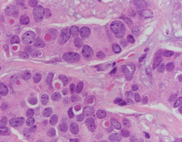
Thanatosomes of various size are surrounded by clear halo (H&E, 1000×).
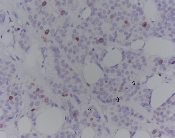
Immunohistochemical expression of Ki-67 in thanatosome rich part of the tumor (some of the thanatosomes are highlighted with arrows) (IHC, 400×).
Hyaline globules have been described in many cell types in association with various nonneoplastic and neoplastic conditions. According to Papadimitriou et al. they are terminal cytoplasmic structures related to cell injury and apoptotic cell death, specifically associated with cytoplasmic blebbing and condensation with secondary plasma protein insudation, resulting in formation of globoid hyaline cellular fragments. They proposed the name thanatosomes for the entire spectrum of hyaline globules to highlight their association to apoptotic cell death [1].
Thanatosomes in breast tumors were rarely reported and investigated [9–11]. D’Alfonso et al. described two cases of malignanat phyllodes tumor with numerous hyaline bodies in the stromal component of the tumors [9]. Panicker et al. described a case of high grade infiltrating NST breast carcinoma with abundant thanatosomes located intracellularly and in the surrounding stroma [10]. Ozerdem and al., reported a case of conventional, spindle cell type mammary myofibroblastoma with intracytoplasmatic hyaline globules [11]. In the report of the cases of phyllodes tumors with thanatosomes D’Alfonso et al. also reported the results of their retrospective search for thanatosomes in 86 high-grade breast lesions. They have found thanatosomes in 16% of high grade breast lesions, mostly in metaplastic spindle cell carcinomas (21% of cases) and malignant phyllodes tumors (20% of cases). The thanatosomes were found focally in only 2 cases of 21 reviewed cases of poorly differentiated invasive NST carcinomas [9].
Histogenetically, thanatosomes are in most cases related to apoptotic cell death. Papadimitriou et al. showed that they represent degenerative phenomenon common to all cell types, which occurs in cells with propensity to apoptosis, in cells with abundant rough endoplasmic reticulum and lysosomes, and in tissues with extracellular protein-rich fluid [1]. According to this theory thanatosomes are composed of apoptotic breakdown products of lysosomal proteolysis, with subsequent plasma protein imbibition, which results in globular shape and eosinophilic color [1,3]. This nonspecific microscopic finding is histological marker of high cell turnover and apoptotic cell death, and therefore in neoplastic conditions is usually related to high grade tumors. Curiously in this report we found numerous intracellular thanatosomes in invasive breast cancer of NST which was moderately differentiated (grade 2). However, the part of the tumor which contained thanatosomes was composed of the less differentiated cells which showed higher proliferative activity expressed by positive Ki-67 immunostain than the rest of the tumor, confirming the relationship between thanatosomes and high turnover of the cells.
In conclusion we reported herein a case of moderately differentiated invasive breast cancer of NST, partially rich with eosinophilic hyaline globules. These globules are related to apoptotic cell death, and therefore called thanatosomes.
Footnotes
Conflict of interest
The author declares no conflict of interest.