
Abstract
The method consists essentially of enclosing the excised stomach in a small volume recorder (a 2-way dog size arterial cannula serves the purpose). A sufficient length of oesophogus and intestine are removed with the stomach to permit introduction of an inflow cannula into the former and an outflow cannula into the latter in a way not to interfere with the valves at either end of the stomach. The stomach is then passed into the “Oncometer,” short segments of rubber tubing serving to make an air tight seal between the cannulae and the “Oncometer.” From the side tube of the latter a rubber tube leads to a delicate tambour which records both peristaltic waves and tone changes. Perfusion of the lumen of the stomach is effected from a constant pressure tank in which the nature of the fluid can be easily altered. The optimum height of the tank is about 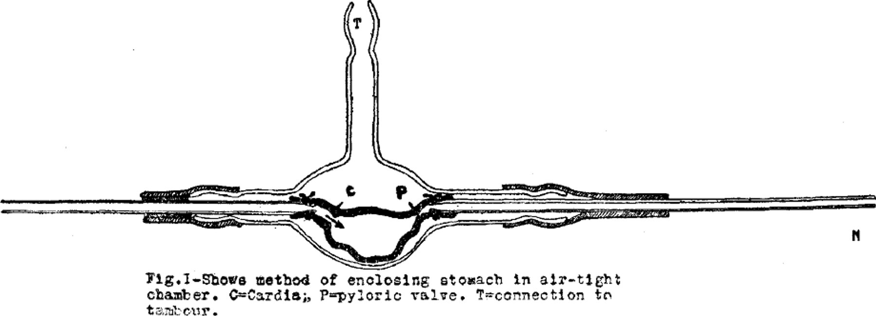 15 cm., the flow ceasing at a height of 8 to 9 cm. A flutter valve is placed between the pressure tank and the stomach to prevent back flow, when peristaltic waves press against a closed pylorus or during antiperistalsis. The operation of the pyloric valve is recorded by placing a drop-counter beneath the tip of the outflow cannula.
The method has the advantages of freedom from indwelling balloons or other similar interferences, and permits direct observations, especially by the aid of a reading glass, at all times.
The feasibility of such an arrangement for the demonstration of normal functions of the stomach and also of the influence of drugs is apparent. A report on such is in preparation.
Get full access to this article
View all access options for this article.