
Abstract
Background
Several population studies demonstrated an increased risk of allergic rhinitis in patients exposed to acetaminophen. However, no histologic studies have been conducted to assess the relationship between acetaminophen exposure and allergic rhinitis.
Objective
In this study, we investigated the association between chronic acetaminophen exposure and the development of allergic rhinitis in a rat model.
Methods
Ten female Sprague-Dawley rats were randomly assigned to either a control (n = 5) or an acetaminophen group (n = 5). The acetaminophen group received 200 mg/kg/day of acetaminophen suspended in yogurt via oral gavage for 120 days. The control group received only the yogurt vehicle. Allergic behavioral responses, including nose rub, eye rub, ear scratching, and neck and/or face scratching, were quantified. The rats were killed, and the noses were harvested. The portion of the nose, including the nasal septum and the inferior turbinates, was embedded in paraffin, sectioned, and stained with hematoxylin and eosin to quantify the inflammatory infiltrate.
Results
The average number of allergic responses per animal was 13.2 in the acetaminophen group versus 6.2 in the control group (p = 0.032). All the rats in the acetaminophen group (100%) had mast cells infiltrating the lamina propria of the inferior turbinate, whereas mast cells were detected in only 40% of the animals in the control group. The average number of mast cells per animal in the acetaminophen group was 134 versus 21 in the control group (p = 0.048).
Conclusions
Our study was the first to demonstrate a histologic association between chronic exposure to acetaminophen and rhinitis. Further research to elucidate the mechanism that underlies these findings is necessary.
Moreover, this effect seems to be dose-dependent. Children exposed to medium and high doses of acetaminophen have an increased likelihood of developing asthma, rhinoconjunctivitis, and eczema. 1 Several mechanisms have been proposed in an attempt to explain this observation. Depletion of glutathione-S-transferase in the upper airway mucosa that leads to increased oxidative stress,1,2 promotion of T-helper 2 (Th2) differentiation pathways, and an immunoglobulin E (IgE) mediated reaction are among the leading hypotheses. 3
Despite the increasing evidence at the population level, to date, there have been no experimental studies that demonstrated a cause-and-effect relationship between acetaminophen exposure and allergic rhinitis. In this pilot study, we examined the relationship between chronic exposure to acetaminophen and the development of allergic rhinitis at behavioral and histologic levels in an animal model.
Materials and Methods
Animal Treatments
Ten female Sprague-Dawley rats (200–300 g body weight) were obtained from breeding stocks maintained at Harlan (Indianapolis, IN). The animals were kept at 22°C with a 12-hour on, 12-hour off light cycle, and were allowed free access to water and food. All procedures that involved the use of the animals were approved by the Edward Hines Jr. Veterans Affairs Hospital.
Institutional Animal Care and Use Committee
The animals were randomly assigned to either a control (n = 5) or an acetaminophen group (n = 5). The acetaminophen group received the 200 mg/kg/day drug suspended in 1 mL of a yogurt vehicle, whereas the control group received 1 mL of the yogurt vehicle alone. Administration was performed daily by oral gavage. The animals were weighed weekly to ensure adequate dosing. The control animals were handled in exactly the same manner as the acetaminophen rats. A period of 120 days was chosen to ensure chronic exposure to the drug and to allow for allergen sensitization to take place. The animals were killed by ketamine overdose and transcardial perfusion with 4% paraformaldehyde at the end of the 120-day period.
Analysis of Behavioral Allergic Responses
At the end of the sensitization period, allergic behavioral responses were assessed. Within 30–60 minutes of acetaminophen administration, the animals were placed in an isolation cage without any external stimulation. A time frame of 30–60 minutes after exposure was selected to encompass the early phase of the allergic response. Two blinded investigators (N.C. and P.C.) observed each animal for 20 minutes. The behaviors were categorized and quantified by the investigators independently of each other. Four main patterns of allergic behavior were quantified: ear scratching, face or neck scratching, eye rub, and nose rub.
Morphologic Evaluation of the Nasal Mucosa
After the behavioral portion of the study was completed, the animals were killed, and the lower jaw and connective tissues over the skull were removed. The snouts were transected along a plane anterior to the second palatal ridge as described by Young, 4 to obtain a specimen that contained the septum and inferior turbinates. The specimens were fixed in formalin and subsequently decalcified and embedded in paraffin. The tissue blocks were then sectioned at 5 μm and stained with hematoxylin and eosin.
To obtain a representative sample of the inflammatory changes, two slides were made per block and two sections were cut per slide. Thus, a total of four samples of tissue were analyzed per animal. The slides were first screened by a pathologist (S.M.) in the Department of Pathology, Loyola Medical Center. In a blinded manner, the pathologist identified the specimens in which an inflammatory infiltrate was present. The inflammatory infiltrate was then independently quantified and qualified by two blinded investigators (N.S. and P.C.). The total number of mast cells and lymphoid aggregates per animal was quantified.
All statistical analyses were conducted by using GraphPad Prism 6 Software, 2013 (San Diego, CA). Differences at p = 0.05 were considered statistically significant. A Mann-Whitney t-test was used to detect statistical significance.
Results
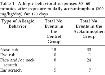
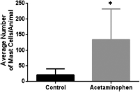
During the 20-minute observation period after acetaminophen administration, the total number of nose rubs for the animals in the control group was 18. The total number of nose rubs in the acetaminophen group was 33. In the control group, there were no eye rubs, whereas one eye rub was detected in the acetaminophen group. Eight face or neck scratches were detected in the control group, whereas, in the acetaminophen group, there were 24 events. There were only five ear scratches in the control group versus seven in the acetaminophen group (Table 1). The average number of allergic responses per animal was 13.2 ± 2.2 in the acetaminophen group versus 6.2 ± 2.8 in the control group (Fig. 1). All data are represented as the average ± standard error of the mean (SEM). This difference was statistically significant (p = 0.032).
Allergic behavioral responses 30–60 minutes after exposure to daily acetaminophen (200 mg/kg/day) for 120 days

The average number of allergic responses per animal after administration of acetaminophen (200 mg/kg/day) dissolved in yogurt in the experimental group versus the yogurt vehicle alone in the control group. Values represent the mean ± SE for five animals in each group (*p = 0.032).
Histologic analysis revealed lymphoid aggregates in one of five rats (20%) in the control group. In the acetaminophen group, three of five rats (60%) had lymphoid aggregates (Table 2). The average number (± SEM) of lymphoid aggregates per animal was 0.2 ± 0.2 in the control group and 0.9 ± 0.5 in the acetaminophen group (Fig. 2). This difference was not statistically significant (p > 0.05).
Type and frequency of inflammatory infiltrate present in lamina propria of the inferior turbinate in acetaminophen rats versus control rats after daily exposure to acetaminophen (200 mg/kg/ day) for 120 days


The average number of lymphoid aggregates detected in the lamina propria of the inferior turbinate of each animal after administration of acetaminophen (200 mg/kg/day) dissolved in yogurt in the experimental group versus the yogurt vehicle alone in the control group. Values represent the mean ± SE for the five animals in each group.
All of the animals in the acetaminophen group had mast cells infiltrating the lamina propria of the inferior turbinate, whereas mast cells were detected in only 40% of the animals in the control group (Table 2). The average number (± SEM) of mast cells per animal in the acetaminophen group was 133.8 ± 98.1 versus 20.8 ± 19.3 in the control group (p = 0.048) (Fig. 3).
The average number of mast cells identified in the lamina propria of the inferior turbinate of each animal after administration of acetaminophen (200 mg/kg/day) dissolved in yogurt in the experimental group versus the yogurt vehicle alone in the control group. Values represent the mean ± SE for the five animals in each group (*p = 0.048).
The morphologic changes in the loose connective tissue of the inferior turbinate observed at the completion of the 120-day period are demonstrated in Fig. 4. The differences between the rats in the control group and those in the acetaminophen group are shown. A lymphoid aggregate and mast cell infiltrate in an acetaminophen-treated rat is demonstrated in Fig. 4 and Fig. 5, respectively. In contrast, the normal architecture of the lamina propria and the lack of an inflammatory infiltrate in a control animal are shown in Figs. 6 and 7.

Histologic changes in the inferior turbinate after daily exposure to acetaminophen. The animals received daily administration of acetaminophen (200 mg/kg/day) via oral gavage. After 120 days, the inferior turbinate and septum were harvested. A transverse section in the region of the inferior turbinate in an acetaminophen-treated rat stained with hematoxylin and eosin is shown. A lymphoid aggregate (denoted by the arrow) in the loose connective tissue of the lamina propria is seen. A 5-μm paraffin-embedded section stained with hematoxylin and eosin, original magnification ×100.
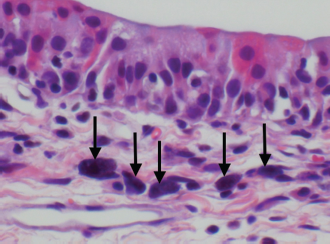
Histologic changes in the inferior turbinate after daily exposure to acetaminophen. The animals received daily administration of acetaminophen (200 mg/kg/day) via oral gavage. After 120 days, the inferior turbinate and septum were harvested. A transverse section in the region of the inferior turbinate in an acetaminophen-treated rat stained with hematoxylin and eosin is shown. Mast cell infiltration in the lamina propria is evidenced (denoted by the arrows), as demonstrated by their round-to-oval cytoplasm and the deep basophilic staining of the cytoplasm, which results from accumulation of histamine granules. A 5-μm paraffin-embedded section stained with hematoxylin and eosin; original magnification ×600).

Normal histology in the inferior turbinate in a control rat. A transverse section in the region of the inferior turbinate stained with hematoxylin and eosin is shown. Note the normal architecture and the lack of lymphoid aggregates. A 5-μm paraffin-embedded section stained with hematoxylin and eosin; original magnification ×100.
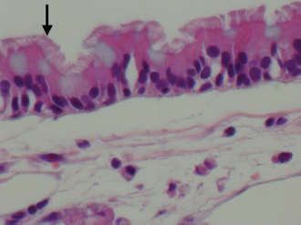
Lack of histologic changes in the inferior turbinate in a control rat. A transverse section in the region of the inferior turbinate stained with hematoxylin and eosin is shown. Note the normal-appearing ciliated respiratory epithelium (arrow). The loose connective tissue of the inferior turbinate is void of mast cells. A 5-μm paraffin-embedded section stained with hematoxylin and eosin, original magnification ×600.
Discussion
In 2000, acetaminophen was first identified as a potential risk factor for the development of atopic diseases. A study by Shaheen et al. 5 reported an association between frequent acetaminophen use and the development of asthma (odds ratio [OR] 1.06) and rhinitis (OR 1.80 weekly use; OR 2.33 daily use) in a case-control study (N = 1574). However, the investigators did not collect detailed information regarding specific dosing of acetaminophen in the study.
Since then, there have been several population-based studies that demonstrate this association and raise concern for acetaminophen as a potential factor that lead to the increase in allergic diseases in children seen over the past 3 decades. In 2004, Cohet et al. 6 reported an increased prevalence of asthma, rhinitis, and eczema symptoms in children 6–7 years old. Karimi et al. 7 studied 3000 children (range, 6–7 years old) and 3000 adolescents (range, 13–14 years old) in Iran. The investigators found that acetaminophen intake resulted in more-frequent symptoms of nighttime dry cough, eczema, and rhinitis. 2 Perhaps the most compelling evidence arose from the Phase Three International Study of Asthma and Allergies in Childhood study by Beasley et al. 1 in 2008. This study included 205,487 children ages 6–7 years from 31 countries, and revealed that acetaminophen use was associated with an increased risk of symptoms of rhinoconjunctivitis (OR 1.33) and eczema (OR 1.30). The exact dose of acetaminophen intake was not reported in the study. The acetaminophen dose was categorized as low, medium, or high in the survey used to collect the data. A follow-up study, which used data from the International Study of Asthma and Allergies in Childhood study, also by Beasley et al. 8 examined if the association was present in adolescents. The investigators found that, among 361,598 patients from 54 countries, ages 13–14 years, there was a significant dose-dependent increased risk of rhinoconjunctivitis (OR 1.33 [medium acetaminophen use] and OR 2.18 [high acetaminophen use]). Furthermore, this association was present across multiple regions of the world. 2 Other epidemiologic studies have since followed, which also revealed an association between acetaminophen and allergic diseases.2,3
These population-based studies, however, have not been able to conclusively demonstrate that the relationship between acetaminophen and atopic diseases is a cause-and-effect one. This has been attributed to several factors. Increased intake of acetaminophen to control symptoms due to viral illnesses in patients with asthma, increased intake to relieve pain or fever caused by asthma itself, and increased intake because acetaminophen has traditionally been considered safe in patients with asthma have been the three leading potentially confounding issues. 2
Our study is the first pilot study to provide some evidence regarding this relationship at histologic and behavioral levels. Our results demonstrated that allergic behavioral responses were increased in animals chronically exposed to high-dose acetaminophen. This observation paralleled the conclusions drawn based on epidemiologic data obtained from adults, children, and adolescents.1–4,6–11
In our study, the inflammatory infiltrate identified consisted mainly of lymphoid aggregates and mast cells. The pattern of mast cell infiltration was markedly different between the acetaminophen group and the control group. One hundred percent of the animals exposed to acetaminophen had mast cells that infiltrated the lamina propria of the inferior turbinate. In contrast, mast cells were detected in only 40% of the animals in the control group. Our study also revealed a statistically significant difference in the number of mast cells per animal when the two groups were compared. The average number of mast cells per animal in the acetaminophen group was 134 versus 21 in the control group. The significant difference detected in the number of mast cells may be indicative of an allergic process because mast cells play a key role in both the immediate and late phases of the allergic response as well as ongoing inflammation.
Within turbinate tissue, lymphocyte aggregates were identified in 60% of the acetaminophen-treated rats group. However, only 20% of the rats in the control group had evidence of lymphocyte aggregates. Although immunohistochemistry was not performed on these lymphocyte aggregates, hematoxylin and eosin staining does indicate that these lymphocyte aggregates consist of either T-cell lymphocytes, B-cell lymphocytes, or both. Although the lymphocytic aggregates were more frequently observed in the acetaminophen group, a comparison of the average number of lymphoid aggregates per animal between the two groups did not achieve statistical significance. This may be explained by the large variability observed within the acetaminophen group.
Nonetheless, detecting lymphocyte aggregates in the lamina propria of the inferior turbinate is a very interesting finding. T lymphocytes play a key role in atopic diseases. Multiple studies indicated that peripheral CD4+ Th2 subsets are increased in asthma and rhinitis, and that Th2-mediated inflammation does occur within the nasal mucosa.12,13 Antigen presentation via major histocompatibility complex type II receptors by antigen presenting cells results in the propagation of CD4+ Th2 cells. In turn, these cells upregulate synthesis and expression of major histocompatibility complex type II receptors.14,15 In addition, Th2 cells can induce immunoglobin type switching in B-cell populations, which results in the production of IgE. Mast cells and basophils may also induce immunoglobulin class switching in B cells as well.16,17 Although our numbers do not indicate that the increased lymphocyte populations within the acetaminophen group are significant, the increased numbers are interesting to note and warrant further investigation.
Our study was limited by several factors. First, the sample size was small and hence decreased our power. This, however, was a pilot study and issues of power would be overcome with further investigation. Increased sample size will also help us determine if the lymphocyte aggregates are significantly more common in rats exposed to acetaminophen. Staining for T-cell and B-cell markers will also help determine whether these lymphocyte aggregates represent a homogenous or a heterogeneous mixture. Second, grooming and allergic behavior in a rat may overlap, which constitutes a potential confounding factor; however, there were statistically increased behaviors in our acetaminophen group, which indicates that allergic reaction was more likely to be the explanation of these behaviors rather than grooming. Third, immunohistochemical staining for IgE was not performed. This will be the focus of future research to validate our findings. Fourth, there are inherent differences between rat and human physiology; such differences must be taken into account when trying to extrapolate our findings from an animal model to humans.
Conclusions
Our findings indicated that chronic exposure to acetaminophen was associated with the development of rhinitis in a rat model. Although our study was conducted in rats, these findings do offer possible insight regarding the reported epidemiologic association between acetaminophen exposure and atopic diseases, particularly allergic rhinitis, in several different populations across the globe. Future research should focus on demonstrating the presence of IgE by immunohistochemical methods in larger animal groups. A better understanding of this relationship may translate into a significant benefit in disease prevention and control in humans.
Footnotes
The authors have no conflicts of interest to declare pertaining to this article