
Abstract
Matrix metalloproteinases (MMPs) are extracellular proteolytic enzymes involved in tumor progression. We present the in vivo detection and quantitation of MMP7 activity using a specific near-infrared polymer-based proteolytic beacon, PB-M7NIR. PB-M7NIR is a pegylated polyamidoamine PAMAM-Generation 4 dendrimer core covalently coupled to a Cy5.5-labeled peptide representing a selective substrate that monitors MMP7 activity (sensor) and AF750 as an internal reference to monitor relative substrate concentration (reference). In vivo imaging of tumors expressing MMP7 had a median sensor to reference ratio 2.2-fold higher than a that of a bilateral control tumor. Ex vivo imaging of intestines of multiple intestinal neoplasia (APCMin) mice injected systemically with PB-M7NIR revealed a sixfold increase in the sensor to reference ratio in the adenomas of APCMin mice compared with control intestinal tissue or adenomas from MMP7-null Min mice. PB-M7NIR detected tumor sizes as small as 0.01 cm 2 , and the sensor to reference ratio was independent of tumor size. Histologic sectioning of xenograft tumors localized the proteolytic signal to the extracellular matrix; MMP7-overexpressing tumors displayed an approximately 300-fold enhancement in the sensor to reference ratio compared with nonexpressing tumor cells. In APCMin adenomas, the proteolytic signal colocalized with the endogenously expressed MMP7 protein, with sensor to reference ratios approximately sixfold greater than that of normal intestinal epithelium. PB-M7NIR provides a useful reagent for the in vivo and ex vivo quantitation and localization of MMP-selective proteolytic activity.
ONE OF THE DEFINING CHARACTERISTICS of malignancy is the ability of cancer cells to metastasize and invade distant organ sites. Proteolysis of the extracellular matrix (ECM) is required to accommodate increased growth, migration, and invasion of tumor cells. Matrix metalloproteinases (MMPs) are a family of extracellular, zinc-dependent proteinases that are capable of degrading most of the multiple components of the ECM. 1 In the tumor microenvironment, host- and tumor-derived MMPs are often misregulated, leading to uncontrolled degradation of the ECM. Of the 23 identified human MMP gene products, MMP7 is one of the smallest members of this family, possessing only the domains necessary for targeting to the secretory pathway, control of latency, and catalytic activity. MMP7 is notably produced by cells of epithelial origin and contributes to tumor formation in a number of epithelium-derived adenocarcinomas.2,3 From a molecular imaging standpoint, MMP7 is a secreted extracellular proteinase that appears to be a promising target for in vivo detection of tumors and quantitative analysis. Thus, MMP7 provides an attractive target for developing an enhanced near-infrared (NIR) proteolytic beacon (PB) to noninvasively assess proteolytic activity.
Quenched fluorescent peptide probes have been used for decades to measure the proteolytic activity of purified enzymes, and quenched fluorescent matrix proteins have been applied to cell culture models to monitor extracellular proteolysis. 4 Small molecule activity-based probes have been used in both in vitro and in vivo studies to monitor cysteine and serine proteinase activities 5 but have not yet proven to be useful for measuring MMP activity. The ability to probe tissue with light for minimal or noninvasive detection of cancer has become feasible mainly owing to the development of fluorescent probes emitting in the NIR spectrum, where tissue has both low absorption and reduced scattering. 6 A number of different fluorescent approaches have demonstrated the capability to detect specific molecular events in vivo.7–10 For optical imaging, Achilefu and colleagues prepared a number of NIR optical contrast agents designed to either bind to or be metabolized by tumors and, together with Chance and colleagues, demonstrated the feasibility of detecting 2 cm-deep subsurface tumors using a metabolism-enhanced near-infrared fluorescent (NIRF) contrast agent and NIRF in vivo imaging.11–13 Weissleder and colleagues used NIR probes to assess proteolytic activity and subsequent inhibition by MMP inhibitors.5,6 Jiang and colleagues described a new strategy using protease-activatable cell-penetrating peptides to deliver fluorescent labels within tumor cells. 14 Probes have been developed to assess proteolytic activity based on Förster resonance energy transfer (FRET): endogenous quenching of closely positioned fluorochromes on peptide substrates that on proteolytic cleavage produce enhanced fluorescence.15–17 In particular, Pham and colleagues developed an MMP7 NIR probe that was capable of imaging protease activity in vitro. 18 Thus, the principle to detect proteolytic activity in vitro and in vivo has been demonstrated.
We previously described a polymer-based fluorogenic peptide substrate that selectively detected MMP7 activity in vitro and in vivo with fluorophores that emit in the visible range. 15 The fluorogenic peptide substrate is built on a Generation 4–polyamidoamine (G4-PAMAM) backbone containing a noncleavable internal reference fluorophore that monitors both the cleaved and the uncleaved reagent. In the present study, we used NIR FRET pair fluorochromes to increase the efficiency of light traveling through tissue. 19 These new reagents, referred to as PB-M7NIR to indicate that they are PBs selective for MMP7 (M7) that emit in the NIR range, extend our previous studies to examining MMP7 activity in the context of multiple in vivo and ex vivo tumor microenvironments, as well as to provide quantitative assessment of relative proteolytic activity.
Materials and Methods
Materials
All chemicals and biochemicals were reagent grade, and solutions were prepared in deionized filtered water (Milli-Q, Billerica, MA). Generation 4 Starburst PAMAM dendrimer (10% w/w in methanol), Brij 35 solution (30% w/v), and Tricine (> 98%) were obtained from Sigma-Aldrich (St. Louis, MO). Polyethyleneglycol PEG5000 was obtained from Shearwater Polymers, Inc (Shearwater, TX). N-Succinimidyl iodoacetate (SIA) was obtained from Pierce Chemical (Rockford, IL). M7 [(AHX)-RPLALWRS-(AHX)-C, where AHX is aminohexanoic acid], a high-performance liquid chromatography-purified peptide that includes two AHX linkers, was obtained from Open Biosystems (Huntsville, AL). The N-hydroxysuccinimidyl (NHS) ester derivatives of Cy5.5 and AF750, two NIR fluorophores, were obtained from GE Healthcare (London, UK) and Molecular Probes, Invitrogen (Carlsbad, CA), respectively. MMP7 was obtained from Calbiochem (San Jose, CA). Low fluorescent (TD-97184) and high-fat chow (TD-5015) were obtained from Harlan Teklad (Madison, WI)
Synthesis of PB-M7NIR
The polymer-based fluorogenic substrate PB-M7NIR was synthesized according to the procedure outlined in Figure 1A.

Synthesis and structure of PB-M7NIR. Panel A, Scheme for the synthesis of PB-M7NIR, a (Cy5-M7)8-PAMAM-(PEG5000) (AF750)6 copolymer. Panel B, The Cy5.5 optical sensor is linked via AHX to the N-terminus of the matrix metalloproteinase-selective cleavable peptide (RPLA*LWRS, cleavage site denoted by an asterisk) that is coupled via a second AHX and cysteine with the PAMAM. The internal reference, AF750, of PB-M7NIR is linked directly to the dendrimer (Starburst PAMAM dendrimer, Generation 4). The structure is not drawn to scale.
Labeling M7 Peptide with Cy5.5
The Cy5.5-labeled peptide Cy5.5-M7 was designed for selective cleavage by MMP7 to yield a relatively insoluble fluorescent labeled N-terminal fragment that includes an AHX linker (scheme 1). In the first step of synthesis, a methanolic solution (5 mM) of the M7 peptide was reacted with 0.8 Eq of Cy5.5-NHS in dimethyl sulfoxide (DMSO) (7 mM) and triethylamine added to 1% (v/v) to fluorescently label the peptide at the N-terminal amine. After overnight reaction at ambient temperature, residual NH2 groups were blocked using NHS-acetate (20 mg/mL in 2.5 Eq/peptide) for 1 hour followed by glycine (2 M in H2O at 2 Eq/acetate) to quench residual NHS-acetate. Dithiothreitol (2 mg/mL in methanol, 0.15 Eq) was then added to the reaction mixture to reduce the sulfhydryl groups on the C-terminal cysteine of Cy5.5-M7.
Coupling Cy5-M7 to G4-PAMAM-PEG
G4-PAMAM-PEG was prepared by reacting an aliquot of the PAMAM stock solution (in methanol) with an equimolar equivalent of NHS-PEG5000 (dissolved at 22 mM in methanol) to generate a heterogeneous product with an average of 1 PEG/PAMAM. The addition of PEG was required to improve the solubility of PB-M7NIR, a modification that was not required in the original PB-M7VIS. 13 To synthesize the thioether-bonded conjugate (Cy5.5-M7)m-PAMAM-PEG5000, the PAMAM-PEG conjugate was first activated by treatment with SIA (8 mg/mL methanol, 20 Eq/PAMAM). After reaction for 20 minutes at ambient temperature, the SIA-activated PAMAM-PEG was immediately reacted with the reduced Cy5.5-M7 peptide in methanolic solution (8 peptides/PAMAM) and allowed to react at ambient temperature (24 hours in the dark) with gentle mixing (scheme 2). The reaction mixture was then diluted 10-fold with aqueous 0.1 mM ethylenediaminetetraacetic acid (EDTA) (pH 8, previously treated with 1 mM phenylmethylsulfonylfluoride) and then concentrated and purified by at least three rounds of diafiltration (CentriPrep YM-10, Millipore, Billerica, MA) followed by concentration. The fraction of Cy5.5-M7 incorporated into the peptide-PAMAM-PEG5000 copolymer (> 95%) was calculated from the relative Cy5.5 concentration in the reaction mixture versus effluents. The product (Cy5.5-M7)8-PAMAM-PEG5000 (≈90% based on the ninhydrin assay) was stored (4°C) at approximately 1 mg PAMAM/mL overnight and subsequently labeled with AF750 as described below.
Coupling of AF750 to (Cy5-M7)8-PAMAM-PEG5000
To label the PAMAM scaffold of (Cy5.5-M7)8-PAMAM-PEG5000 with AF750 (scheme 3), the peptide-PAMAM copolymer was made 50 mM in Na2CO3 (pH 9) and reacted with up to 8 Eq of AF750-NHS (7 mM in DMSO). After overnight incubation at ambient temperature (22–24°C in the dark), the reaction mixture was diluted 10-fold with aqueous 0.1 mM EDTA (pH 8), and the product (Cy5.5-M7)8--PAMAM-(PEG5000)(AF750)n was separated from unincorporated AF750 by diafiltration (as above) using a total of four washes with the same buffer. Incorporation of AF750 was calculated from absorbance at 750 nm, with a concentration determined to be 6 AF750/PAMAM. The final product (Cy5.5-M7)8--PAMAM-(PEG5000)(AF750)6, now referred to as PB-M7NIR, was stored in the dark at 4°C in 0.1 mM EDTA (pH 8) until administration after dilution into sterile saline.
Fluorescence Spectroscopy
Fluorescence excitation and emission spectra of PB-M7NIR, diluted with deionized water (0.05–0.1 µM) to obtain an absorbance of < 0.05 OD, were measured in 4 mm × 4 mm quartz cuvettes at 25°C using an L-format Quanta Master QM-9 photon counting fluorimeter (1 nm steps, 2 nm slits) operated with Felix software (Photon Technology International, Lawrenceville, NJ). Fluorescence spectra of PB-M7NIR and subsequent controls were recorded before and after treatment with MMP7 and are illustrated with the same amplitude after cleavage to account for differences in the fluorescence concentration of each sample. MMP cleavage was determined as previously described. 15 The activity of purified human liver cathepsins B and L, both from Athens Research and Technology (Athens, GA), toward the peptide in PB-M7NIR was tested using 0.5 mM PB-M7VIS in 50 mM sodium citrate buffer (pH 5.0), 5 mM EDTA, and 10 mM dithiothreitol. 20 Both cathepsins B and L were active in this buffer toward DQ-collagen (10 µg/mL).
Cell Culture
SW480 human colon cancer cells were obtained from American Type Culture Collection (Manassas, VA). Stable clones expressing the neomycin selection cassette (SW480neo) or neomycin and MMP7 (SW480mat) were isolated and characterized as reported previously. 21 Cells were maintained in Dulbecco's Modified Eagles's Medium containing 10% (v/v) fetal calf serum at 37°C in a 5% CO2 environment.
In Vivo Imaging of Xenograft Model
SW480neo control and MMP7-expressing SW480mat colon cancer cells (1 × 10 6 cells) were seeded on the flanks of athymic nude mice (n = 13) (Harlan, Indianapolis, IN). Tumors of approximately 5 to 10 mm in size developed after 3 to 4 weeks of growth. Mice were placed on a low-fluorescence diet 1 week postinjection of cells to reduce autofluorescence, particularly from the intestinal region. After 4 weeks of tumor growth, mice were anesthetized using 2% isofluorane and imaged using a cryogenically cooled charge-coupled device (CCD) camera, IVIS 200 Imaging System (Xenogen, Alameda, CA), in the Cy5.5 (Sensor-Cy5.5) and ICG (reference-AF750) channels. PB-M7NIR (1.0 nmol in 100 µL of sterile 0.9% saline) was retro-orbitally injected, and animals were imaged for up to 30 minutes postinjection. Additional image sets of animals were recorded every hour for up to 4 hours postinjection of PB-M7NIR. In vivo results were from 13 animals, each bearing control and MMP7-expressing xenograft tumors with ex vivo data from tumors removed from four of the same animals. All animal experiments were in accordance with Institutional Animal Care and Use Committee (IACUC) regulations. The imaging data sets were analyzed using Living Image software (Xenogen) in the Cy5.5 and ICG channels that predominantly measure Cy5.5 (sensor) and AF750 (reference) fluorescence, respectively. Regions of interest were created to measure the average radiance (photons/s/cm2/ steradian) both pre- and postinjection of PB-M7NIR in the tumor-bearing regions and in the hind leg of the mouse (muscle tissue) for use as normal tissue control. Sensor and reference signal is measured either as signal above preinjection background or signal minus background postinjection as indicated.
Ex Vivo Imaging of Intestinal Adenomas
Four congenic C57Bl/6-Min (Min/+) mice positive for the ApcMin allele (Jackson Laboratory, Bar Harbor, ME), three congenic C57Bl/6-Min-MMP7-null mice, 22 and four C57Bl/6 normal control mice (Jackson Laboratory) were placed on a high-fat diet (Harlan Teklad) for 15 weeks and then on low-fluorescence chow (TD-97184, Harlan Teklad) for 2 weeks to reduce tissue autofluorescence. Mice were anesthetized using 2% isofluorane and retro-orbitally injected with PB-M7NIR (1.0 nmol in 100 µL sterile saline 0.9%). Approximately 1 hour postinjection, animals were sacrificed using CO2 asphyxiation, at which point their small intestine (duodenum, jejunum, ileum), cecum, and colon were removed, rinsed with ice-cold 1× phosphate buffered saline (50 mL), and opened longitudinally to reveal adenomas on the luminal surface. Images were taken in the Cy5.5 (Cy5.5-sensor) and ICG (AF750-reference) channels using a cryogenically cooled CCD camera (IVIS 200 Imaging System) to examine fluorescence signal from MMP7 cleavage. The tumor and nontumor image data sets were analyzed using Living Image software to measure fluorescence intensities in regions of interest. Signal was recorded as average radiance (photons/second/cm2/steradian) for both channels and calculated as signal minus background intensity.
Histologic Imaging of Proteolytic Activity
Xenograft tumors or intestinal adenomas were each resected, placed in optimal cutting temperature (OCT) embedding medium, snap-frozen in liquid nitrogen, and stored at −20°C until sectioned (5–10 µm) using a cryomicrotome. For histologic analysis, OCT medium was removed from the samples by immersing the slides in H2O followed by 70% EtOH. Slides were aqueously mounted (Biomeda, Foster City, CA) in medium containing 4′,6-diamidino-2-phenylindole (DAPI, 2 µM). Quantitative fluorescence imaging was done by taking digital pictures with a full-frame, black and white CCD camera (MicroMax 1317-K1, Princeton Instruments, Trenton, NJ) coupled to a fluorescence microscope with a variety (10×, 20×, and 40× oil immersion) of Plan-Neofluar objective lenses (Axiophot; Carl Zeiss, Thornwood, NY). Camera control, image acquisition, and analyses were performed using Metamorph imaging software (Universal Imaging, Downingtown, PA). Fluorescence signal was linear with camera exposure time, and the exposure conditions were optimized for maximum dynamic range. White light and DAPI images were used to focus and orient the specimen field before fluorescence excitation. At least two images were then acquired for optimal excitation. Light was collected using the 40× objective lens under oil immersion. Fluorescence sensor signal (peak emission 694 nm) was discriminated using a Cy5.5 NIR bandpass filter set (Chroma Technology Corp., Brattleboro, VT) from reference fluorescence signal (peak emission 776 nm) using a Cy7 bandpass filter set (Chroma Technology Corp.). The intensity in each channel was calculated as the average counts/pixel after subtraction of background signals from control samples (non-tumor-bearing C57Bl/6 intestine).
Immunohistochemistry
Spontaneously occurring intestinal polyps were harvested from Min/+ and Min-7-null mice, along with normal intestinal tissue harvested from C57Bl6 normal mice, frozen in OCT medium, and used to generate 5 µm sagittal sections. Sections were rinsed in a series of graded alcohols to remove the OCT medium, rehydrated, incubated in 0.6% hydrogen peroxide for 30 minutes to remove endogenous peroxidase activity, and heated to 95°C for 3.5 minutes in 10 mM citrate buffer, pH 6.0, to reveal antigenic epitopes. Blocking was performed by incubating for 1 hour at room temperature in a solution containing 5% goat serum, 1% bovine serum albumin, 0.5% Tween 20, and 0.1 M MgCl2 in 10 mM Tris-HCl, pH 7.4. Sections were then incubated with a rat monoclonal antibody raised against MMP7 23 diluted 1:100 in blocking solution overnight at 4°C. The slides were washed three times in tris-buffered saline (TBS) containing 0.05% Tween 20 before incubating for 1 hour at room temperature with biotinylated rabbit antirat immunoglobulin G (Vector Laboratories, Burlingame, CA) diluted 1:1000. After another series of washes in TBS containing 0.05% Tween 20, the sections were processed using the Vectastain Elite ABC kit (Vector Laboratories) according to the manufacturer's instructions. Positive signal was detected using diaminobenzidine (Sigma, St. Louis, MO) as a chromogen, and the sections were counterstained with Mayer hematoxylin (Sigma).
Statistical Analysis
All data generated using PB-M7NIR ex vivo imaging assays were analyzed with one-way analysis of variance (ANOVA) (Newman-Keuls multiple comparison test) for three sample sections or for two sample sets using a nonparametric (Mann-Whitney) method. Data generated using the xenograft and Min models were analyzed using a nonparametric (Mann-Whitney) method (GraphPad Software, San Diego, CA).
Results
Preparation and Fluorescence Properties of PB-M7NIR
PB-M7NIR was constructed on a pegylated (PEG5000) dendrimeric scaffold (Starburst; G4-PAMAM) with both an optical protease sensor (Cy5.5-M7) and an internal reference, AF750 (see Figure 1A). The peptide sequence of Cy5.5-M7 is based on a fluorogenic peptide that was optimized for MMP7 cleavage. 21 Based on the results of preliminary studies, PB-M7NIR reagent was prepared to have maximum incorporation with similar equivalents of sensor and reference fluorophores for optimal self-quenching and fluorescence resonance energy transfer quenching of the sensor (Cy5.5) fluorescence. In the synthesis, efficient coupling of Cy5.5-M7 peptide was achieved at up to ≈8 mol of peptide/mol PAMAM. The incorporation of the reference fluor, AF750, was tested at various ratios with respect to PAMAM-PEG5000 (not shown); the addition of 6 Eq/PAMAM-PEG5000 yielded a reagent with an average of ≈6 AF750/PAMAM-PEG500, reflecting about 75% efficiency in the coupling reaction and a final product, (Cy5.5-M7)8-PAMAM-PEG5000(AF750)6 (Figure 1B). After treating PB-M7NIR with active MMP7, the relative increase in Cy5.5 fluorescence is approximately fivefold compared with an EDTA-treated control (Figure 2). In contrast, the AF750 reference fluorescence signal increases minimally on treatment, remaining virtually the same signal as the EDTA-treated control. Comparison of the amplitudes of the emission spectra of PB-M7NIR before and after treatment with MMP7 shows that the Cy5.5 fluorescence signal functions as an optical sensor to detect proteolysis of the Cy5.5-M7 peptide, whereas the AF750 fluorescence serves as an internal reference to monitor the total (cleaved and uncleaved) concentration of the reagent, consistent with the original design of this PB. The peptide in PB-M7NIR is identical to that in PB-M7VIS, which was shown to be 56-fold more active with MMP7 compared with MMP2 and 13-fold more selective for MMP7 than MMP3. 15 PB-M7VIS was not a substrate for cathepsin B or L when tested at pH 5.0 (data not shown).
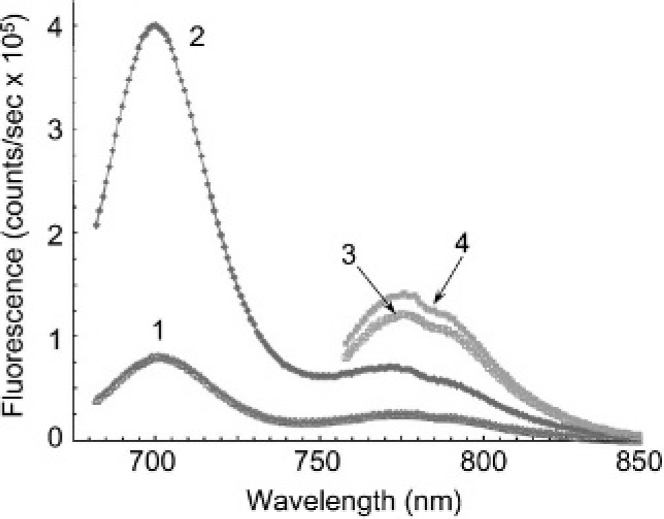
Fluorescence spectra for PB-M7NIR before and after matrix metalloproteinase 7 (MMP7) treatment. Spectra 1 represent the untreated PB-M7NIR Cy5.5-M7 peptide and spectra 3 the untreated AF750 internal reference. Spectra 2 demonstrate an approximately fivefold increase in fluorescence of the Cy5.5 sensor following MMP7 treatment, whereas spectra 4 demonstrate a minimal increase in fluorescence following MMP7 treatment.
In Vivo Imaging of Subcutaneous Xenograft Tumors
To evaluate the selectivity of PB-M7NIR, athymic nude mice were subcutaneously injected on either flank with a human colon cancer cell line that does not express MMP7 (SW480neo) and a cell line derived from the same parental cells following transfection with the human MMP7–complementary DNA (SW480mat). Both cell lines express several MMPs, including MMP9, 24 and to our knowledge differ only in the expression of MMP7. Once tumors were established, 1 nmol of PB-M7NIR was injected intravenously, resulting in a final concentration of approximately 0.5 µM, which was calculated to be below the dissociation constant of 2 µM kD to reduce nonspecific cleavage and optimize for MMP7 selectivity. Mice were imaged at various time points up to 4 hours postinjection in the Cy5.5 channel to measure the Cy5.5 sensor signal and in the ICG channel to measure the AF750 reference signal (Figure 3, A-D). Signal in the reference channel was highest at the first measurement after injection (1 hour) and decreased thereafter, presumably owing to the clearance of the cleaved and uncleaved PAMAM-AF750 core (Figure 3E). In contrast, the sensor signal markedly increased over the first 3 hours, consistent with MMP7 cleavage, resulting in enhanced fluorescence in the Cy5.5 channel. Sensor signals varied in intensity from mouse to mouse; however, signal in the control tumor was always less than in the MMP7-expressing tumors in the same mouse (Table 1). To further evaluate the effective cleavage of PB-M7NIR, sensor to internal reference ratios were calculated to assess the cleaved (sensor) to total substrate (reference) retained in the tumor; the sensor to reference ratio increased over time (see Figure 3E). Using data from the 4-hour time point, sensor to reference ratios were calculated for the control tumor and the MMP7-expressing tumor (Table 1). The sensor to reference ratio in the MMP7-expressing tumors ranged from 24 to greater than 1,000, with an average of 275 and a median of 60.8. The control neomycin tumor in the same animal consistently showed a lower sensor to reference ratio (Figure 3F), but the numbers were highly variable and ranged from 1 to 293 (mean = 69.7 and median=39.2). Despite mouse-to-mouse variation, the sensor to reference ratio in the MMP7-expressing tumors divided by the sensor to reference ratios in the control tumor in the same mouse gave an average 6.8-fold increase and a median of 2.2 (see Table 1).

In vivo imaging of mouse subcutaneous xenograft tumors with PB-M7NIR. Dorsal, caudal view of a nude mouse 4 weeks following subcutaneous injection of SW480neo (Neo) or SW480mat (Mat) cells. Tumor areas (approximately 57 mm2 each) are shown in white light (A), the Cy5.5 sensor channel (B), and the ICG reference channels (C) 4 hours following retro-orbital intravenous injection of 1.0 nmol PB-M7NIR. Encircled areas represent regions of interest for quantitative assessment. Note accumulation of PB-M7NIR in the kidneys of the mouse as detected with the ICG reference channel (C) but selective accumulation of sensor signal in the Mat tumor indicative of proteolytic activity (B). Sensor signal on the spine and tail are presumed to be due to low levels of circulating, activated probe that become detectable when they are close to the surface of the mouse. D, Higher magnification of the sensor signal images of the Neo and Mat tumors of the same mouse at the 1-, 2-, 3-, and 4-hour time points. E, Sensor (red squares) and reference (green triangles) signals plotted over time in a Mat tumor showing increased signal in the sensor channel starting at the 2-hour time point, reaching a maxima at the 3- to 4-hour imaging point. The sensor to reference ratio (blue inverted triangles) increases with time. Mean and standard deviation, n = 3. F, Sensor signal in the SW480neo and SW480mat tumors for each mouse is indicated by the linked symbols (n = 13).
Fluorescence Imaging Detection of PB-M7NIR in Vivo

Fluorescence intensities (photons/s/cm2/steradian) measured in the Cy5.5 (signal) and ICG (reference) channels (n = 13) of images containing anesthetized live mice carrying bilateral xenograft tumors (SW480neo and SW480mat). Regions of interest were drawn around the tumors, and the results are expressed in average radiance after correction for background signal. The results were taken at the 4-hour time point postinjection of PB-M7NIR, with sensor to reference ratios (S/R) determined for each tumor with an average (mat tumor to neo tumonratio) of 6.87 and a median of 2.2.
Quantitative Ex Vivo Fluorescence Imaging of PB-M7NIR in Histologic Sections
To localize MMP activity in the tumor microenvironment at the cellular level, subcutaneous xenograft tumors were removed 4 hours postinjection of PB-M7NIR and snap-frozen prior to cryostat sectioning (z= 5 µm). Sections were aqueously mounted with DAPI and analyzed by fluorescence microscopy (Figure 4, A-H). In the MMP7-positive xenograft tumors, PB-M7NIR sensor signal was predominantly localized at the tumor-stroma interface, with high fluorescence being detected in the ECM (see Figure 4, A-D). Sensor signal was not localized in the control tumors (see Figure 4, E-H) and remained lower than the MMP7-positive tumor signal for all mice. Sensor to reference ratios were calculated for each sample in three different regions of the tumor (Figure 4I). The sensor to reference ratio in the MMP7-expressing tumors ranged from 600 to over 1,000, with an average of 692 and a median of 629. The control neomycin tumor in the corresponding animal consistently showed a lower sensor to reference ratio, with an average of 2.3 and a median of 1.9. On average, the mat tumor sensor to reference ratio divided by the neo tumor sensor to reference ratio gave an average 355-fold increase and a median of 360 (see Figure 4, I and J). It should be noted that the sensor and reference signals measured using the fluorescent microscope are in counts/second, differing from the average radiance measurements using the IVIS instrument. However, the dramatic increase in sensor to reference ratios in the MMP7-positive tumors compared with the control tumors confirms the ability of PB-M7NIR to effectively localize and assess MMP7 activity in the tumor microenvironment.

Histologic imaging of PB-M7NIR in xenograft tumors. SW480mat (A-D) and SW480neo (E-H) xenograft tumors were harvested postinjection of PB-M7NIR at 4 hours and frozen in optimal cutting temperature, sectioned, and stained with DAPI. Images taken with the Axiophot fluorescent microscope, 40×, white dash = 50 microns. A and E, The DAPI channel depicts cell nuclei in blue. B and F, Sensor channel (peak emission 694 nm) demonstrating activated PB-M7NIR in red. C and G, Reference channel (peak emission 776 nm) showing PB-M7NIR in green (reference signal presumed to be due to macrophage uptake of PB-M7NIR). D and H, Merge of the images in A to C and E to G, respectively. The dotted lines indicate the boundry between the highly cellular tumor and the less cellular stromal, extracellular matrix (ECM)-rich region. I, Sensor, reference, and sensor to reference (S/R) ratio values from the integrated background-corrected intensities for neo and mat tumors from four individual mice. Images from each tumor were taken in triplicate, and mean ± SD are shown. J, Plot of the S/R ratios for the neo and mat tumors from each mouse.
Ex Vivo Imaging of MMP7 Activity in APCMin Mice
The sensitivity of PB-M7NIR was tested by identifying intestinal adenomatous polyps in APCMin mice. APCMin mice have a mutation in the adenomatous polyposis coli gene that predisposes them to develop multiple benign adenomas throughout the entire intestinal tract at an early age, mimicking the human condition familial adenomatous polyposis coli. 25 The adenomas express the messenger ribonucleic acid (mRNA) for several MMP family members, most notably MMP7, which localizes to the epithelial component of the tumors.22,26 MMP7 expression has been reported in early-stage, benign tumors of the gastrointestinal tract, 27 and reduction in MMP7 activity specifically 22 or MMP activity generally 28 reduces the number of polyps, suggesting that MMP7 represents an appropriate target for prevention strategies. 29
PB-M7NIR was administered to 15-week-old APCMin mice by retro-orbital injection, and the mice were sacrificed 1 hour postinjection, the time of maximum sensor and reference signal in this model system. The intestines and colon were removed and opened longitudinally, displaying luminal adenomas. Adenomas were imaged under white light, Cy5.5 sensor, and ICG reference channels (Figure 5, A-D, and data not shown). Adenomas in APCMin mice demonstrated robust sensor signal, with sensor to reference ratios averaging approximately 100 (Figure 5, F and G). In contrast, the intestines of wild-type mice demonstrated very little fluorescence in the sensor channel, with sensor to reference ratios less than 2 (see Figure 5G). PB-M7NIR was capable of detecting polyps of all sizes, with sensor to reference ratios remaining independent of tumor size and efficient cleavage, resulting in the detection of adenomas as small as 0.01 cm2 (see Figure 5, B, C, and H). To determine the selectivity of the signal, APCMin mice genetically deficient in MMP7 were generated, 17 and the intestines were imaged with PB-M7NIR (Figure 5E). Although these mice develop less tumors owing to the deficiency in MMP7, the tumors that do develop have a much lower sensor signal than wild-type APCMin mice and an average sensor to reference ratio of 7 (see Figure 5, F and G). Statistical analysis was performed on this data using one-way ANOVA, with a Newman-Kuels multiple comparison test showing significant differences between Min and wild-type (p < .001) and Min and Min-7-null (p < .001) data sets, and an approximately sixfold sensor to reference enhancement (Min vs Min-7-null) that can be specifically attributed to MMP7.

Ex vivo imaging of PB-M7NIR in APCMIN intestinal adenomas. Explanted mouse intestine from an APCMIN mouse with spontaneous polyps 60 minutes postinjection of 1 nmol of PB-M7NIR beacon. A and B, White light images. C and D, Near-infrared fluorescent image in the Cy5.5 (sensor) channel. B and C, Higher magnification of region of interest (ROI) 5 (red circle in panels A–C). E, Sensor channel image of intestines from APCMin/MMP7-null mouse 1 hour postinjection of 1 nmol of PB-M7NIR beacon. Polyps are circled in red. F, Table of data from individual mice showing the number of polyps/mouse, the polyp size (mean ± SD), the sensor signal (mean ± SD), and the sensor to reference (S/R) ratios (mean ± SD) (n = the number of detectable polyps). G, S/R ratios for random ROIs from the intestine of two wild-type mice (WT, n = 4) and adenomas from two individual APCMin/MMP7-null mice (Min-7-null, n = 4) and two APCMin mice (Min-WT, n = 4); p < .001, one-way analysis of variance. H, S/R ratios are plotted versus tumor size. The correlation coefficient rxy, < 0.1 (n = 4 mice), is considered to show no evidence of a correlation between the × and the y axis.
Ex Vivo Histologic Imaging of Adenomas
To determine on a cellular level where PB-M7NIR has been activated by MMP7 in the APCMin model, both whole-mount and sagittal sections of intestinal adenomas were imaged in the Cy5.5 (sensor) and Cy7 (reference) channels by fluorescence microscopy at 10× and 40× magnification. Fluorescence in the sensor channel localized around the adenomas in the epithelial compartment in the intestines of the APCMin mice, with no signal in the lumen or in the connective tissue (Figure 6, A-I). Sensor to reference ratios between seven and greater than 25 were observed in the adenomas of APCMin mice (Figure 6J). As a control, normal wild-type C57Bl6 mice treated with PB-M7NIR had their intestines removed and imaged in the same manner, showing limited activation of the beacon with sensor to reference ratios less than 3 (see Figure 6J). To confirm the presence and localization of MMP7, histochemical analysis was performed using a monoclonal antibody for MMP7. Images are shown at 40× magnification for the same sectioned adenoma, verifying that MMP7 localizes to the epithelial compartment of the adenomas (see Figure 4, K and L). Similar to the results with ex vivo IVIS imaging, the adenomas displayed an approximately sixfold enhancement in signal that could be specifically attributable to MMP7.

Histologic imaging of PB-M7NIR in APCMIN adenomas. Images of a single, intact adenoma (A–D, 10× objective) and a sagittal frozen section (E–I, 40× objective) of the same intestinal adenoma from an APCMin mouse, showing two distinct glandular structures within the adenoma, each with a central lumen. A frozen section from a single glandular structure in the same adenoma at higher power shows a ring of epithelial cells around a central lumen (K and L). Staining with a matrix metalloproteinase 7 (MMP7) antibody (K) shows that the enzyme is concentrated in an intracellular structure on the luminal side of the epithelial cells. A and E, White light. B and F, False-red coloring in the Cy5.5 (sensor) channel. C and G, Cy7 (reference in green) channel. D, Merge of images from both Cy5.5 and Cy7 channels. H, DAPI (blue) signal showing the nuclei of cells, in particular, the nuclei at the base of the epithelial cells that form adenoma glandular structures. I, Merge of images from the Cy5.5, DAPI, and Cy7 channels. J, Quantitative fluorescent images were analyzed using Metamorph software, with intensities recorded in counts/second. Sensor to reference ratios were calculated after background subtraction. K and L, Immunohistochemical staining of the same adenoma with a specific anti-MMP7 monoclonal antibody (K) or an IgG control antibody (L); detector = brown precipitate. A to D white line =100 microns; E to I and K to L white line = 50 microns.
Discussion
Noninvasive imaging techniques provide an extraordinary opportunity for the molecular imaging of cancer. PB-M7NIR is an enhanced version of a previously developed proteolytic nanobeacon built on a commercially available dendrimer scaffold with peptide switches selective for cleavage by MMP7 and optical sensors based on FRET. 15 PB-M7NIR improves on this previous work by using NIR sensor and reference fluorochromes to increase the efficiency of light traveling through tissue. 19 The inclusion of an internal reference fluorophore also allows for the detection of both the uncleaved and the cleaved reagent, facilitating quantitative analysis and providing a much needed means to evaluate protease activity. We have demonstrated the ability of the proteolytic nanobeacon to selectively assess MMP activity using models that overexpress or eliminate endogenous MMP7 activity, whereas the efficiency of PB-M7NIR at detecting the relatively small areas of protease activity in the APCMin model demonstrates the applicability of this technology for the early detection of premalignant lesions.
The PB-M7NIR reagent described in the present study is comparable to protease-activated NIR probes commercially available as MMPSense (VisEn Medical, Woburn, MA). PB-M7NIR differs from the graft copolymer-based reagents in the PAMAM dendrimer scaffold used as the vehicle for circulation, the specificity of the peptide sequence, and the use of both sensor and reference fluorophores. PAMAM dendrimers have well-defined structures that facilitate flexibility to improve the overall bioavailability of the fluorogenically labeled peptide substrates and enhance its sensitivity. 30 PB-M7NIR relies on quenching of sensor fluorophores by both homotransfer self-quenching and FRET with the internal reference fluorophores. The internal reference fluorophore provides quantitative analysis of the concentration of the substrate and is useful to evaluate the pharmacokinetics of PB-M7NIR. We achieved an approximately fivefold switch in PB-M7NIR, but the amplitude of the optical sensor might be further increased by altering the sensor to reference ratio and/or Cy5.5-M7 to PAMAM ratio of the reagent. We showed that the sensor to reference ratios of PB-M7NIR is a useful indicator of relative MMP7 activity in vivo, but agents such as this may be able to assess absolute levels of MMPs in normal and diseased tissue. Our further studies focus on absolute quantitative measurements of MMP activity by agents such as PB-M7NIR that could be applied to human tumors.
In vivo imaging of subcutaneous xenograft tumors using PB-M7NIR showed selective imaging of MMP7 activity. In these studies, both xenograft tumors were derived from the same SW480 human colon cancer cell line expressing several MMP family members 24 and differ only in the overexpression of MMP7 in the SW480mat cells. They were injected on opposite sides of the same animal to maintain similar tumor environments and minimize animal-to-animal variation. Accumulation of biocompatible macromolecules has been reported for a variety of solid tissue tumors owing to the enhanced permeability and retention effect of probes in the tumor owing to leaky and constricted vasculature.31,32 The SW480mat and SW480neo tumors develop with consistent sizes and masses and have been shown by histologic analysis to show no significant differences in vasculature. 33 Therefore, the SW480neo xenograft tumors serve as an appropriate control for the SW480mat xenograft tumors from a physiologic perspective. It is noted that although the SW480neo control cells express undetectable levels of MMP7 mRNA and protein in culture, 33 MMP7 can be contributed by infiltrating inflammatory cells. 34 Variable levels of these host bone marrow-derived inflammatory cells in the SW480neo tumors may account for the variability in sensor to reference ratios seen in tumors from the present study and the modest average enhancement of signal in the expressing versus nonexpressing xenograft tumors compared with the much larger difference in APCMin mice with and without MMP7 expression. Host MMP7 activity can be eliminated by the use of MMP7-deficient mice, as in the APCMin-7-null mice, but become more complicated for the xenograft studies that would require concomitant inactivation of the immune system to accept the human tumor xenografts.
When evaluating PB-M7NIR cleavage by MMP7 in the xenograft tumors, sensor to reference ratios were much higher in the ex vivo sections than the sensor to reference ratios seen in vivo, most likely owing to the lack of the absorption properties of tissue and attenuation of signal owing to the depth of the source. The sensor to reference ratios seen in the sections serve to localize the activation of PB-M7NIR at the cellular level, although only a small fraction of the tumor can be imaged at one time. MMP7 was found to be localized at the tumor boundary, corresponding to previous data showing MMP7 activity in macrophages,34,35 vascular endothelial cells adjacent to the tumor, 36 and mononuclear phagocytes. 37 Although some MMP7 activity was found within the xenograft tumors, results suggest that MMP7 predominantly cleaves PB-M7NIR at the tumor-stroma interface, with high activity being detected in the dermal ECM. The results suggest that MMP7 physically associates with ECM components in an active form. MMP7 may be associated with its substrates, such as the proteoglycan versican or tenascin-C, 2 both of which can be found in non-basement membrane matrices.
The APCMin mouse provides an ideal model for evaluating the sensitivity of PB-M7NIR owing to the large number of adenomas that develop in the intestinal tract at various sizes. PB-M7NIR was found to show significant sensor signal in ex vivo imaged adenomas as small as 0.01 cm2, with sensor to reference ratios being independent of tumor size. This size of tumor is very difficult to detect with white light, suggesting that PB-M7NIR could be applied to enhance the detection of very small intestinal adenomas by colonoscopy. The specificity of PB-M7NIR was also demonstrated by the significant reduction in signal in all polyps of APCMin-MMP7-null mice compared with APCMin mice. It is not currently clear if MMP-based PBs can be sufficiently quantitative to be useful for prognostic significance, for example, in distinguishing benign from malignant tumors. This application may require an absolute difference, the presence or absence of a specific MMP, rather than an alteration in the levels of expression and/or activation to be feasible.
Sagittal sections of intestinal adenomas from APCMin mice found effective localization of MMP7 in the epithelial compartment of the adenomas that correspond to histochemical analysis using an MMP7 antibody. At the subcellular level, we observed MMP7 staining in the basolateral region of the tumor cells. However, MMP7 activity appeared to be present throughout the cell. It is not clear if this represents the distribution of active MMP7 and/or the distribution of the product throughout the cell. It is interesting that the signal did not localize to the basement membrane or ECM between glands in the adenomas, in contrast to the abundant ECM staining in the stroma surrounding the subcutaneous tumors. This may be a result of the extreme loss of polarity with the SW480 colon carcinoma cells compared with the benign adenomas, since in cultured polarized epithelial cells MMP7 activity is primarily localized to the apical compartment. 38 It is also interesting that the MMP7-attributable signal is enhanced approximately 300-fold in the xenograft tissues compared with the 6-fold enhancement in the intestinal tissues. Factors that may contribute to this difference include the overexpression of MMP7 in the SW480mat tumor cells compared with the endogenous levels of MMP7 in the adenomas, the more abundant ECM in the dermis compared with the intestine, and the possibility that there is selective retention of either the enzyme or PB-M7NIR proteolytic products by an ECM component that is differentially expressed in these tissues. Additional studies using high-resolution multiphoton microscopy are currently under way to assist in addressing these possibilities.
In summary, PB-M7NIR is the first of the dendrimeric NIRF probes designed for the in vivo detection and imaging of MMP7 activity. The intravenous administration of PB-M7NIR allowed for the sensitive and selective visualization and localization of MMP7 activity in whole animals and in tissue sections. The ability to image the tumor microenvironment may help distinguish the specific functions of MMPs at various stages of tumor progression and in different tumor models. Considerable hurdles to the application of this technology to human disease remain, including the safety and regulatory issues surrounding the development of contrast agents for human use, the need for quantitation of absolute as opposed to relative enzyme levels, and the lack of instrumentation that is sufficiently sensitive, quantitative, and widely available for clinical use. However, these contrast agents show promise for superficial tumors or those that are accessible by endoscopy and may be useful for the detection and prognosis of these tumors, as well as in assessing response to treatment. Reagents of this type can easily be modified to image a wide variety of diseases associated with enhanced proteolytic activity.
Footnotes
Acknowledgment
Imaging was performed at the Vanderbilt University Institute for Imaging Science small animal imaging research program, supported by the National Institutes of Health grant U24 CA 126588.