
Abstract
Gastrin-releasing peptide (GRP) receptors are overexpressed on several types of human cancer cells, including breast, prostate, small cell lung, and pancreatic cancers. Bombesin (BBN), a 14–amino acid peptide that is an analogue of human GRP, binds to GRP receptors with very high affinity and specificity. The aim of this study was to develop a new fluorescent probe based on BBN having high tumor uptake and optimal pharmacokinetics for specific targeting and optical imaging of human breast cancer tissue. In this study, solid-phase peptide synthesis was used to produce H2N-glycylglycylglycine-BBN[7–14]NH2 peptide with the following general sequence: H2N-G-G-G-Q-W-A-V-G-H-L-M-(NH2). This conjugate was purified by reversed-phase high-performance liquid chromatography and characterized by electrospray-ionization mass spectra. The fluorescent probe Alexa Fluor 680-G-G-G-BBN[7–14]NH2 conjugate was prepared by reaction of Alexa Fluor 680 succinimidyl ester to H2N-G-G-G-BBN[7–14]NH2 in dimethylformamide (DMF). In vitro competitive binding assays, using 125I-Tyr4-BBN as the radiolabeling gold standard, demonstrated an inhibitory concentration 50% value of 7.7 ± 1.4 nM in human T-47D breast cancer cells. Confocal fluorescence microscopy images of Alexa Fluor 680-G-G-G-BBN[7–14]NH2 in human T-47D breast cancer cells indicated specific uptake, internalization, and receptor blocking of the fluorescent bioprobe in vitro. In vivo investigations in SCID mice bearing xenografted T-47D breast cancer lesions demonstrated the ability of this new conjugate to specifically target tumor tissue with high selectivity and affinity.
Research investigations into bombesin (BBN) and specific receptors of BBN have provided valuable insight into this tetradecapeptide since its initial isolation from the skin of the frog Bombina bombina in 1971 by Anastasi and colleagues. 1 Gastrin-releasing peptide (GRP) is the mammalian counterpart of BBN. GRP is a 27–amino acid peptide first isolated from porcine stomach by McDonald and colleagues in 1979. 2 GRP is structurally and functionally similar to BBN in many ways. For example, GRP and BBN share amidated C-terminus sequence homology of 7 amino acids, -Trp-Ala-Val-Gly-His-Leu-Met-NH2. Furthermore, each of these peptides functions as gastrointestinal hormones and neurotransmitters, exerting a variety of physiologic and pharmacologic effects in various human systems. 3 Studies have also shown that BBN and GRP may act as autocrine growth stimulators in a variety of human neoplasms. 4
The BBN receptor family is composed of the following receptor subtypes: (1) neuromedin B receptor (BB1), (2) GRP receptor (BB2, BB3), (3) the orphan receptor subtype (BB3), and (4) the BBN receptor subtype (BB4).5–12 Reports demonstrate that a variety of human tumors and tumor cell lines overexpress the GRP (BB2) receptor, including prostate, pancreatic, gastric, and small cell lung cancers, making this a viable approach for development of new single-photon emission computed tomography, positron emission tomography, or therapy-based agents.13–17 Reports also indicate the expression of the GRP receptor subtype in estrogen receptor (ER)-positive and ER-negative immortalized breast cancer cell lines.18–23 For example, studies using primary human breast carcinoma and axillary metastastic tissue have confirmed the presence of the GRP receptor subtype and the expression of the GRP receptor gene in a large percentage of the tissues sampled.23,24 Therefore, the design and development of new diagnostic or therapeutic site-directed conjugates based on BBN for targeting mammalian GRP receptors overexpressed on human breast cancers have some impetus.25–36 For example, development of noninvasive diagnostic strategies for specific human cancers is a new and exciting approach toward prognosis and monitoring progression of disease. Studies show that the high-throughput signal afforded by fluorescence imaging might offer an alternative to traditional tomographic imaging by alleviating many of the imaging artifacts seen in imaging systems of this general type.37–39 Therefore, design and development of new site-directed, fluorescent, targeting vectors for dynamic optical imaging of human cancers hold some significance. In this study, we developed a conjugate of BBN holding an N-terminal fluorescent tag that might be useful in determining the diagnosis and disease progression of ER-positive breast cancer. We conjugated Alexa Fluor 680 succinimidyl ester to the N-terminal primary amine of H2N-Gly-Gly-Gly-Gln-Trp-Ala-Val-Gly-His-Leu-Met-NH2. We herein demonstrate the high affinity and selectivity of this new conjugate for the GRP receptor overexpressed on ER-positive T-47D human breast cancer cells. The potential to use radiolabeled BBN analogues to effectively target ER-positive breast cancer cells has recently been reported. 40 The idea of targeting ER-positive breast cancer cells via a targeting vector bearing a fluorescent label is a viable alternative to traditional radiolabeled conjugates of this general type. In vitro and in vivo molecular imaging experiments targeting GRP receptor–positive T-47D human breast cancer cells are described.
Materials and Methods
Materials
All solvents were either American Chemical Society (ACS) certified or high-performance liquid chromatography (HPLC) grade and were obtained from Fisher Scientific (Chicago, IL) and used as received. Fmoc-amino acids, coupling reagents, and resins were purchased from Calbiochem-Novabiochem Corp. (San Diego, CA). Alexa Fluor 680 succinimidyl ester was purchased from Molecular Probes, Inc. (Eugene, OR) and used without further purification. BBN[1–14] was purchased from Sigma-Aldrich (St. Louis, MO) and used without further purification. All other reagents were purchased from Aldrich Chemical Co. (St. Louis, MO), Fisher Scientific, or ACROS Chemicals (Morris Plains, NJ)and used without further purification. Electrospray-ionization mass spectral (ESI-MS) analyses were performed at the University of Missouri Proteomics Center Biomolecular Research Facilities, University of Missouri-Columbia. Confocal fluorescence microscopy images were obtained at the University of Missouri Life Sciences Center, University of Missouri-Columbia. 125I-Tyr4-BBN was obtained from NEN Life Science Inc. (Boston, MA). T-47D human breast cancer cells were purchased from American Type Culture Collection (Manassas, VA) and provided to the investigators by the Cell and Immunobiology Core, University of Missouri-Columbia. In vivo fluorescence imaging of T-47D tumor-bearing rodent models was performed using a Xenogen IVIS imaging system, which is housed within the Biomolecular Imaging Center at the University of Missouri-Columbia School of Medicine-Harry S. Truman Memorial Veterans' Hospital.
Solid-Phase Peptide Synthesis
Peptide synthesis was performed on an Applied Biosynthesis (Foster City, CA) Model 432 automated peptide synthesizer employing traditional Fmoc chemistry according to methods previously described. 25 Briefly, the reaction of 4-methylbenzhydrylamine (HBTU)-activated carboxyl groups on the reactant with the N-terminal amino groups on the growing peptide provided for stepwise amino acid addition. Rink amide O-benzotriazole-N,N,N′,N′-tetramethyl-uroniumhexafluorophosphate (MBHA) resin (25 μmol), Fmoc-protected amino acids with side-chain protections (75 μmol) were used for solid-phase peptide synthesis of the parent BBN conjugate. The final product was cleaved by a standard procedure using a cocktail containing thioanisol, water, ethanedithiol, and trifluoroacetic acid in a ratio of 2:1:1:36 and precipitated into methyl-tert-butyl-ether. The crude peptide was purified by HPLC (vide infra), and the solvents were removed on a LABCONCO (Kansas City, MO) CentriVap concentrator. Typical yields of the crude peptide were ≈80%. ESI-MS was used to characterize the parent H2N-G-G-G-BBN[7–14]NH2 peptide conjugate.
Conjugation of Alexa Fluor 680 Succinimidyl Ester to H2N-G-G-G-BBN[7–14]NH2 Peptide Conjugate
A stock solution of H2N-G-G-G-BBN[7–14]NH2 peptide was prepared to a concentration of 10 mg/mL using 0.15 M NaHCO3 (pH = 8.5). For reasons of solubility, 100 μL of absolute ethanol was added to the stock peptide solution. To approximately 2 mg of stock solution peptide was added 2.5 mg of Alexa Fluor 680 succinimidyl ester in 250 mL of dimethylformamide with stirring. The reaction was allowed to proceed for 5 hours at room temperature under zero-light conditions (Figure 1). Reaction progress was monitored by reversed-phase high-performance liquid chromatography (RP-HPLC). The crude Alexa Fluor peptide conjugate was purified by RP-HPLC (vide infra), and the solvents were removed on a LABCONCO CentriVap concentrator. The yield of the puified peptide conjugate was ≈60% based on RP-HPLC. ESI-MS was used to characterize the new Alexa Fluor 680-BBN[7–14]NH2 peptide conjugate.

High-performance liquid chromatographic profile of Alexa Fluor 680-BBN conjugate, Alexa Fluor 680-G-G-G-BBN[7–14]NH2, retention time (tR) = 13.0 minutes.
HPLC Purification and Analysis
HPLC analysis and purification of the peptide conjugate were performed on a Waters 600s controller equipped with a 626 pump and 2487 dual-wavelength absorption detector, an Eppendorf CH-30 column heater, and Hewlett-Packard HP3395 integrators. HPLC solvents consisted of H2O containing 0.1% trifluoroacetic acid (solvent A) and CH3CN containing 0.1% trifluoroacetic acid (solvent B). A Phenomenex Jupiter C-18 (5 μm, 4.6 × 250 mm) column was used with a flow rate of 1.5 mL/min. The HPLC gradient system began with a solvent composition of 95% A and 5% B and followed a linear gradient of 30% A, 70% B from 0 to 25 minutes and 30% A, 70% B to 5% A, 95% B from 25 to 30 minutes.
In Vitro Cell Binding Affinity Studies
The inhibitory concentration 50% (IC50) of Alexa Fluor 680-G-G-G-BBN[7–14]NH2 peptide conjugate was determined by a competitive displacement cell binding assay using 125I-Tyr4-BBN as the radiolabel. Briefly, 3 × 10⁶ T-47D human breast cancer cells (suspended in Dulbecco's Modified Eagle's Medium/F-12K media containing 0.01 M Minimum Essential Medium and 2% bovine serum albumin [BSA] [pH5.5]) were incubated at 37°C for 1 hour in the presence of 20,000 cpm 125I-Tyr4-BBN and increasing concentration of Alexa Fluor 680-BBN[7–14]NH2 conjugate. At the end of the 1-hour incubation period, the reaction medium was aspirated and the cells were washed four times with media. Cell-associated radioactivity was determined by counting in a Packard Riastar gamma counting system.
Uptake, Internalization, and Blocking Studies of Alexa Fluor BBN Conjugate in Human T-47D Breast Cancer Cells Using Fluorescence Microscopy
In vitro cell binding studies were performed to determine the efficiency of internalization and residualization of Alexa Fluor 680-G-G-G-BBN[7–14]NH2 in T-47D human breast cancer cells. These studies were performed in a method similar to that previously reported.25,34 Briefly, 3 × 10⁶ T-47D cells were suspended in RPMI 1649 medium at pH 7.4 containing 2.4 mg/mL HEPES, 0.1 μg/mL bacitracin, and 2 mg/mL BSA in the presence of conjugate for a period of 40 minutes at 37°C (5% CO2). After incubation, the reaction medium was centrifuged (1 minute, 8,000 rpm) and aspirated and cells were washed with the incubation media. The degree of cell-associated activity was determined by confocal fluorescence microscopy at the University of Missouri Life Sciences Center. Briefly, these studies were performed using a Zeiss LSM 510 M-200 Axiovert Microscope (Carl Zeiss, Inc., Thornwood, NY). The microscope was equipped with a 63/1.4 oil immersion objective and a filter set. The images were acquired in two steps. Initially, the cell image was acquired under illumination of a 542 nm Ar2− ion laser. The fluorescence image was acquired using a 632 nm He-Ne laser and a bandpass filter covering 650 to 710 nm. On image acquisition, the two images were overlaid onto one another to examine the precise location of fluorescence signal using Zeiss LSM Image Examiner software version 3.2.0.115 (Carl Zeiss, Inc., Thornwood, NY).
The degree of Alexa Fluor conjugate trapped in the cells was determined by removal of surface-bound molecules using a pH 2.5 (0.2 M acetic acid and 0.5 M NaCl) buffer wash following the wash with incubation media. Internalized, cell-associated Alexa Fluor conjugate was also determined by confocal fluorescence microscopy imaging of this human tumor cell line. To assess the degree of receptor specificity of these conjugates in human breast T-47D cancerous cells, a blocking study was performed. Briefly, 5 μg of native BBN[1–14] peptide was incubated with the cells prior to incubation with Alexa Fluor conjugate. The degree of blocking was assessed by confocal fluorescence imaging of the cellular media as previously described.
In Vivo Fluorescence Imaging of Alexa Fluor-Beta Alanine BBN Conjugate in Tumor-Bearing Rodent Models Using a Xenogen IVIS Imaging System
In vivo fluoresence imaging studies were performed in SCID (severely compromised immunodeficient) mice bearing human T-47D human breast cancer xenografts. Four- to 5-week-old female Institute of Cancer Research (ICR) SCID outbred mice were obtained from Taconic (Germantown, NY). The mice were housed five animals per cage in sterile microisolator cages in a temperature-and humidity-controlled room with a 12-hour light/12-hour dark schedule. The animals were fed autoclaved rodent chow (Rawlston Purina Company, St. Louis, MO) and water ad libitum. All animal studies were conducted in accordance with the highest standards of care as outlined in the National Institutes of Health guide for care and use of laboratory animals and the policy and procedures for animal research at the Harry S. Truman Memorial Veterans' Hospital. For assessment of normal uptake of conjugate, 50 μL of phosphate-buffered saline was injected via the tail vein into a SCID mouse bearing T-47D human breast cancer tumors on the left and right flanks, followed by administration of 50 μg of Alexa Fluor 680-G-G-G-BBN[7–14]NH2 in 100 μL of isotonic saline. To further assess the degree of specificity and selectivity this conjugate has for the GRP receptor, an in vivo blocking experiment was performed in which 50 μg of commercially available BBN[1–14] was injected into the tail vein of a SCID mouse bearing T-47D xenografted tumors 15 minutes prior to injection of Alexa Fluor 680-G-G-G-BBN[7–14]NH2 conjugate. Mice were euthanized by CO2 euthanasia 15 minutes following intravenous administration of the Alexa Fluor 680-G-G-G-BBN[7–14]NH2 conjugate. All mice were immediately imaged using a Xenogen IVIS 200 system (Xenogen Corp., Alameda, CA). Briefly, the mice were placed in the temperature-controlled imaging chamber under zero-light conditions. A low-level illumination reference image was taken followed by fluorescence imaging of the subject. All images were acquired over 0.05 seconds with a cy5.5 filter set, a low-fluorescence tungsten-halogen lamp level, medium (sensitivity) and small (high resolution) binning, and a 12.7 cm of field of view. Images were analyzed using Living Image software (Xenogen Corp.).
Magnetic resonance imaging (MRI) of tumor-bearing animals was performed following optical imaging experiments to correlate the relative fluorescence degree of uptake to the anatomic features (ie, xenografted tumors) of the mice. Diffusion-weighted spin echo multislice images were acquired on a 7 T/210 mm Varian Unity Inova MRI system equipped with a quadrature-driven birdcage coil (38 mm internal diameter) (Varian Inc., Palo Alto, CA). The following parameters were used: repetition time/echo time = 2,000 ms/37 ms, b factor = 1,700 s/mm2, diffusion gradients in x, y, and z directions, 7 to 11 slices, slice thickness = 1 mm with no gap, image matrix size = 256 × 256, field of view = 30 × 30 mm, number of averages = 2.
Results
In this study, we developed a new Alexa Fluor-BBN conujugate with very high affinity for the GRP receptor (see Figure 2). This new Alexa Fluor 680-G-G-G-BBN[7–14]NH2 conjugate was conveniently synthesized by solid-phase peptide synthesis of the parent BBN ligand followed by N-terminal conjugation of the active succinimidyl ester of the Alexa Fluor molecule. Coupling to the less reactive amidated C-terminus was not observed. This new conjugate was purified by RP-HPLC (see Figure 1) and characterized by ESI-MS. Yields of HPLC-purified conjugate were approximately 60%. ESI-MS analysis was consistent with the molecular weight calculated for the new Alexa Fluor conjugate. For example, Alexa Fluor 680-G-G-G-BBN[7–14]NH2 showed a molecular ion of 1953.2. The calculated molecular ion for this conjugate was 1952.8.
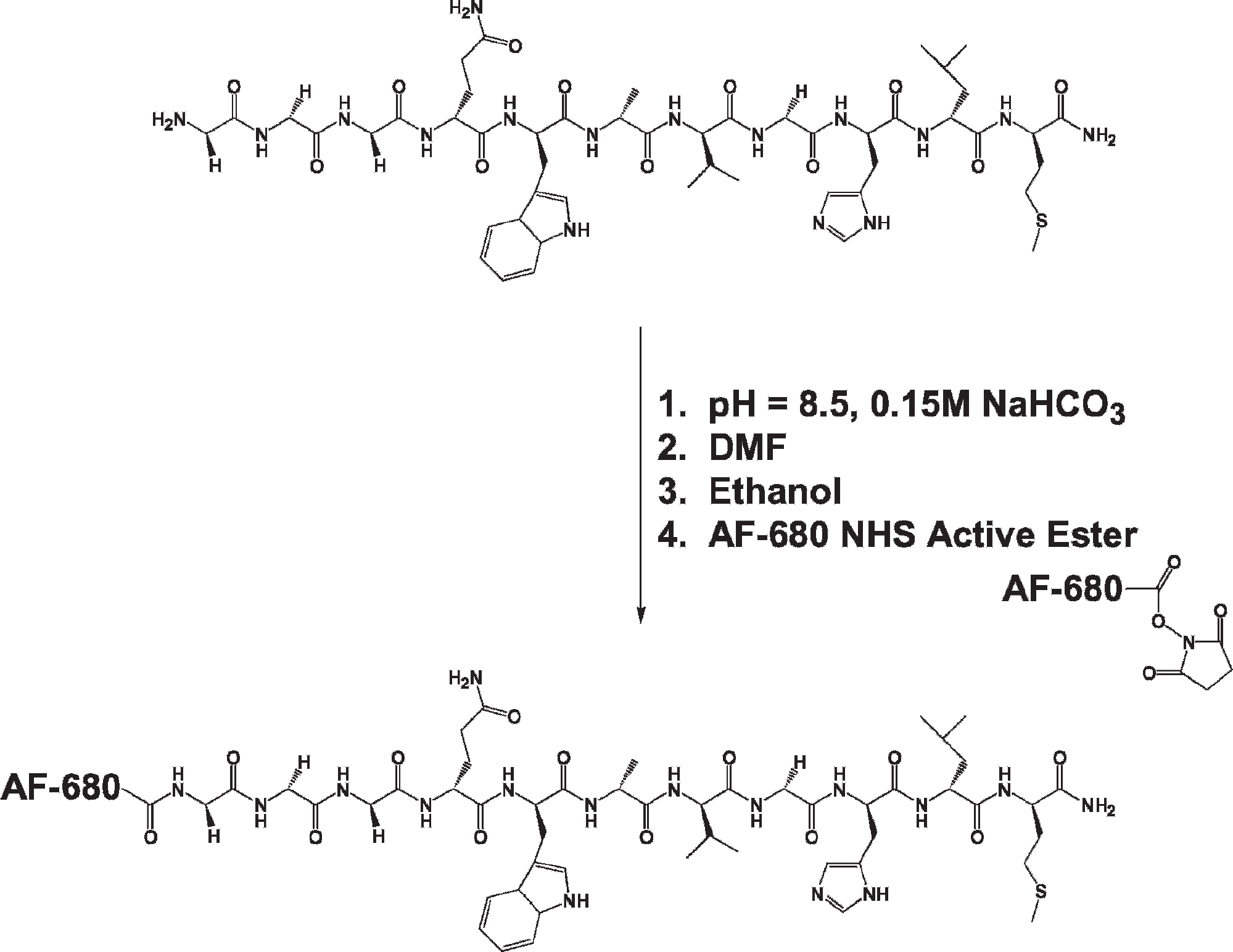
Synthesis of AF 680-G-G-G-BBN[7–14]NH2 conjugate.
To assess the binding affinity (IC50) of the Alexa Fluor-G-G-G-BBN[7–14]NH2 conjugate for the GRP receptor, in vitro competitive cell binding assays were performed using human T-47D breast cancer cells, a cell line known to express the GRP receptors in very high numbers.18–23 Alexa Fluor 680-G-G-G-BBN[7–14]NH2 conjugate of known concentration was assayed against the radiolabeling gold standard 125I-Tyr4-BBN. This new peptide conjugate demonstrated high specificity and affinity for the GRP receptor in T-47D breast cancer cells. For example, the IC50 value in the T-47D cell line was determined to be 7.7 ± 1.4 nM (Figure 3).

In vitro cell binding affinity (inhibitory concentration 50%) (IC50) of Alexa Fluor (AF) 680-G-G-G-BBN[7–14]NH2 in human, T-47D breast cancer cells (7.7 ± 1.4 nM).
Figures 4 to 6 summarize the results of studies to assess the degree of uptake, internalization, and blocking of the Alexa Fluor-G-G-G-BBN[7–14]NH2 conjugates in T-47D breast cancer cells via confocal fluorescence microscopy. Assessment of the degree of Alexa Fluor 680-G-G-G-BBN[7–14]NH2 conjugate associated with the cells after a 40-minute incubation period was evaluated following a cell wash with pH 7.4 incubation media (see Figure 4). The results of these studies clearly indicate the effectiveness of Alexa Fluor 680-G-G-G-BBN[7–14]NH2 to specifically target the GRP receptor. To assess receptor-mediated internalization of Alexa Fluor 680-G-G-G-BBN[7–14]NH2 conjugate, surface-bound conjugate was removed using pH 2.5 (0.2 M acetic acid and 0.5 M NaCl) buffer. After washing the cells with pH 2.5 buffer, much of the conjugate remained internalized within the cells (see Figure 5). In vitro blocking studies, in which high levels of BBN[1–14] were administered to the cells prior to the Alexa Fluor 680-G-G-G-BBN[7–14]NH2 conjugate, reduced the uptake/retention in normal GRP receptor–expressing T-47D cells, as shown in Figure 6. The results demonstrate the high affinity and selectivity of this conjugate for GRP receptors overexpressed on T-47D breast cancer cells. In fact, there is little or no indication of fluorescent signal associated with the cells following the blocking experiment.

Confocal fluorescence photomicrograph of normal in vitro cell binding affinity of Alexa Fluor 680-G-G-G-BBN[7–14]NH2 in human, T-47D breast cancer cells.

Confocal fluorescence photomicrograph of internalized uptake of Alexa Fluor 680-G-G-G-BBN[7–14]NH2 in human, T-47D breast cancer cells.

Confocal fluorescence photomicrograph of blocked uptake of Alexa Fluor 680-G-G-G-BBN[7–14]NH2 in human, T-47D breast cancer cells by wild-type BBN[1–14].
To assess the in vivo uptake of the Alexa Fluor conjugates, we evaluated Alexa Fluor 680-G-G-G-BBN[7–14]NH2 in rodents bearing human T-47D cancer cell xenografts. T-47D human breast cancer cells overexpress the GRP receptor and are a good model for these investigations. In this study, 50 μg (25 nmol) of conjugate was injected into the tail vein of a SCID mouse bearing T-47D xenografted tumors. The animal was euthanized 15 minutes following intravenous injection and immediately imaged using a Xenogen IVIS 200 imaging system. Figure 7A clearly demonstrates the in vivo effectiveness of this new conjugate for targeting the GRP receptor with very high selectivity and affinity. Studies in three additional mice (ntotal = 4) also showed specific uptake of conjugate comparable to Figure 7A. In vivo molecular specificity of this conjugate is further demonstrated by blocking experiments in which 50 μg of commercially available BBN[1–14] was injected into the tail vein of a SCID mouse bearing T-47D xenografted tumors 15 minutes prior to injection of Alexa Fluor 680-G-G-G-BBN[7–14]NH2 conjugate. Figure 7B shows the relative uptake of Alexa Fluor 680-G-G-G-BBN[7–14]NH2 conjugate in GRP receptor–expressing tumors on the left and right flanks of this rodent model. Figure 7, C and D, correlates the anatomic features of the xenografted flank tumors by micro-MRI analysis to the qualitative images of Alexa Fluor 680-G-G-G-BBN[7–14]NH2 tumor uptake acquired via the Xenogen IVIS 200 imaging system.
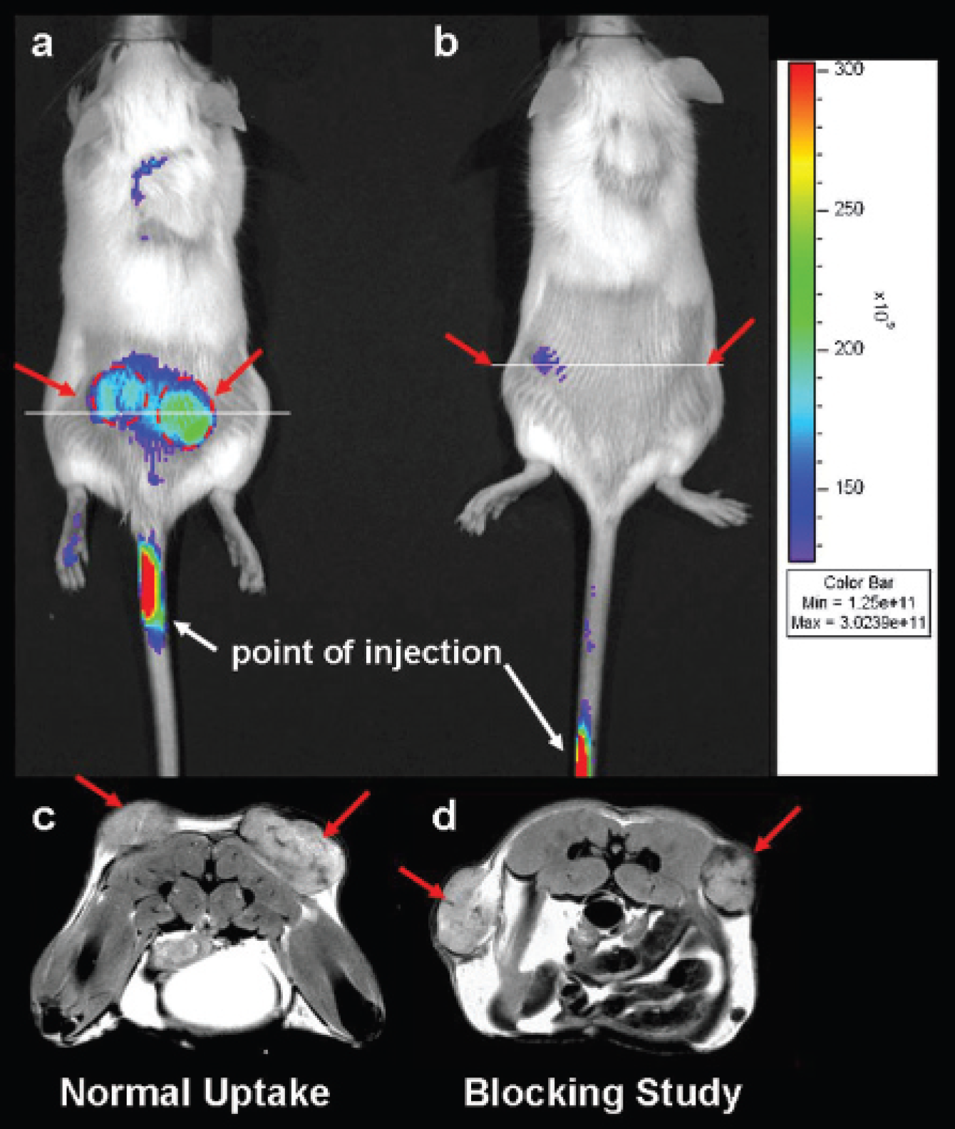
In vivo uptake and blocking experiment of Alexa Fluor 680-G-G-G-BBN[7–14]NH2 in SCID mice bearing human T-47D breast cancer cell xenografts. Xenogen fluorescence images of mice with (A) normal uptake and (B) blocking assay. Magnetic resonance images of cross sections through tumors for (C) normal uptake and (D) blocking assay.
Discussion
Optical imaging technologies such as fluorescence molecular tomography are emerging as new and exciting molecular imaging tools for diagnosis of human disease. In addition, fluorescence molecular tomography shows advantages as a relatively low-cost, noninvasive procedure that uses highly sensitive, nonionizing probes for tissue targeting. In this study, we demonstrated the effectiveness of using Alexa Fluor 680 succinimidyl ester to conjugate the N-terminal primary amine of H2N-G-G-G-BBN[7–14]NH2, a GRP receptor–specific peptide that has demonstrated high specificity and selectivity for the GRP receptor subtype 2. 25 This conjugation technology provides a mechanism for appending large molecular weight molecules with inherent fluorescent properties to either the N-terminal primary amine or the epsilon primary amines of lysine-containing peptides or antibodies to produce stable conjugates for dynamic in vivo optical imaging. Less reactive amidated C-termini, however, do not readily react with succinimidyl esters, making this a very selective conjugation method. In our hands, Alexa Fluor 680-G-G-G-BBN[7–14]NH2 conjugate is stable at a temperature of −80°C for periods extending 6 months. Other Alexa Fluor 680 compounds of this general type designed and developed in our laboratory have demonstrated similar stability profiles. Alexa Fluor 680-G-G-G-BBN[7–14]NH2 conjugate was isolated and purified by RP-HPLC, producing an intense blue product in very high yield. The chemical identity of the new conjugate was verified by ESI-MS.
Maximizing uptake and residualization of the fluorescent conjugate in tumors on receptor binding is an important factor in optimizing the diagnostic potential of the conjugate for dynamic in vivo optical imaging. In vitro binding experiments and internalization and blocking assays of Alexa Fluor 680-G-G-G-BBN[7–14]NH2 in human T-47D breast cancer cells were performed to evaluate the potential efficacy of this new conjugate for further in vivo investigations. Alexa Fluor 680-G-G-G-BBN[7–14]NH2 conjugate showed very high affinity for the GRP receptor in T-47D breast cancer cells with an IC50 of 7.7 ± 1.4 nM.
The mechanism of internalization for conjugates of this type is receptor-mediated endocytosis. Traditional experiments to study the mechanism of internalization often involve a ligand binding experiment using a radiolabeled targeting vector followed by an acid wash (ie, pH = 2.5, acetic acid) procedure to distinguish cell surface receptor binding from internalization. 34 Grady and colleagues observed endocytosis in KNRK epithelial cells transfected to express the GRP receptor using fluorescent probes cyanine 3.18–labeled GRP and GRP receptor antibodies. 4 In this study, they used confocal microscopy to show rapid internalization of GRP and its receptor by a clathrindependent mechanism into early endosomes. 4 In the current study, we demonstrated the ability of Alexa Fluor 680-G-G-G-BBN[7–14]NH2 to effectively target GRP receptors overexpressed on human T-47D breast cancer cells. This agonist ligand is effectively endocytosed by the cells as demonstrated in Figure 5. Washing the cells with pH 2.5 buffer removed much of the surface-bound conjugate, leaving only internalized Alexa Fluor 680-G-G-G-BBN[7–14]NH2 within the cells. Lack of fluorescent signal in T-47D breast cancer cells (see Figure 6) following an effective block with BBN[1–14] demonstrates the high affinity and selectivity of this new conjugate for GRP receptors. To our knowledge, these confocal microscopic images are the first examples of receptor-mediated uptake of fluorescent GRP analogues in human cancer cell lines.
Dynamic optical imaging studies of T-47D breast cancer tumor xenografts in rodent models demonstrates the effectiveness of Alexa Fluor 680-G-G-G-BBN[7–14]NH2 to specifically target GRP receptor–expressing cells in vivo. These studies indicate specific uptake of conjugate in tumor tissue (see Figure 7A). Some degree of uptake was observed in collateral tissue of the abdomen. This is not unexpected owing to the hydrophobic nature of the high molecular weight targeting vector. Blocking investigations (see Figure 7B), in which BBN[1–14] was used to saturate the GRP receptor prior to administration of Alexa Fluor 680-G-G-G-BBN[7–14]NH2, showed little or no fluorescent signal associated with tumor tissue. These studies further demonstrate the high degree of selectivity and affinity of Alexa Fluor 680-G-G-G-BBN[7–14]NH2 conjugate for the GRP receptor. In vitro confocal microscopy studies of this conjugate in GRP receptor–expressing PC-3, human prostate cancer cells, a cell line also known to overexpress the GRP receptor, have further demonstrated the effectiveness for conjugates of this type to specifically target the BBN receptor subtype 2.
In this study, we have not made an attempt to quantify the degree of uptake in tumor or surrounding tissue. Recently, we reported the synthesis and characterization of a dual-probe (ie, Alexa Fluor 680 and 1,4,7,10-tetraazacyclododecane-1,4,7,10-tetraacetic acid [DOTA]) targeting vector based on BBN for each optical and nuclear or MRI-based imaging that might provide valuable biodistribution data for conjugates of this type while at the same time allowing for dynamic optical imaging of tumor tissue via fluorescence-based targeting probes. 41 Furthermore, we have since developed specific bioprobes of BBN conjugated to Alexa Fluor 750 (excitation wavelength = 749 nm; emission wavelength = 775 nm) that may prove valuable for optical imaging of tumors deeply embedded in tissue compared with other conjugates of this type.