
Abstract
Breast MRI is more sensitive than mammography in detecting breast cancer. However, MRI as a screening tool is limited to high-risk patients due to cost, low specificity and insufficient evidence for its use in intermediate-risk populations. Nonetheless, in the past decade, there has been a dramatic increase in the use of breast-screening MRI in the community setting. In this review, we set to describe the current literature on the use of screening MRI in high- and intermediate-risk populations. We will also describe novel applications of breast MRI including abbreviated breast MRI protocols, background parenchymal enhancement and diffusion-weighted imaging.
This activity has been planned and implemented in accordance with the Essential Areas and policies of the Accreditation Council for Continuing Medical Education through the joint providership of Medscape, LLC and Future Medicine Ltd. Medscape, LLC is accredited by the ACCME to provide continuing medical education for physicians.
Medscape, LLC designates this Journal-based CME activity for a maximum of 1.0 AMAPRA Category 1 Credit(s)™. Physicians should claim only the credit commensurate with the extent of their participation in the activity.
All other clinicians completing this activity will be issued a certificate of participation. To participate in this journal CME activity: (1) review the learning objectives and author disclosures; (2) study the education content; (3) take the post-test with a 75% minimum passing score and complete the evaluation at www.medscape.org/journal/whe; (4) view/print certificate.
LEARNING OBJECTIVES
Upon completion of this activity, participants will be able to:
Distinguish recommendations for the use of breast MRI
Evaluate the risk for breast cancer and the use of MRI among women with a BRCA mutation
Assess the potential role of breast MRI among women with a moderate increased risk for breast cancer
Assess the role of breast density in the risk for breast cancer and the use of breast MRI
In the US, breast cancer is the second most common cause of cancer death in women. In 2013, there were approximately 232,340 new cases of invasive breast cancer, 64,640 new cases of carcinoma in situ and 39,620 breast cancer-related deaths in US women [1]. The average lifetime risk for developing breast cancer is 12.4%, or approximately one in eight women [2]. For average-risk women, mammography is the only recommended imaging modality to screen for breast cancer in women older than 40 years old.
Although the age of onset and frequency of screening mammography is controversial, in the 50–69-year age group screening mammography has proven to be efficacious, reducing breast cancer-related deaths by 14–32% [3]. The sensitivity and specificity of digital screening mammography are 70–77% and 92–97%, respectively [4,5]. The use of mammography is limited in high-risk patients due to lower sensitivity (32.6–50.0%) [6,7]. In particular, BRCA1-associated cancers are less likely to have ductal carcinoma in situ (DCIS)-related micro-calcifications or irregular margins, further limiting the use of mammography in these patients [8,9]. Furthermore, it can be difficult to distinguish breast cancer from normal fibroglandular tissue on mammography in patients with dense breast tissue or in patients with lobular carcinoma [10]. Contrast-enhanced MRI is more sensitive compared with mammography; therefore, in populations with an increased lifetime risk of breast cancer, MRI may be an appropriate screening tool [11–13].
Dynamic contrast-enhanced breast MRI (DCE-MRI) includes a multi-phase T1-weighted series of pre-contrast images, early-phase post-contrast images and delayed-phase post-contrast images. The analysis of contrast uptake allows MRI to assess kinetic characteristics of lesions, in addition to morphology. Furthermore, sequences can be modified for fat suppression to improve contrast between lesions and background fat. With these techniques, a 3D high-quality evaluation of the breasts can be made, regardless of breast density. In high-risk patients, MRI has improved sensitivity of 91% [6,7].
In this article, we will discuss the literature evaluating MRI as a screening tool for women at high and intermediate risks of developing breast cancer, the current guidelines for breast MRI screening and novel ways of evaluating breast MRI including abbreviated breast MRI protocols, background parenchymal enhancement (BPE) and diffusion-weighted imaging (DWI).
Current MRI screening guidelines
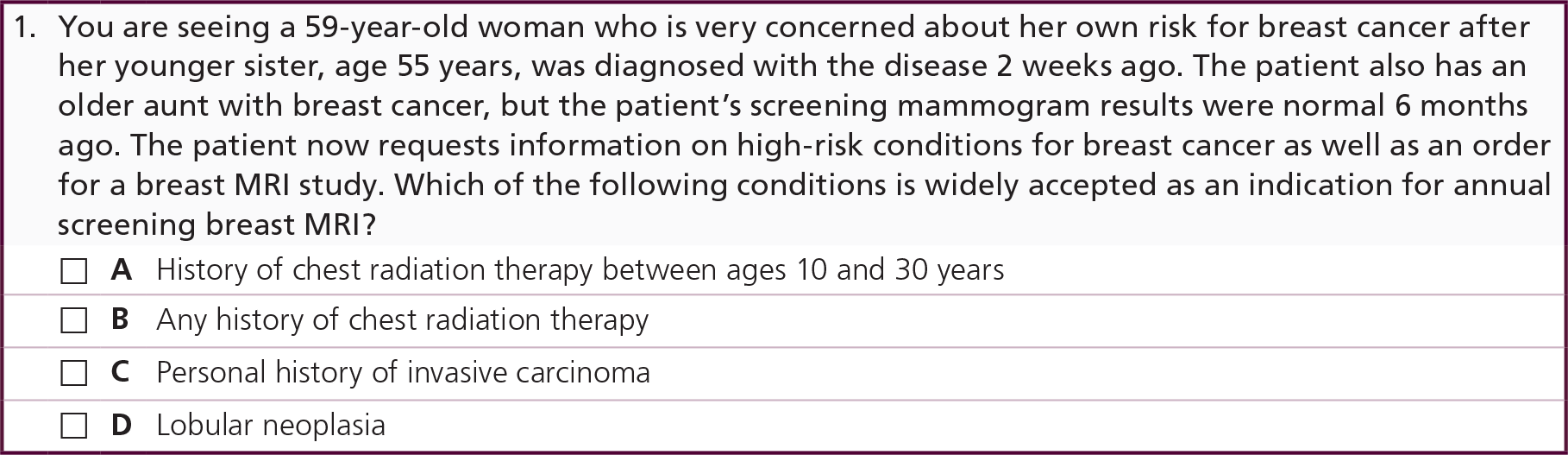
The current breast cancer screening guidelines for the American Cancer Society (ACS), National Comprehensive Cancer Network (NCCN), American College of Radiologists (ACR), American College of Obstetricians and Gynecologists and The American Society of Breast Surgeons state that an annual screening MRI is appropriate for patients at high-risk of developing breast cancer, in addition to an annual mammogram [11–15]. High-risk patients include those with a known or suspected BRCA1 or BRCA2 gene mutation, a history of chest radiation therapy between the ages of 10 and 30 years old, genetic syndromes associated with breast cancer, or a ≥20–25% lifetime risk of developing breast cancer.
For patients at intermediate risk (defined as 15–20% lifetime risk, or a personal history of invasive/in situ carcinoma, lobular neoplasia or atypical hyperplasia), the ACS and ARC both recognize that the use of screening MRI is still under investigation. There is not enough evidence to clearly recommend for or against the use of complementary MRI in these patients, though we will review the current literature on MRI use in intermediate-risk patients.
All of the above national organizations agree that a screening MRI should not be recommended in average-risk women with a lifetime risk of <15%.
Regardless of these national guidelines, over the past decade there has been a 20-fold increase in the use of breast MRI. According to a new study by Stout et al., screening MRI, which was a rare indication in 2000, made up 57.6% of MRI use in 2011, even though only 21.0% met ACS criteria for MRI screening [16].
Breast cancer risk calculation
As described above, calculating a patient's lifetime risk of breast cancer is important for selecting which patients require additional screening with MRI. Commonly used risk prediction models include the BRCApro, Gail model, Tyrer–Cuzick model and the Claus model, each using different personal or familial predictor variables to calculate risk.
Though the Gail model is commonly used, its sensitivity and specificity vary widely, it has poor discriminatory accuracy in internal and external validation, and it has been shown to underestimate breast cancer risk compared with the Tyrer–Cuzick model [17–20]. Though there are modest improvements in the Gail model when breast density is taken into account [17,21–22], a review by Berg et al. specifically states the Gail model should not be used for selecting patients for MRI screening. This is because the Gail model does not consider breast cancer in second-degree relatives or the age of diagnosis of breast cancer in first degree relatives [23]. The Tyrer–Cuzick model has been found to be the most consistent model, as it factors in age, BMI, hormonal factors, as well hereditary factors such as family history of either breast or ovarian cancer in first- and second-degree relatives [19,24].
A new study by Hallingsworth and Stough took a new approach in patient selection using the inclusion criteria for the ACRIN 6666 trial to identify high-risk patients for supplemental high-risk screening. In addition to lifetime risk, this study also considered breast density, 5-year risk calculation, past history of breast cancer, lobular carcinoma in situ (LCIS) and atypical hyperplasia. Using this method, 33 patients (2%) were diagnosed with mammographically occult breast cancer. Based on the lifetime risk of ≥20–25% used to justify MRI screening by the ACS, NCCN and ACR, 27.3, 3 and 36.4% would have received MRI screening based on the Gail, Claus and Tyrer–Cuzick models, respectively [25]. This demonstrates the limitations of the available risk calculators, and further research is needed to examine the effectiveness of tailoring breast cancer screening based on based on personal and familial risk factors.
High-risk patients
According to the ACS, NCCN and ACR, patients who are at high risk of developing breast cancer should get an annual breast MRI in addition to their yearly mammogram [11–13]. These patients include BRCA1 or BRCA2 mutation carriers or those with a first-degree relative who has a BRCA mutation, patients treated with chest radiation between the ages of 10 and 30, patients with other genetic mutations such as the TP53 mutations (Li-Fraumeni syndrome), or PTEN mutations (Cowden syndrome and Bannayan-Riley-Ruvalcaba syndrome), or those with a lifetime risk for breast cancer of ≥20–25%. Although there are no studies specifically looking at the use of breast MRI screening in patients with PTEN and TP53 mutations, these patients are at increased risk of early-onset breast cancer and supplemental screening with breast MRI is recommended.
Breast MRI screening has been shown to be cost effective in high-risk patients, and only a small percentage of US women above the age of 30 are considered to be at high risk based on the current guidelines. Based on a study by Graubard et al. using data from the National Health Interview Survey from 2000 and 2005 and a modified Gail model risk calculator, 1.09% of women in the US between the ages 30 and 84 (approximately 880,063) meet the criteria for being high risk [26].
The majority of the large prospective trials of screening MRI were performed in high-risk patients. In high-risk patients, MRI has greater sensitivity than mammography (77–91% and 32.6–50.0%, respectively), but lower specificity (81.0–97.2% and 93.0–99.8%, respectively) (Tab le 1).
BRCA1 & BRCA2 mutation carriers
Approximately 10% of breast cancer cases are associated with a hereditary germline mutation (the majority of cases are spontaneous), with BRCA1 and BRCA2 being the most common mutations. The BRCA gene is a tumor suppressor gene involved in gene repair, and women who inherit a mutated allele have a 60–80% lifetime risk of developing breast cancer. Women with a BRCA1 gene mutation also have a 33% risk of developing ovarian cancer. Genetic screening and counseling are available for women with a family history of breast or ovarian cancer, and for women with a known BRCA mutation in the family, particularly Ashkenazi Jewish women in whom the BRCA mutation is more common [31].
BRCA1 breast cancers differ in molecular and clinical characteristics compared with BRCA2 breast cancers. BRCA1 tumors are more likely to be estrogen receptor (ER) negative, progesterone receptor (PR) negative and HER2/neu receptor-negative (triple negative). These cancers are more aggressive, often high grade and tend to be larger at diagnosis compared with other high-risk women (Figure 1) [32,33]. Furthermore, BRCA1-associated tumors are less likely to be associated with DCIS, and tend to have smooth margins [8,9]. Therefore, these cancers often have a more benign appearance on mammography, without DCIS-associated micro-calcifications or irregular borders. In contrast, BRCA2 tumors are often ER positive, PR positive, smaller at diagnosis (80% found to be DCIS or <10 mm, compared with 49% in <i>BRCA1 patients) and grow more like sporadic breast cancers [34].

Surgical treatment with prophylactic bilateral mastectomy and/or bilateral salpingo-oophorectomy are alternatives to annual screening in BRCA patients. Mastectomy in mutation carriers has been shown to decrease breast cancer incidence and mortality. Furthermore, bilateral salpingo-oophorectomy in these women decreases breast cancer incidence, ovarian cancer incidence, as well as all-cause mortality [35]. A risk-reducing alternative to surgical treatment is chemoprevention with selective ER modulators (such as tamoxifen) or aromatase inhibitors, which decrease the risk of receptor-positive breast cancer in BRCA carriers. Newer chemopreventive agents targeting receptor-negative breast cancer are currently being studied [36].
Of the seven studies evaluated in Tab le 1, five were conducted in patients who were either BRCA mutation carriers or at elevated risk based on family history, and two were conducted only in BRCA1 and BRCA2 mutation carriers [28,30]. Some screening trials show that when BRCA mutation carriers are separated and analyzed as a unique subset distinct from other high-risk women, there may be an even greater relative increase in MRI sensitivity compared with mammography. For example, Kuhl et al. showed that their overall sensitivity for cancer detection was 91 and 33% in MRI and mammography respectively, but in the cohort of women who were mutation carriers the difference was more pronounced, with MRI and mammography sensitivities of 100 and 25%, respectively [6]. However, it is important to note that there were only eight patients in this cohort. Similarly, Leach et al. found that contrast-enhanced MRI was particularly sensitive for BRCA1 cancers relative to mammography (77% for MRI vs 40% for mammography sensitivity for all patients and 92% versus 23% for BRCA1 carriers (13 cancers), likely due to the lower sensitivity of mammography for BRCA1 cancers [29]. Kriege et al. divided their cohort into mutation carriers (BRCA1, BRCA2, PTEN and TP53 mutations, 50–85% lifetime risk), high-risk women (15–29% lifetime risk) and intermediate-risk women (15–29% lifetime risk), and found the highest cancer detection rate (26.5/1000) to be in women with known mutations as opposed to women in the non-mutation carrier high-risk group (5.4/1000) or moderate-risk group (7.8/1000) [27].
Chest irradiation
Screening breast MRI is currently recommended for women with a history of chest irradiation between the ages of 10 and 30 years old, and screening should begin 8 years after the radiation therapy but not before the age of 25. This is because approximately 13–20% of women treated with chest irradiation for pediatric cancer, particularly Hodgkin lymphoma, are diagnosed with breast cancer by age 40–45 years. Of these women, the earliest reported cases of breast cancer developed 8 years after radiation therapy [37].
Fewer studies have been dedicated to evaluating the sensitivity of breast MRI in women with a history of chest irradiation. Sung et al. performed a retrospective review of 247 screening exams in 91 women who had a history of chest irradiation. During their study period 10 cancers were detected, 4 were detected on MRI alone (all invasive carcinomas) and 3 with mammography alone (DCIS and DCIS with microinvasion). They reported a 4.4% incremental breast cancer detection rate when using supplemental MRI in addition to mammography, and the two imaging modalities had equivalent sensitivities in this patient population (66.7%). The authors concluded that MRI is a useful adjunct to mammography [38].
A 2013 prospective study by Ng et al. further explores the use of supplemental breast MRI screening in women treated with chest irradiation for Hodgkin lymphoma. Over a 3-year study period, the sensitivity of mammography versus MRI was not significantly different, with 68 and 67%, respectively. Combining screening with mammography and supplemental breast MRI increased the sensitivity to 94%, further promoting the current ACS guidelines [39].
High-risk screening intervals
Annual screening MRI and mammography in high-risk patients can either be performed concurrently or staggered. The advantage of simultaneous screening with MRI and mammography is easy cross-referencing between modalities. By contrast, alternating 6-month MRI and mammography regimens allows for more consistent surveillance. It is very difficult to perform randomized controlled trials to address the optimal screening frequency and regimen. To this end, Lowry et al. recently performed a comparative effectiveness analysis using computer modeling to determine the optimal multimodality strategy for screening. They analyzed film mammography (FM), digital mammography (DM), as well as FM and MRI or DM and MRI in contemporaneous and alternating regimens initiated at either 25, 30, 35 or 40 years of age. Furthermore, they studied annual MRI with delayed/alternating DM/FM versus clinical follow-up. They found that annual MRI starting at age 25 years and delayed/alternating DM at age 30 years was the most effective screening strategy in BRCA mutation carriers [40].
Le-Petross et al. conducted a retrospective chart review focusing on BRCA mutation patients who were screened with alternating mammograms and breast MRI every 6 months. Of these 73 women, 13 cancers were detected in 11 women, 12 of which were detected on MRI but not the mammogram performed 6-months prior [41]. Although prospective studies have not been performed, a study by Cott Chubiz et al. shows that alternating can be cost effective, particularly in BRCA1-mutation patients [42].
Intermediate risk
Although the ACS has found insufficient evidence to recommend for or against the use of MRI screening in women with a 15–20% lifetime risk of breast cancer, it is worth evaluating the current literature of MRI use in these patients. This group also includes women with a personal history of invasive cancer or DCIS, patients with a history of atypical ductal/lobular hyperplasia (ADH, ALH) or LCIS, and women with heterogeneously dense or extremely dense breasts on mammography. The role of a supplemental screening ultrasound instead of screening MRI for this subset of women is also being explored. To date, there is no consensus about screening ultrasound in this setting [13].
Personal history
Given the advances in systemic chemotheraphy and targeted radiation therapy, the 10-year risk of a local breast cancer recurrence is approximately 5–10%. The lifetime risk of breast cancer recurrence in these patients depends on their age at diagnosis, oftentimes putting them in an intermediate-risk group. There is currently minimal data on the use of screening MRI in this patient population, however supplemental imaging with MRI or ultrasound is sometimes used in these patients when it is difficult to rule out recurrence due to post-surgical changes. Houssami et al. found that the sensitivity of mammography is significantly lower in patients with a personal history compared with a matched cohort of women without a personal history (65.4 and 76.5%, respectively; p < 0.001). Patients with a personal history also had a greater interval cancer rate (3.6 per 1000 screens vs 1.4 per 1000 screens) [43]. Surgical scarring and changes secondary to radiation therapy may compromise early detection of breast cancer recurrence when screening with mammography alone.
Several recent studies suggest potential value in screening women with a personal history of breast cancer. Brennan et al. conducted a retrospective review of screening MRIs in 144 women with a personal history of breast cancer and no family history. Cancer was detected in 12% of these women (18 cancers in 17 women), and MRI screening had a positive predictive value (PPV) of 39%. Of these 17 women, 10 had cancers that were only detected on MRI, which were more likely to be DCIS (4/10) or minimal breast cancers (7/10, defined as DCIS or node-negative invasive cancer <1 cm in size), but these differences in stage were not significant [44].
This high detection rate by Brennan et al. has not been replicated to the same degree by others. Schacht et al. conducted a retrospective review of screening breast MRI examinations of 702 women, of whom 345 had a genetic or family history of breast cancer, 208 had a personal history of treated breast cancer and 97 had both risk factors. The absolute risk of detecting breast cancer in patients with a personal history of breast cancer was 2.8%%, which was not significantly greater than that for the genetic/family history group (2.0%). Of note, the relative risk of breast cancer for women with both personal and familial/genetic risk was 3.04 (95% CI: 1.05–8.86), when compared with familial/genetic risk alone [45]. Patients with both a personal and familial risk of breast cancer may represent a unique high-risk group, though this does not change their management, as screening breast MRI would be recommended on the basis of their familial/genetic risk factors.
The patients with a personal history studied in these retrospective studies do not necessarily represent all patients with a personal history, as patient selection was based on referral patterns. The ACS has decided that there is currently not enough evidence for or against the use of screening breast MRI in patients with a personal history of breast cancer alone. Prospective studies are needed to better evaluate the use of screening MRI in this patient population.
LCIS, ALH & ADH
Compared to the general population, women diagnosed with LCIS are at increased risk for breast cancer, with their risk increased by 1% every year after their diagnosis (6–10-fold risk compared with the general population) [11,46]. Despite the elevated risk associated with LCIS, studies examining the use of screening MRI in these patients have garnered mixed results, and there are relatively few studies focusing on this population. King et al. performed a retrospective review of 776 patients with LCIS who were nonrandomly screened with annual mammography and clinical exams either with or without supplemental MRI. Cancer was detected in approximately 13% of patients in both cohorts after a median of 58 months, with no difference in clinical staging. Although these patients are clearly at an increased risk for developing breast cancer, screening MRI provided no increase in breast cancer detection [46].
Two relatively large retrospective studies show that MRI screening in patients with LCIS can identify breast cancer occult to mammography. Friedlander et al. performed a retrospective review of 307 screening MRI exams in 133 patients with a diagnosis of LCIS. Cancer was detected in 5/133 patients (3.8%), none of whom had abnormal findings on mammography, with a PPV for screening studies for which biopsy was recommended in 18.5% [47]. Similarly, Sung et al. retrospectively evaluated 670 screening MRI exams in 220 women with a history of LCIS and found 17 cancers in the study period, 12 of which were identified MRI only (nine invasive; three DCIS) and five of which were identified by mammography only (two invasive; three DCIS) [48]. Both authors conclude that MRI appears useful as an adjunct screening method for women with a history of LCIS, similar to high-risk populations. However, these were both retrospective one-arm studies, and we are not aware of any existing prospective studies assessing the use of screening MRI in this patient population.
While there are very few studies specifically studying the use of MRI in patients with LCIS, there are even fewer studies focusing on its use in other high-risk lesions, such as ADH and ALH. Nonetheless, there is evidence that multiple centers perform screening MRIs on women with a history of high-risk lesions regardless of ACS recommendations or the lack of research [49]. The risk of developing breast cancer with ADH is not as high as the risk associated with LCIS, but is still fourfold to fivefold the risk of the general population [11]. Port et al. retrospectively compared screening results in patients with either a history of LCIS or a history of ADH or ALH. All patients were screened with mammography, and MRI screening was used at the discretion of the providing physician. They found only a slight benefit in using MRI to screen for LCIS over mammography, and no benefit for using MRI versus mammography to screen patients with ALH or ADH [50]. Of note, this study categorized women with a history of either ALH or ADH in the same group without distinction; there could be room for future MRI studies to tease out the differences between these two entities. In addition, the patients recommended to undergo supplemental MRI screening were more likely to have a strong family history of breast cancer which could confound any benefit seen by MRI in this study.
Dense breasts
Increased mammographic density is known to be an independent risk factor for breast cancer and not secondary to delayed detection. It has been shown that women with extremely dense breast have a fourfold to sixfold increased odds of developing breast cancer [51,52]. Currently there are no large or prospective studies directly evaluating the use of screening MRI for patients with dense breasts on mammography. According to a 2004 study by Sardanelli et al., in patients with increased breast density (defined as scattered fibroglandular, heterogeneously dense or extremely dense patterns on mammography), MRI was more sensitive than mammography (81% for MRI vs 60% for mammography), with no significant difference in PPV (71 and 78%, respectively). In patients with predominantly fatty breast tissue, there was no significant difference in either sensitivity or PPV [53]. Currently, the ACR does not recommend screening MRI for patients with dense breast tissue on mammography, due to the cost and high rates of both imaging recall and false positives.
Berg et al. studied the additive value of annual ultrasound or a single screening MRI in asymptomatic women with heterogeneously dense or extremely dense breast tissue and at least one other high- or intermediate-risk factor. Of the 2662 women who had undergone three rounds of screening with mammogram and ultrasound screenings, a subset of 612 women were assessed with an additional contrast-enhanced MRI screening exam. Out of 111 cancers found, 30% were detected on mammography only, 29% by ultrasound only and 8% by MRI only. Supplemental MRI yielded 14.7 additional cancers/1000 screening exams while the addition of ultrasound resulted in 4.3 additional cancers/1000 screening exams. However, MRI resulted in a recall rate of 20% of which 43/612 (7%) received a biopsy. Of this group, 8/43 (19%) had cancer. Ultrasound, on the other hand, resulted in a 7% recall rate of which 242/4814 (5%) needed biopsy; of this group, cancer was found in 18/242 (7%) [54]. Similarly, Kuhl et al. found that MRI alone yields 14.9 additional cancers/1000 screening exams [55]. Nonetheless, it remains unclear if the increased yield and associated with breast MRI screening provides a survival benefit beyond earlier detection. Given the cost associated with breast MRI and the anxiety associated with the high recall rates, these results do not justify in the use of breast MRI screening provides a survival benefit beyond earlier detection. Given the cost associated with breast MRI and the anxiety associated with the high recall rates, these results do not justify the use of breast MRI screening in intermediate-risk women with dense breasts.
Average risk
Currently there is no data supporting the use of breast MRI to screen women with a less than 15% lifetime risk of breast cancer, and therefore is not recommended by either the ACS or ACR [11,13]. Screening breast MRI in average-risk women is not cost effective, with high false-positive rates resulting in unnecessary additional imaging, biopsies and anxiety.
Potential limitations of breast MRI
Mortality benefit
Although studies show that MRI is more sensitive than mammography, and able to detect mammographically occult breast cancers (Table 1) [54], including smaller cancers [56] with node-negative disease [6], it remains unclear what mortality benefit this will have [57]. To date, no studies have definitively demonstrated decreased breast cancer-specific mortality in high-risk patients [58]. Studies have found improved survival, but survival measurements could be confounded by lead-time bias of earlier detection [59].
Prospective studies evaluating MRI screening of the breast.

PPV was analyzed per BI-RADS categories or level of suspicion. Numbers are taken from the BI-RADS 5 or suspicious CBE category.
51 cancers detected, one of which was non-Hodgkin's lymphoma. Five cancers were excluded from analysis of the modalities.
PPV was analyzed per screening year. % are given for year 1.
BI-RADS: Breast Imaging-Reporting and Data System; CBE: Clinical breast examination; MG: Mammogram; NA: Not applicable; NR: Not reported; PPV: Positive predictive value; Sn: Sensitivity; Sp: Specificity; US: Ultrasound.
Warner et al. attempted to address this question in their 2011 prospective study examining breast cancer incidence in women with BRCA1 or BRCA2 mutations [56]. The authors assessed 1275 women of whom 445 were screened with MRI and 830 with conventional screening over a mean of 3.2 years. They hypothesized that there would be a lower incidence of advanced-stage breast cancer in the women undergoing MRI screening. There was a cumulative incidence of 13.8% DCIS and 1.9% stage II–IV breast cancer in the MRI group, with 7.2% DCIS and 6.6% stage II–IV breast cancer in the comparison group. In addition, there was a statistically significant decrease (70%) in incidence of large or node-positive invasive disease in the 6-year period after MRI screening was initiated with an adjusted MRI screening hazard ratio of 0.3 (95% CI: 0.12–0.72; p = 0.008) for stage II–IV breast cancer. While MRI is associated with a lower incidence of advanced disease, Warner and his colleagues acknowledge that more studies are necessary before a mortality benefit can be proven.
Cost–effectiveness
There have been several studies evaluating the cost effectiveness of screening MRI, particularly in high-risk populations, such as BRCA1 and BRCA2 mutation carriers. The cost per quality-adjusted life-year (QALY) less that US$100,000 is deemed cost effective. The UK MARIBS study enrolled 649 women, and found the most cost-effective screening regimen was targeted toward BRCA1 and BRCA2 subgroups compared with other high-risk groups [60]. Plevritis et al. found that for women ages 35–54 years, the cost per QALY terms is US$55,420 for BRCA1 carriers and US$130,695 for BRCA2, when performing MRI in addition to mammography. When the analysis was performed specifically in women with dense breasts, the cost decreases to US$41,183 for BRCA1 carriers and US$98,454 for BRCA2 carriers [61]. This difference between BRCA1 and BRCA2 is attributed to the fact that BRCA1 tumors tend to be mammographically occult. BRCA2 tumors more frequently will present with suspicious calcifications on mammography. Lee et al. compared the QALY and lifetime costs of three screening strategies (mammography, MRI and combined mammography–MRI screening) in 25-year-old BRCA1 mutation carriers. They found that annual combined screening using MR and mammography was the most effective (44.62 QALYs vs 44.50 for MRI alone and 44.46 for mammography alone), but was also more expensive (US$110,973 combined vs US$108,641 for MRI and US$100,336 for mammography alone). In this study, screening with both MRI and mammography in BRCA1 carriers was cost effective, costing US$69,125 per QALY [62].
A study by Taneja et al. also found the use of screening breast MRI to be effective with BRCA1/2 women, with a cost per QALY of US$25,277. This study also performed a cost-effective analysis on women with other high-risk characteristics, defined as ≥20% lifetime risk, generally related to strong family history. In doing this, they found that the cost per QALY varies widely with the estimated prevalence of disease in the population. Populations with a 3% prevalence are considered cost effective (cost per QALY of US$45,566), but with the lowest estimate of 0.5% prevalence, the cost per QALY is approximately US$310,616 [63].
It is difficult to determine the true cost–effectiveness of screening MRI for women at intermediate lifetime risk of developing breast cancer given the difficulty in determining the true risk and prevalence of this group [64]. In intermediate-risk patients it is unclear if supplemental screening is needed beyond mammography. Therefore, it is important to study other less expensive imaging modalities including ultrasound and abridged breast MRI.
Future perspective
Abridged Breast MRI (AB-MRI) screening protocols
Although DCE-MRI demonstrates improved sensitivity for detecting invasive breast cancers and DCIS, widespread use of breast MRI is limited by the cost, as well as the time need to acquire and interpret the exams. The use of an AB-MRI screening protocol, consisting of mainly the first post-contrast subtracted images, is a new area of investigation. This protocol could potentially increase access to breast MRI by significantly reducing the cost and time associated with the exam.
Kuhl et al. recently presented preliminary data on AB-MRI screening at the 2013 American Society of Clinical Oncology. They studied 443 women at increased risk of breast cancer who had normal mammograms and underwent breast MRI screening. They then compared the accuracy and diagnostic yield of the AB-MRI protocol compared with the results of the full protocol. A total of 11 breast cancers were diagnosed (18.2 additional cancers/1000 screens), and the AB-MRI and full protocols were both positive in 10/11 cases (91%). With an average reading time of 28 s, the AB-MRI protocol's sensitivity and specificity were not statistically different from that of the full protocol [65]. Similarly, an abstract presented by Heacock et al. at the 2013 Radiological Society of North America conference studied the use of AB-MRI to detect known breast cancer lesions. Using an AB-MRI protocol (including the following images: pre-contrast TI/T2, first post-contrast T1 and first subtraction post-contrast TI), they retrospectively reviewed 100 breast MRI exams from patients with known lesions (blinded to the lesion location and other clinical history). The sensitivity of the AB-MRI protocol was 98%, and two cases of DCIS were missed (subsequently seen on the second post-contrast images) [66]. This study is limited in that each patient had a known biopsy-proven cancer lesion.
Though this is a promising new area of research, these are preliminary studies. Further examination of AB-MRI is needed before it can be implemented clinically.
Background parenchymal enhancement
BPE reflects the amount of fibroglandular tissues seen on the first post-contrast series of a standard DCE-MRI examination. It is described as minimal, mild, moderate or marked enhancement, and reflects the vascularity of the breast parenchymal tissue and is sensitive to hormonal changes [67]. BPE has been shown to vary with the menstrual cycle, increase with hormone replacement therapy and decrease with menopause, anti-estrogen therapies and bilateral salpingo-oopherectomy [68–72].
Although BPE has not been found to directly correlate with breast density on mammography [73], similar to the effect of breast density on mammography, moderate or marked BPE provides background noise which could potentially decrease the sensitivity of breast MRI. This has been demonstrated in two studies by Hambly et al. and DeMartini et al., showing a positive correlation between follow-up recommendations, biopsy rates and BPE [74,75]. Although these two studies do not show an increased incidence of cancer with increased BPE, King et al. have suggested a relationship between BPE and breast cancer risk [67].
BPE is an area of continued research and the evaluation of BPE has not yet been included in the Breast Imaging-Reporting and Data System lexicon. Nonetheless, BPE has important clinical implications in terms of exam outcome and risk stratification, and a standard and accurate assessment of BPE will be an important aspect of future clinical practice.
Diffusion-weighted imaging
DWI quantifies the mobility of water molecules in tissue on MRI. Water transport is most commonly quantified as an apparent diffusion coefficient (ADC), a model using the principle that tissue-confined water behaves similarly to free water, but with reduced diffusivity. In imaging oncology, restricted diffusion (a reduction in ADC) is a marker of cellularity, as it represents a decrease in extracellular space relative to the more viscous intracellular fluid of proliferating cells [76]. Studies by Kul et al. and Bogner et al. show the potential of DWI to increase the specificity of breast MRI, as differences in ADC values may be able to distinguish DCIS from both normal tissue and invasive ductal carcinoma. ADC values in DCIS are lower compared with normal parenchyma, but significantly higher than the ADC seen in invasive ductal carcinoma [77,78]. This intermediate ADC value is not specific to DCIS, and may overlap with other benign and malignant (invasive lobular carcinoma) lesions.
Unlike the evaluation of BPE, DWI does not require the use of intravenous contrast. Therefore, DWI-MRI could be a useful alternative for patients with poor renal function or an allergy to gadolinium. Though DWI-MRI has not been found to be more accurate than DCE-MRI, it is still significantly more accurate than mammography [79]. Further research into the application and standardization of breast DWI are needed.
Conclusion
The current literature shows that MRI has improved sensitivity compared with mammography in the high-risk populations as described above, particularly in BRCA1 and BRCA2 mutation carriers. More prospective and randomized controlled studies are needed to demonstrate both the value of screening MRI in the moderate-risk population and the overall mortality benefit of screening MRI.
Executive summary
The current guidelines recommend annual screening MRI in addition to mammography in high-risk patients, including BRCA-mutation carriers and women with a >20% lifetime risk of breast cancer.
Despite the current guidelines, there has been a 20-fold increase in breast MRI use over the last decade, and a relatively small percentage meets the above criteria.
Breast MRI has significantly improved sensitivity over mammography in women with BRCA mutations, and annual MRI screening is cost effective in this population.
Recent studies evaluating concurrent versus staggered annual MRI/mammography screening suggest that alternating the two screening modalities every 6 months is optimal and cost effective.
Studies have yet to determine whether screening breast MRI in intermediate-risk populations is advantageous, particularly patients with a personal history of breast cancer or lobular carcinoma in situ.
Currently, screening breast MRI is not specifically indicated for patients with increase breast density on mammography.
More prospective and randomize trials are required to study the value of screening MRI, and other less expensive imaging modalities in intermediate-risk patients.
Abridged MRI protocols are currently being studied to improve the costs and time associated with breast MRI. More research is necessary before these protocols can be implemented clinically.
Moderate or marked background parenchymal enhancement on dynamic contrast enhanced breast MRI, like mammographic breast density, is believed to contribute to breast cancer risk and hinders exam interpretation.
Diffusion-weight imaging may increase the specificity of MRI in distinguishing ductal carcinoma in situ from invasive ductal carcinoma or normal breast tissue.
The role of dynamic contrast-enhanced screening breast MRI in populations at increased risk for breast cancer
To obtain credit, you should first read the journal article. After reading the article, you should be able to answer the following, related, multiple-choice questions. To complete the questions (with a minimum 75% passing score) and earn continuing medical education (CME) credit, please go to www.medscape.org/journal/whe. Credit cannot be obtained for tests completed on paper, although you may use the worksheet below to keep a record of your answers. You must be a registered user on Medscape.org. If you are not registered on Medscape.org, please click on the “Register” link on the right hand side of the website. Only one answer is correct for each question. Once you successfully answer all post-test questions you will be able to view and/or print your certificate. For questions regarding the content of this activity, contact the accredited provider,


Footnotes
No writing assistance was utilized in the production of this manuscript.