
Abstract
Electrospun poly(vinyl alcohol) (PVA) nanowebs treated with honey were prepared to exploit the high surface area of nanofibers and the many different beneficial properties of honey. Nanowebs fabricated from biocompatible polymers like PVA, and treated with natural substances like honey, may find use in various biomedical applications such as wound bandages. Fourier transform infrared spectroscopy was used to confirm the incorporation of honey into electrospun PVA nanowebs. Treated nanowebs were characterized by evaluating their antimicrobial properties, breathability characteristics, and tensile properties. PVA nanowebs treated with honey demonstrated adequate breathability characteristics for potential use in wound dressings. In this study, no antibacterial activity was observed after treatment with honey.
Keywords
Introduction
A wound is generally defined as an injury to the surface of the skin due to physical, chemical, mechanical, and thermal factors. In more scientific terms, it is a disruption of the normal anatomic structure and function of the skin. 1 Wound healing is a complex biological process that is often aided by use of wound dressing materials in vitro. Wound dressing materials are expected to have the following properties to facilitate effective wounds healing: 1) absorb wound exudates, 2) have antimicrobial properties, 3) be breathable to enable gaseous and fluid exchanges, and 4) be sterile, nontoxic, non-allergenic, and non-scaring. 2 Electrospun nanofibers are increasingly being considered for use as wound dressings owing to their unique properties (e.g., high surface area to volume ratio and high porosity).3–8 More importantly, the presence of small pores in nanofibers may likely ensure protection from invading exogenous microorganisms. 9
Antimicrobial activity is an important requirement for nano-fibers to be used in wound dressings as they should prevent the colonization of microbes that may lead to subsequent infections. Treatment of electrospun nanofibers with antibiotics, such as rifampicin and paclitaxel, 10 mefoxin, 11 and metal oxide nanoparticles such as silver, 12 has been discussed in the literature. However, the possibility of microorganisms developing antibiotic resistance, 13 and the ambiguity of nanoparticle toxicity, 12 resulted in the need for safe and environmentally friendly alternatives. Merrell et al. 14 have reported the treatment of poly(ϵ-caprolactone) nanofibers with natural substances such as curcumin. They proposed the use of these nanofibers in diabetic wound dressings owing to the antioxidant and anti-inflammatory properties of curcumin. 14
Another naturally available substance that is currently being investigated is honey.15,16 Honey is a supersaturated solution primarily composed of sugars such as glucose and fructose. Other constituents of honey include enzymes and amino acids, vitamins, minerals, organic acids, and aromatics.15,16 Theenzymatic components of honey include invertase, amylase, glucose oxidase, catalase, and acid phosphorylase.15 Theantibacterial, antioxidant, antitumor, anti-inflammatory, and antiviral properties of honey are well documented in the literature.17,18 The antimicrobial properties of honey can be attributed to the following: 1) the inherently low pH of honey does not favor microorganism growth, 2) the osmo-larity or high sugar content prevents availability of moisture/ water for the growth of microorganisms, and 3) hydrogen peroxide generated by the activity of glucose oxidase enzyme when honey is diluted. Hydrogen peroxide is the major source of antimicrobial activity in honey. Glucose oxidase is sensitive to heat and light, therefore, heating honey may decrease its antimicrobial activity. In some cases, honey itself contains substances such as catalase, 15 which destroys hydrogen peroxide generated by glucose oxidase, and other floral components. 15
Treatment of electrospun nanofibers with honey to exploit the various properties of honey is also being studied. Wang and He
19
have electrospun a mixture of poly(vinyl alcohol) (PVA) and Acacian honey to investigate the influence of thermal effects on the spinning process. In another study, Maleki et al.
20
have electrospun different weight ratios of PVA and Iran-Tabriz honey. They observed that fiber diameter decreased with increasing concentrations of honey in the mixture. The release characteristics of dexamethasone sodium phosphate and the applicability of PVA nanowebs treated with honey and other anti-inflammatory drugs as potential wound dressings were also studied.
20
Furthermore, Puttamayutanon et al.
21
have attempted to study the antimicrobial activity of PVA nanowebs treated with honey collected from
All the aforementioned studies attempted to discuss the applicability of PVA nanowebs treated with honey for wound dressing and other biomedical applications. How-ever, nanowebs need to be thoroughly characterized to investigate their applicability in wound dressings. 2 The objective of the present study was to develop and characterize honey-treated PVA nanowebs as a potential candidate for use in wound dressing applications. Fourier transfer infrared (FTIR) spectroscopy was used to confirm the incorporation of components of honey into PVA nanowebs. Also, emphasis was placed on understanding the antibacterial properties and breathability characteristics of PVA nanowebs treated with honey.
Breathability of a wound dressing governs the loss of water and other exudates from the wound. 2 Studies have seldom reported the breathability of honey-treated PVA nanowebs, an important consideration for evaluating their effectiveness in next-to-skin biomedical applications. Honey-treated PVA nanowebs used in this study were characterized by 1) testing their antimicrobial activity, 2) evaluating their moisture vapor transmission rate (MVTR) in terms of their breath-ability, and 3) assessing their tensile properties. These tests are included in a battery of tests for characterizing wound dressings used by official compendia such as British and US Pharmacopoeia, national test standards, such as British and American Standards for testing, and accredited laboratories such as the Surgical Materials Testing Laboratory. 2 Studies, to a very limited degree, discussed such a thorough characterization of honey-treated PVA nanowebs.
Experimental
PVA (MW 89,000-98,000 and 99+% hydrolyzed) was obtained from Sigma-Aldrich. Mueller Hinton agar and Mueller Hinton broth were procured from Sigma-Aldrich.
Preparation of Electrospinning Solution
A 12% PVA solution in 90:10 deionized water and honey was used as the electrospinning dope and was prepared in the following fashion. Initially, 12 g of PVA was dissolved in 15 mL of deionized (DI) water at 80 °C for 3 h with intermittent stirring. Honey (2 mL) was mixed with 3 mL of DI water separately (honey concentrations from 10% to 50% were tested, with 10% chosen for this study). Finally, both solutions were mixed using a magnetic stirrer after bringing the PVA solution to room temperature (RT). The purpose of bringing PVA solution to RT was to minimize any effects of high temperature on enzymes and other constituents of honey. Also, a 12% PVA solution in DI water was prepared to develop native PVA nanowebs for use as a control.
Electrospinning Set-Up
A syringe equipped with a 20 gauge needle and containing the dope (PVA solution and PVA solution with honey) was loaded onto a Harvard Apparatus PHD 2000 infuse/withdraw pump. A polymer flow rate of 0.02 mL/min was maintained. Nanofibers were drawn by charging the needle of the syringe to a voltage of 25 kV using a Gamma High Voltage Power Supply unit (ES 30P-5W, Gamma High Voltage Research). An aluminum collector was placed at a distance of 15 cm from the tip of the syringe.
Henceforth in this paper, nanowebs electrospun from PVA dope alone are referred to as native PVA nanowebs. Nanowebs prepared from a solution of PVA and honey are referred to as treated PVA nanowebs.
SEM Characterization
Morphology of treated PVA nanowebs was characterized using a Hitachi S-4300SE/N scanning electron microscope (SEM). An accelerating voltage of 2 kV was used with no coating on the nanowebs. Average fiber diameter was calculated by measuring the diameters of 80 fibers from various locations of the web.
FTIR Spectroscopy Measurements
A Bruker Vertex 70 spectrophotometer was used to obtain the attenuated total internal reflection (ATR) mode FTIR spectra of all samples. The spectrophotometer was equipped with a liquid nitrogen cooled mercury-cadmium-telluride (MCT) detector. Spectra were recorded in the range of 4000 to 800 cm−1. An average of 128 interferograms measured at a resolution of 4 cm−1 and apodized with a Blackman-Harris 3-Term function were used to compute the spectra. Dry air from a purge gas generator (Parker Balston Model 75-52, Parker-Hannifin Corp.) was used to continuously purge the spectrophotometer bench and sample compartment. All measurements were carried out at 23 °C.
Antimicrobial Activity Tests
The antibacterial activity of treated PVA nanowebs was tested against a Gram-positive (
MVTR Evaluations
British Standard Evaporative Dish method (BS 7209:1990) was used to measure the water vapor permeability or breathability of the treated PVA nanowebs. The breath-ability of nanowebs, measured in terms of moisture vapor transmission rate (MVTR) (g/m2/day), was calculated using Eq. 1.
Tensile Properties
Tensile properties of treated PVA nanowebs were measured using an Instron 5569 tensile tester. A modified version of ASTM D638-10 (Standard test method for tensile properties of plastics) was used to carry out the measurements. A 2.5-Newton load cell at a crosshead speed of 10 mm/min was used for all measurements. The maximum load (
Results and Discussion
Morphology
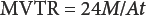
The SEM images and fiber diameter distribution histogram of treated PVA nanowebs are provided in Fig. 1. The average fiber diameter of treated nanowebs was 655 ± 256 nm. Treated nanowebs were observed to have fiber diameters in the range of 200 nm up to 1 μm. It was observed in our previous studies that native PVA nanowebs had an average fiber diameter of 245 ± 62 nm. 22 Hence, it was inferred that the addition of honey to the electrospinning dope resulted in an increase in the average fiber diameter of the nanowebs. This is in contrast to the observations of Maleki et al., 20 who proposed that the presence of honey increased the conductivity and charge densities of electrospinning dope, thereby resulting in a fiber diameter decrease. Nevertheless, the increase in fiber diameter could be attributed to an increase in viscosity of electrospinning dope 23 due to addition of honey. Honey is a highly-viscous solution of sugars, the addition of which increased the viscosity of the dope. Nanofiber morphology was also affected due to the increase in viscosity of the dope, which resulted in the formation of bead-like structures (Fig. 1). 24 A decreased percentage of honey in the dope resulted in smoother fibers without the bead-like structures. Many experimental runs were conducted to determine the optimal concentration of honey used in this study.
SEM images of (a) honey treated PVA nanowebs and (b) fiber diameter distribution histogram.
FTIR Analysis
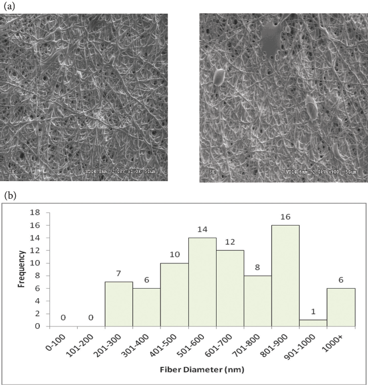
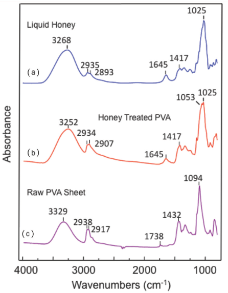
FTIR results comparing native and treated PVA nanowebs are presented in Fig. 2. The spectra indicate that treatment with honey incorporated carbohydrates into the PVA matrix. The spectrum of the pure honey dopant (Fig. 2a) had a strong peak near 1025 cm−1 that contained weaker features extending toward higher energy characteristics of the C-O stretching and bending vibrations common to honey and its simple sugars.25–27 The spectrum of the treated PVA sample (Fig. 2b) was similar to the spectrum of the pure honey dopant, but with additional components arising from the PVA matrix. The shoulder toward the high energy in Fig. 2b was in the region of the strong 1094 cm−1 peak associated with C-O vibrations in the PVA nanoweb (Fig. 2c).27–29
ATR FTIR spectra of (a) pure honey, (b) treated PVA nanowebs, (c) native PVA nanowebs, and (d) heat crosslinked native PVA nanowebs. The dashed line indicates the spectral region dominated by vibrations common to honey and its simple sugars.
Antimicrobial Activity
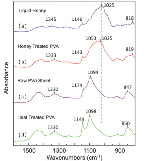
Figs. 3a and b present the results of antibacterial assays performed with PVA nanowebs.
Antimicrobial activity of treated PVA nanowebs. (a) activity of nanowebs on
Treatment with honey did not confer any antimicrobial activity to electrospun PVA nanowebs (Fig. 3a). No zones of inhibition were observed for any of the PVA nanowebs treated with honey (S1, S2, and S3). More importantly, nei-ther the positive control nor pure honey demonstrated any antimicrobial activity against
The dependence of honey antimicrobial activity on geo-graphical region and the type of plants the bees feed on 30 seems to be the main reason the pure honey used in this study failed to exhibit any inherent antimicrobial activity.
Breathability
Breathability of wound dressings is an important criterion that governs the loss of water and other exudates from the wound. Breathability of normal skin was observed to be 279 g/m2/day. 2,31 On the other hand, breathability of injured skin was evaluated to be in the range of 279-5138 g/m2/day, depending on the nature and extent of the skin injury.2,31Based on these observations, it was suggested that wound dressing breathability should be in the range of 2000 to 2500 g/m2/day to prevent excessive wound dehydration, as well as build-up of wound exudates, thereby supporting the process of wound healing.2,31 In the present study, the breathability of treated PVA nanowebs was 1894.1 g/m2/ day (n = 5, se = 10.33, where n is the number of replicates and se is the standard error). Our previous studies indicated that breathability of native PVA nanowebs electrospun under similar conditions was 1209.5 g/m2/day. 22
Treatment with honey resulted in a significant increase (p < 0.05) in the breathability of PVA nanowebs. The increase in the MVTR of treated nanowebs was attributed to several factors. The hygroscopicity of honey, which is the ability of a substance to absorb and hold moisture from its surrounding environment, was one factor. 32 FTIR studies suggested an increase in the breathability of the treated PVA nanowebs. The spectral features in Fig. 2b demonstrate that honey components became incorporated within the PVA matrix through the electrospinning process. These infer-ences corroborate with the observations of Arslan et al., 26 who treated polyethylene terephthalate (PET) with honey to investigate its potential for use as wound dressings. ATR-FTIR analysis of honey treated PET webs also revealed peaks characteristic of carbohydrates and other sugars. 26 The carbohydrates added hydrophilic functional groups to the polymer matrices, which likely enhanced the breathability of the PVA nanowebs.
Breathability of a substrate is known to be strongly influenced by the presence of surface functional groups 33 and an increase in the average fiber diameter of treated nanowebs. Examination of treated nanoweb morphology suggests that treatment with honey resulted in an increase in average fiber diameter. This often results in a low fiber packing density, which eventually increases the porosity of nanowebs. 34 A low fiber packing density and larger nanoweb pore size would provide one explanation for an increase in the MVTR of treated nanowebs.
The relative hydrophilic nature of treated nanowebs was also evident in the high energy region of the infrared spectra (Fig. 4). The 1645 cm−1 band in the spectra of honey and treated PVA nanoweb (Figs. 4a and b) was a signature of the H-O-H bending vibrational mode of water. 28 The band was absent in the spectrum of the native PVA nanoweb (Fig. 4c), indicating the native sample resisted water uptake. The 3329 cm−1 band in the spectrum of the native PVA nanoweb arose from the O-H stretching vibration of the alcohol group in the polymer.27–29 The band was shifted considerably to higher energy in the spectra of the honey and the treated PVA nanoweb. The shift reflected the contribution from the O-H stretching vibrations of water in the samples and the expected hydrogen bonding interactions 35 likely between water molecules and -OH groups in the sugars. As the breathability values of treated PVA naonwebs (1894.1 g/m2/ day) were observed to be very close to the breathability of ideal wound dressings (2000–2500 g/m2/day), they may play a crucial role in accelerating the wound healing process.
ATR FTIR spectra of (a) pure honey, (b) treated PVA nanowebs, and (c) native PVA nanowebs.
Tensile Properties
Wound dressings must possess certain mechanical properties such as tensile strength, flexibility, bending, and elastic properties. 2 Important tensile properties of treated PVA nanowebs are depicted in Table I.
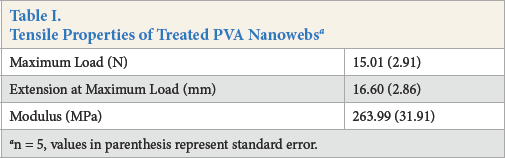
Results from FTIR analysis of native and treated PVA nanowebs showed a peak at 1143 cm−1 in the spectrum of the latter (Fig. 2b). Although the feature coincided with a weak band in the pure honey sample, its intensity and sharpness were characteristic of increasing PVA crystallinity.22,29 Such a peak was also observed in an earlier study that dealt with heat crosslinking of electrospun PVA nanowebs. 22 A representative spectrum is shown in Fig. 2d. Heat cross-linking resulted in an increase in PVA polymer crystallinity and PVA nanoweb strength. 22 The onset of sharpness in the 1143 cm−1 peak following honey treatment may result from ordering within the PVA matrix facilitated by interactions between carbohydrates and segments of the PVA polymer. Further characterization is needed to trace the origin of this effect. Nevertheless, it can be inferred that treatment with honey may result in an increase in tensile properties of PVA nanowebs.
Conclusions
We demonstrated the preparation and characterization of electrospun PVA nanowebs treated with honey. The incor-poration of honey components into the PVA nanowebs was confirmed by FTIR spectroscopy studies. Neither pure honey, nor the honey-treated PVA nanowebs, exhibited any inhibitory effects on tested microorganisms. Treatment with honey was found to increase the breathability of PVA nanowebs. Also, treatment with honey added additional hydrophilic functional groups into the PVA matrix, resulting in enhanced moisture vapor transmission characteristics. Honey-treated PVA nanowebs displayed enhanced mechanical properties. Treatment of PVA nanowebs with honey from other sources may confer antimicrobial activity to these nanowebs, as the antimicrobial properties of honey depend on many different factors. A prudent choice of honey that can impart antibacterial activities to PVA nanowebs is of critical importance. The antimicrobial and anti-inflammatory properties of honey could be effectively exploited by incorporating them into nanofiber webs to facilitate their potential use in biomedical applications such as wound dressings.
Footnotes
All the authors contributed to the work. UT and SR conceived the idea behind the project and UT carried out the experimental work. The authors declare no competing financial interest.
Acknowledgements
Seshadri Ramkumar gratefully acknowledges the financial support from The