
Abstract
Stress-inducible Hsp70i and constitutively expressed Hsc70 are highly related heat shock proteins. Aberrant expression levels and intracellular localization of these proteins has been suggested as a potential marker in certain tumors. The aim of our study was to work out a reliable, immunohistochemical detection of the stress-inducible Hsp70i protein and enabling discrimination between Hsp70i and Hsc70 proteins in paraffin-embedded human tissues. We tested the effect of several fixative procedures and antigen retrieval on the effectiveness of the Hsp70i detection in murine cells cultured in vitro and in liver of rats subjected to heat shock. For cells grown in vitro, specific Hsp70i immunoreactivity was obtained with all fixatives used. However, samples fixed in 10% formalin and 4% paraformaldehyde required antigen retrieval. In liver tissue embedded in paraffin, regardless the fixative used, positive Hsp70i staining could be visible only if antigen retrieval was applied. We applied this procedure for detection of Hsp70i in routine sections of breast and lung cancers fixed with 10% formalin and found that the application of thermal antigen retrieval significantly enhanced the SPA810 immunoreactivity and reduced background staining. This procedure enabled also the differential detection of Hsp70i and Hsc70 in routine histopathological preparations.
T
Since the studies of Brown et al. (1993) on the properties of the inducible Hsp70 and constitutive Hsc70, these proteins have been essentially considered to be equivalent, functionally similar chaperones. As a consequence, in a number of studies to detect the HSP70 proteins, the antibodies which recognize both Hsc70 and Hsp70i proteins (e.g., Sigma BRM-22, DAKO anti-HSP70) have been used. However, recent studies showed significant differences between both forms. A search for a relationship between the chaperone function and the oligomerization state of Hsp70s revealed that Hsp70i is not aggregated at physiological temperature, but aggregates after heat shock of cells, whereas Hsc70 already exists in an aggregated form at 37C (Angelidis et al. 1999). It has been also suggested that Hsc70 and Hsp70i can be expressed in a regional and cell type–specific manner at physiological temperature in vivo (Dean et al. 1999). Especially important seems to be the observation that stress-inducible Hsp70i, but not the constitutive Hsc70, cosegregates with immunogenicity of tumor cells in vivo and that the association with peptides is more pronounced for Hsp70i than for Hsc70 in various conditions (Menoret et al. 2002). According to these authors, the inducible Hsp70i can be more efficient in discriminating the intracellular “non-self polypeptides” than can the constitutive Hsc70.
The subtle but possibly significant structural and functional differences between Hsc70 and Hsp70i proteins raise the problem of a reliable, differential immunohistochemical method of detection of these stress proteins in paraffin-embedded human tissues used for immunohistopathological analysis. This is especially important for the detection of the Hsp70i protein, which has been reported to be highly and constitutively expressed in various tumors. The relation of the expression level of this protein to prognosis and efficiency of cancer immunotherapy has been a subject of intense study.
Some commercial antibodies specifically recognize Hsc70 and Hsp70i proteins, but it has been recently found that the fixative and the postfixation procedure can significantly influence immunohistochemical detection. For the SPA810 antibody (StressGen Biotechnologies; Victoria, BC, Canada), which specifically recognizes the inducible Hsp70i protein, Tytell et al. (1998) obtained significantly better results for rat brains and postmortem human brains if tissues were fixed in methacarn instead of formalin. Conversely, Trieb et al. (2001), using the same antibody to study Hsp70i expression in transplanted human kidney, reported that Carnoy's fixative (nonaldehyde, ethanol-based fixative) gave negative results, whereas formalin with thermal antigen retrieval yielded positive staining.
The aim of our study was to determine the conditions of immunohistochemical detection of Hsp70i with use of SPA810 antibody depending on different immunostaining methods. We believe that this study will be helpful to establish a standard immunohistochemical procedure enabling detection of Hsp70i in histological/paraffin embedded sections for routine immunohistopathological evaluation.
Materials and Methods
Cell Cultures
Mouse melanoma cells B16 were grown in DMEM. Subconfluent cell cultures were heated at 42.5C for 3 hr. Control and heated cells were fixed by immersion in the following fixatives: (a) methanol for 10 min at −20C and 30 sec postfixation in cold acetone, (b) methacarn for 10 min at 4C, (c) 1% formaldehyde in PBS for 30 min at 4C, (d) 10% formaldehyde in PBS for 30 min at 4C, (e) 4% paraformaldehyde in PBS for 30 min at 4C.
After aldehyde fixation, the slides were rinsed in PBS; after fixing in methacarn, they were rehydrated in decreasing methanol concentrations. For all fixatives, deionized H2O was used for final washing. The slides were then air dried and stored at −20C.
Animals
Male Wistar rats 2.5–3 months old weighing 250–300 g were used. They were bred in the onsite Animal Facility of the Centre of Oncology in Gliwice. Rats were housed in a temperature-controlled environment (20–22C) with a 12-hr photoperiod and provided with a laboratory fodder and water ad libitum. All animal procedures conformed to institutional regulations and European regulations concerning the protection of animals.
Rats received an intraperitoneal injection of Vetbutal (Biowet Pulawy Ltd; Pulawy, Poland) for narcosis, and were vertically immersed in a water bath to the level of the xiphoid process. Experimental rats were subjected to heat shock for 1 hr either at 42C with 3 hr of recovery or at 41C with 90 min of recovery. Control animals were kept in Vetbutal narcosis for corresponding periods. Thioacetamide (TAA; Sigma, St Louis, MO) was administered intraperitoneally in a single dose of 50 mg/kg for 48 hr. Liver specimens, not larger than 5 mm each dimension, were fixed by immersion at 4C without mixing. Specimens were fixed for 24 hr in formaldehyde and paraformaldehyde and then washed in PBS (overnight, with at least three changes). Specimens were fixed in methacarn for 20 hr, and subsequently rehydrated in decreasing methanol concentrations (two changes 30 min each in 100%, 80%, 50%, and 30% methanol and deionized H2O for final washing).
Human Carcinomas
Human breast (5 cases) and non–small-cell lung carcinoma specimens (10 cases) were either fixed in 10% formalin in PBS or in methacarn, as described for animal material. Archival specimens of breast carcinoma from Department of Pathology, Institute of Oncology, were fixed in 10% formalin, but the fixation time and formalin pH were not standardized.
Immunohistochemistry
Immunohistochemical staining was performed on cell cultures on slides, and 7-μm thick paraffin sections. Slides (both cell cultures and sections) either proceeded without or were submitted to antigen retrieval. Antigen retrieval was performed by two, 5-min cycles in boiling 0.01 M citrate buffer in a microwave oven or 0,02% pronase E in PBS digestion for 4 min at 37C (according to Gillet et al. 1994).
The endogenous peroxidase activity was blocked with 1% H2O2 in PBS. For detection of inducible HP70i, the slides were incubated overnight with monoclonal antibody SPA810, clone C92 (StressGen Biotechnologies), at a final concentration of 10 μg/ml. Biotinylated secondary antibody, an avidinbiotin complex (Vector Laboratories; Burlingame, CA) and 3,3′-diaminobenzidine (Sigma Biochemicals; St Louis, MO) as a chromogen were applied for visualization of the immunoreaction. Omission of the primary antibody was considered as a negative control. Histopathological alterations were assessed on hematoxylin and eosin stained sections.
Results
We tested several of the most commonly used fixatives based on methanol (methanol/acetone, methacarn) or aldehyde (formalin, paraformaldehyde). For detection of the heat-inducible Hsp70i protein, we selected the SPA810 antibody, which in our hands caused less background problems than other commercially available anti-Hsp70i antibodies. The specificity of the antibody has been confirmed by a routine Western blotting as demonstrated previously (Zborek et al. 2002). In B16 cells submitted to hyperthermia, a positive Hsp70i immunoreaction was visible in cytoplasm, nuclei, and nucleoli of the majority of cells. As shown in Table 1, reproducible results were obtained with all fixatives used, but 10% formalin and 4% paraformaldehyde required thermal antigen retrieval in citric buffer. Specimens fixed in 1% formalin did not require antigen retrieval to reveal Hsp70i protein.
HSP70i localization in B16 cells after heat shock (+ denotes arbitrary assessed intensity of immunostaining)
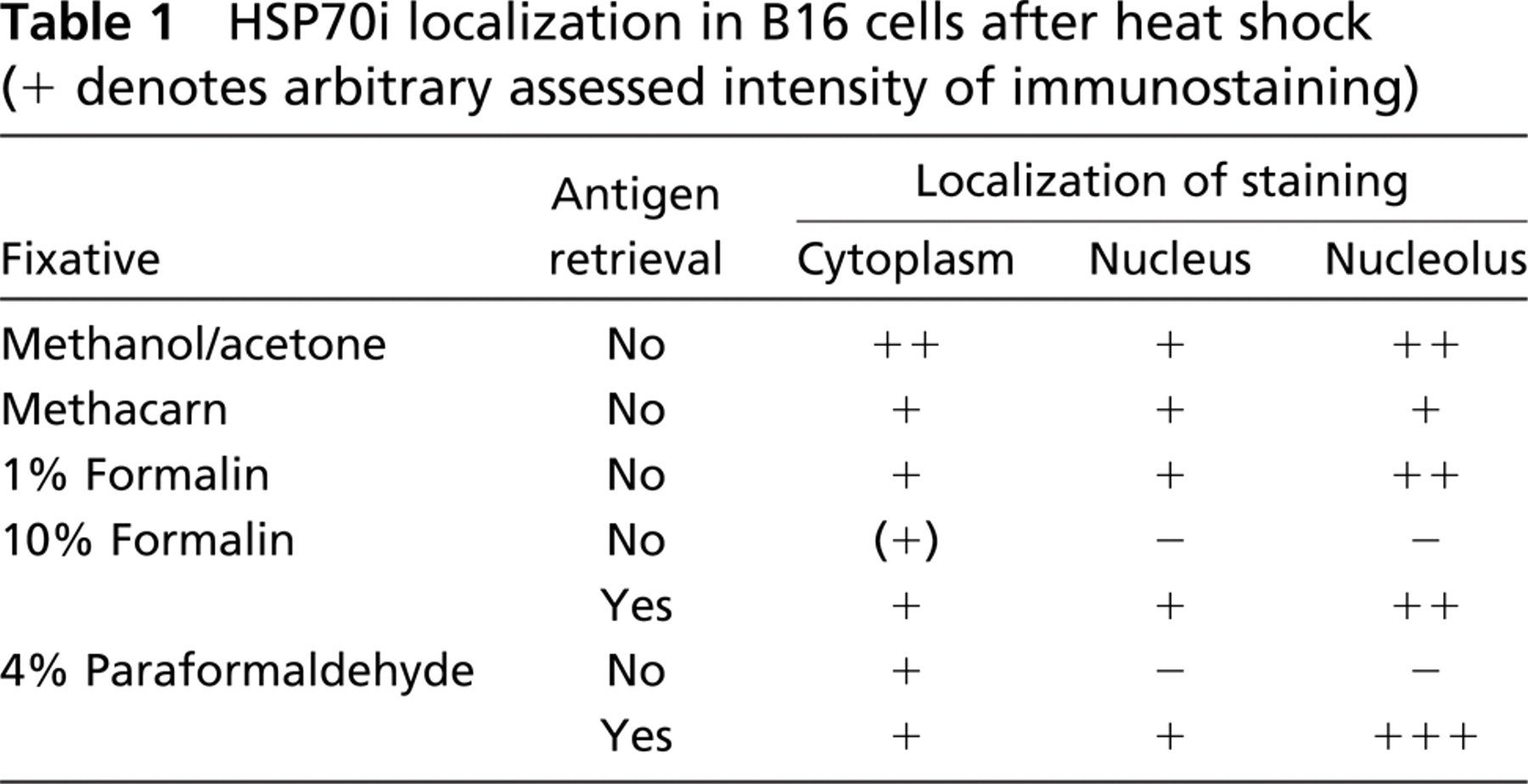
The immunostaining pattern was analogous to the one established by Pelham (1984) for heat shocked cells grown in culture. Thus cytoplasm and nucleoplasms were only weakly stained, whereas a majority of positive Hsp70i immunoreaction was highly concentrated in nucleoli (Figure 1). In control cells, only a weak cytoplasmic background staining could be observed (Figure 1).
Next we examined Hsp70i immunostaining pattern on paraffin-embedded rat liver sections with the same set of fixatives except the methanol/acetone one. As a control tissue, we used the liver of untreated animals or animals injected with a single dose of the hepatotoxicant, TAA. We chose the latter as a control because we had previously shown by Western blot analysis (Zborek et al. 2002) that treatment of rats with TAA did not induce Hsp70i expression. Rats were subjected to hyperthermia in the conditions in which a Western blot analysis (Zborek et al. 2002) confirmed strong expression of the Hsp70i protein. Each immunohistochemical analysis was performed with or without antigen retrieval (Table 2).
Expression of HSP70i in the rat liver after hyperthermia (+ denotes positive immunoreaction, — denotes no staining; +/− in samples fixed with 1% formalin denotes some positivity observed in sinusoids and in cytoplasm of single hepatocytes; see text)
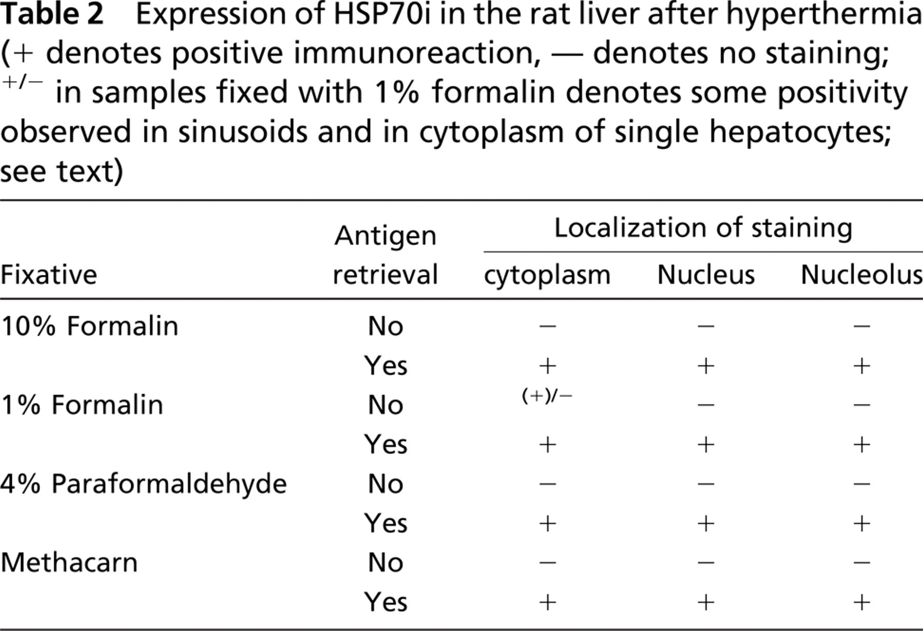
In livers of non–heat-shocked rats and animals treated with TAA, no positive staining for Hsp70i could be observed, and application of antigen retrieval did not affect the result (Figure 2). In livers of heat-shocked rats, a positive immunostaining for Hsp70i was obtained only after antigen retrieval. The distribution of the Hsp70i-positive hepatocytes was basically independent of the fixative used. They could be seen mainly in the regions of central veins (Figure 2). It has to be noted that in samples fixed with 1% formalin some staining could be observed in sinusoids and in cytoplasm of single hepatocytes dispersed in liver lobuli (not shown). The data presented above seem to indicate that for successful detection of the Hsp70i protein in paraffin-embedded tissue specimens, the mode of fixation may not be decisive, whereas the treatment of samples by antigen retrieval seems essential.
Here we should mention that two of five batches of SPA810 antibody gave false-positive nuclear and cytoplasmic immunostaining in livers of TAA-treated rats when microwave retrieval was omitted. The pattern of staining resembled the one obtained with the antibody against both forms of Hsp70 (described in Zborek et al. 2002). Including antigen retrieval into the IHC procedure resulted in disappearance of this nonspecific staining.
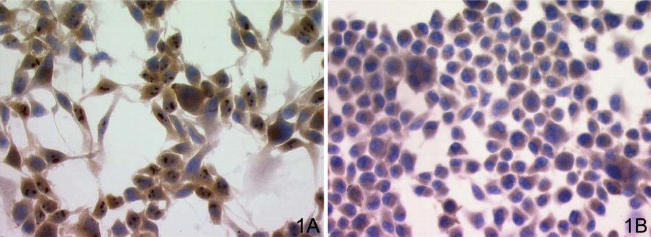
Immunoreaction for Hsp70i (SPA810) in B16 cultured cells. (
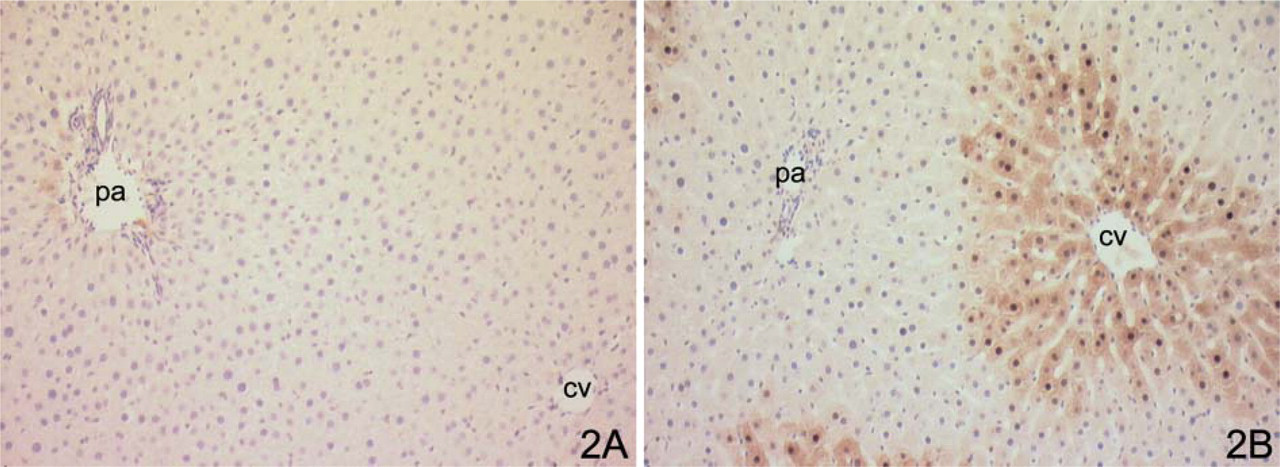
Immunoreaction for Hsp70i (SPA810) in rat liver. (
Finally, we analyzed the Hsp70i expression in paraffin-embedded specimens of human non—small-cell lung and breast carcinomas. Samples tested were fixed by us either in 10% formalin in PBS or methacarn, or alternatively, provided by the pathology department where they were routinely fixed in 10% formalin (fixation time and formalin pH were not standardized; archival sections). We have chosen these two types of tumors to illustrate the possible complexity of the Hsp70i detection in tissue/tumor samples.
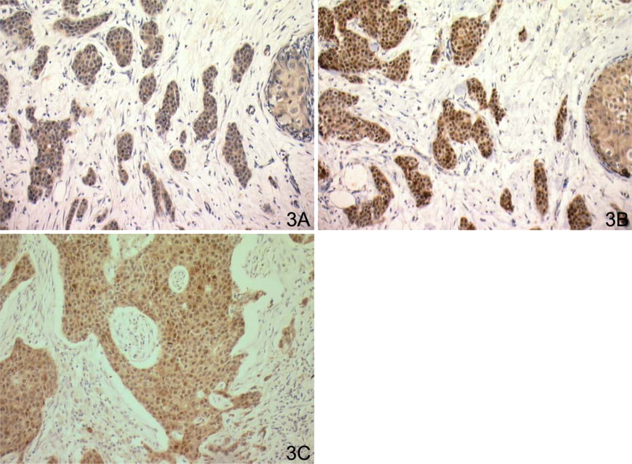
Immunoreaction for Hsp70i (SPA810) in breast cancer. (
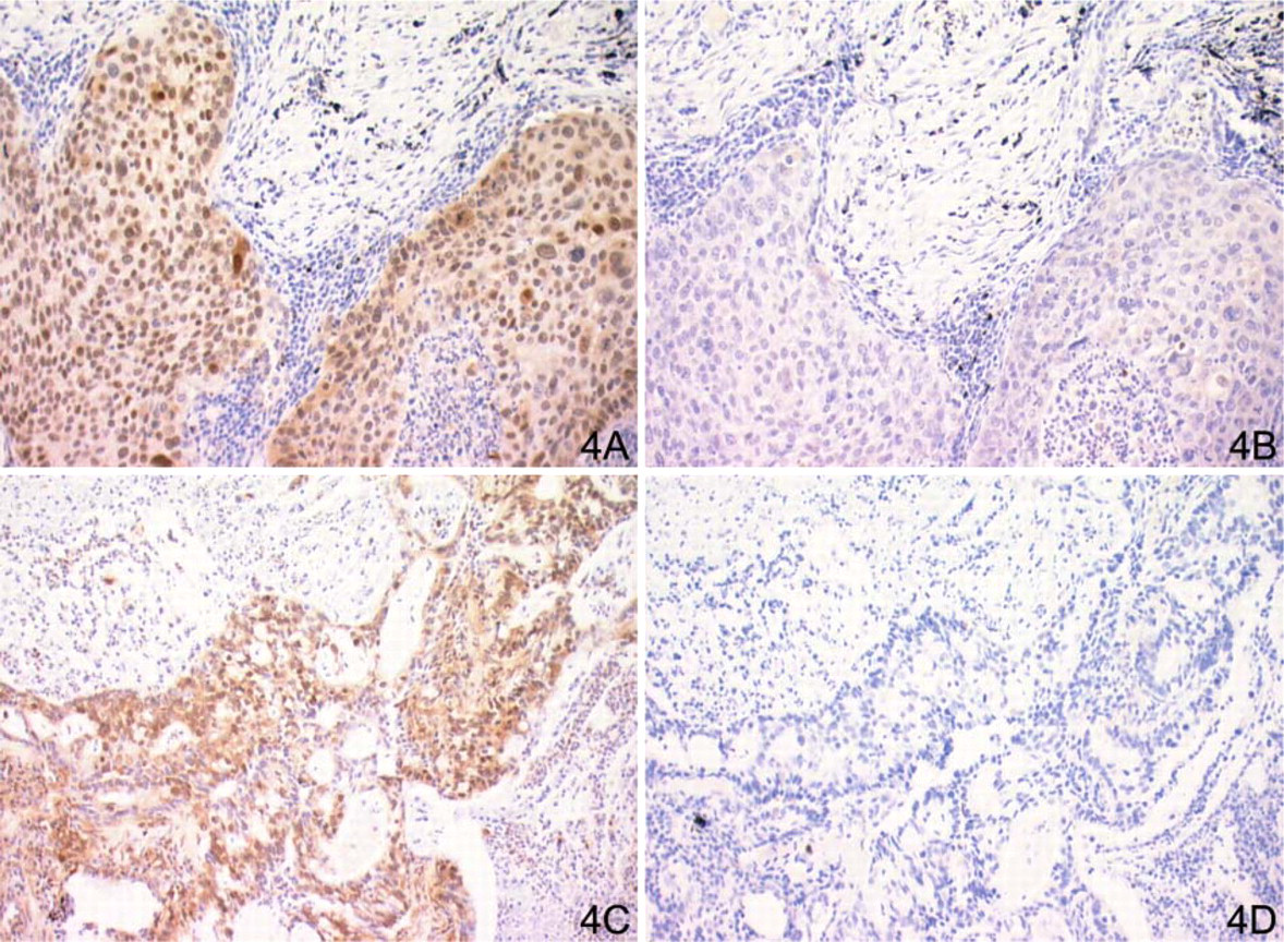
We found that the use of methacarn fixation was ineffective for Hsp70i detection. SPA810 staining was negative in all cases despite the thermal or enzymatic antigen retrieval. In formalin-fixed material, the presence of Hsp70i can be convincingly demonstrated only if antigen retrieval is applied (Figure 3 and Figure 4A and 4B). If this step was omitted, no immunostaining in the tumor area could be observed. To determine selectivity of the detection of the Hsp70i in antigen retrieval, we selected the non–small-cell lung cancer specimens in which Hsc70 was overexpressed. Then we performed immunohistochemical reaction with anti-Hsp70i antibody (SPA810). It can be seen from the Figures 4C and 4D that in the tumor area the anti-Hsc70 antibody gave strong immunostaining, whereas the reaction with anti-Hsp70i antibody was negative.
In archival sections of breast carcinomas, processed without antigen retrieval, a faint Hsp70i nuclear and cytoplasmic staining was frequently visible (Figure 3). When sections were subjected to microwave retrieval, a significant enhancement of nuclear immunoreaction (Figure 3) was observed. A comparison of immunoreaction performed on specimens fixed in 10% formalin in PBS or in 10% unbuffered formalin (Figure 3) demonstrated that SPA810 antibody staining was not dependent on the formalin-fixation procedure (standardized vs not standardized formalin fixation) but on the type of fixative used (formalin vs methacarn).
Discussion
Multiple data from the in vitro and animal studies indicate that the stress inducible Hsp70i protein can influence the phenotype of cancer cells (reviewed in Jolly and Morimoto 2000; Ciocca and Calderwood 2005). Cancer cells, which overexpress this protein, are more resistant to apoptosis and tolerate higher doses of anticancer drugs (reviewed in Garrido et al. 2003; Sreedhar and Csermely 2004). Also, Hsp70i seems to facilitate proliferation of cells with damaged DNA or mitotic spindle, thus being a possible selecting factor contributing to evolving cancer phenotypes (Karlseder et al. 1996; Wang et al. 2004). On the other hand, overexpression of Hsp70i in primary cancer can indicate the more efficient application of HSP-associated autologous anticancer vaccines (reviewed in Manjili et al. 2002; Hoos et al. 2004). These features of the Hsp70i protein constitute a rationale for search for a relationship between the expression level of this stress protein and other molecular markers, clinical parameters, and patient's survival, because a new prognostic relationship may be uncovered.
Immunoreaction for Hsp70i (SPA810) in case of squamous cell carcinoma. (
Comparison of Hsp70 expression, fixation types, and antibody description in various types of cancer
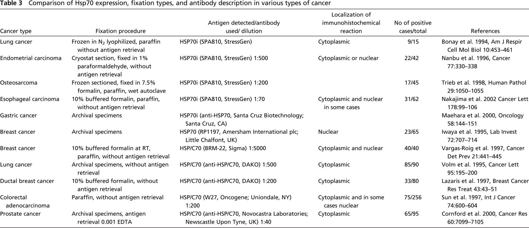
Unambiguous detection of the Hsp70i protein by immunohistochemistry is not easy because of the possible interference from structurally and antigenically related, constitutively synthesized cognate stress protein, Hsc70 (see Introduction), and the possibility of variable, sample-dependent accessibility of Hsp70i epitopes. A survey of the published results shows that various methods of sample processing and various antibodies had been used to study expression pattern of the Hsp70i and Hsc70 in primary cancers. Selected examples illustrating diversity of the methodological approaches are shown in the Table 3.
It can be assumed that difficulties in Hsp70i recognition, together with differences in tissue processing and immunohistochemistry protocols, influences the results of Hsp70i detection. In our hands, when using the SPA810 antibody after paraffin embedding, the thermal antigen retrieval step was critical for detection/visualization of Hsp70i.
Our results are consistent with data obtained by Trieb et al. (2001). In the transplanted human kidney, these authors demonstrated that formalin fixation is superior to that with Carnoy's (ethanol-based fixative) and showed the importance of antigen unmasking step in effective Hsp70i detection. Antigen retrieval with wet autoclaving performed in 0.5M citrate buffer was more effective than microwave treatment. The authors ascribe the failure in detection of Hsp70i protein with Carnoy's fixative to low pH (although they did not test effect of pH separately). The antigen retrieval technique was also proven necessary for the Hsp70i detection, with SPA810 antibody, in ischemic gerbil brain fixed with 4% paraformaldehyde (Nisino and Nowak 2004).
Tytell et al. (1999) compared influence of fixation method (10% formalin vs methacarn) on Hsp70i with SPA810 antibody in human postmortem brain. The authors showed that fixation with 10% formalin in contrast to diluted formalin (1%) or noncrosslinking fixative (methacarn) masked antigen feasibility and affected the immunohistochemical reaction. However, they did not perform antigen retrieval.
Because formalin is used as a fixative of choice in pathological laboratories, we think that our standardization of immunostaining procedure for Hsp70i detection in paraffin-embedded material is important and can help in routine histopathological practice. It should be mentioned that our studies are more systematic than any other reported so far on the differential immunodetection of the Hsp70i and Hsc70i proteins.
Footnotes
Acknowledgements
This work was supported by the grant awarded to Prof. Zdzislaw Krawczyk by the Foundation for Polish Science.
We thank Ms. A. Kyrcz and Ms I. Peick for their excellent technical assistance.